MOJ
eISSN: 2379-6162


Case Report Volume 13 Issue 2
1Head and Neck Surgery Service, Instituto de Cancerología – Clínica Las Américas AUNA, Medellín, Colombia
2Medical Doctor, Universidad CES, Medellín, Colombia
3Head and Neck Surgeon, Fundación Universitaria de Ciencias de la Salud (FUCS), Bogotá, Colombia
4Medical Intern, Fundación Universitaria San Martín, Sabaneta, Colombia
5General Surgeon, Universidad de Caldas, Manizales, Colombia
Correspondence: Julián Jaramillo Martinez, Instituto de Cancerología - Clínica Las Américas Auna, Medellín, Colombia
Received: April 17, 2025 | Published: May 1, 2025
Citation: Martinez JJ, Rendón LMG, Herrera CG, et al. Cervicomediastinal abscess due to occult esophageal perforation with spontaneous sealing: a case report of an unusual presentation of boerhaave syndrome. MOJ Surg. 2025;13(1):35-38. DOI: 10.15406/mojs.2025.13.00290
Boerhaave syndrome is a rare but potentially fatal condition resulting from spontaneous esophageal perforation, typically associated with a characteristic clinical triad of vomiting, chest pain, and subcutaneous emphysema. However, in some cases, symptoms may be nonspecific, and initial diagnostic studies inconclusive, which can lead to delayed recognition and treatment. We report a case of a healthy young adult who developed cervical pain, odynophagia, and systemic symptoms after multiple episodes of vomiting. Despite normal findings on endoscopy, further imaging studies were conducted due to ongoing clinical suspicion, which revealed a cervicomediastinal abscess that required surgical drainage. This case highlights the importance of maintaining a high index of suspicion for Boerhaave syndrome and the need for advanced imaging in patients with suggestive symptoms, even when initial evaluations appear normal. It is important to emphasize that some cases of rupture may resolve spontaneously, which can complicate the diagnostic process. Timely diagnosis and intervention are key to improving outcomes in this condition.
Keywords: transmural esophageal rupture, mediastinitis, abscess, complication, spontaneous sealing, boerhaave syndrome
Boerhaave syndrome, first described by Hermann Boerhaave in 1724, refers to a spontaneous transmural perforation of the esophagus most commonly occurring after forceful emesis. Although rare, it is associated with high morbidity and mortality, especially when diagnosis and treatment are delayed. The left posterolateral wall of the distal third of the esophagus is the most frequent site of rupture. The classical presentation, known as Mackler’s triad—vomiting, chest pain, and subcutaneous emphysema—is only present in a minority of cases, making clinical suspicion essential. In some instances, the perforation may seal spontaneously, further complicating diagnosis. This case report presents a patient with an unusual clinical evolution: initial negative endoscopic findings, followed by the development of a cervicomediastinal abscess, in the context of suspected esophageal rupture with spontaneous sealing.
A 26-year-old male with no relevant medical history, except for psychoactive substance use and THC addiction, presented with a six-day history of progressive emetic episodes. Symptoms initially began with recurrent vomiting, increasing in both frequency and intensity. On the second day, the patient reported a sensation of air tracking into the neck following an intense vomiting episode. Over subsequent days, he experienced general deterioration, weight loss, and remained afebrile.
Upon arrival to the emergency department, he was clinically stable. Initial assessment focused on an emetic and constitutional syndrome. The initial lab workup revealed elevated acute-phase reactants, without any other significant abnormalities. Upper endoscopy showed no neoplasms or mucosal lesions, and a water-soluble contrast esophagogram demonstrated no signs of rupture.
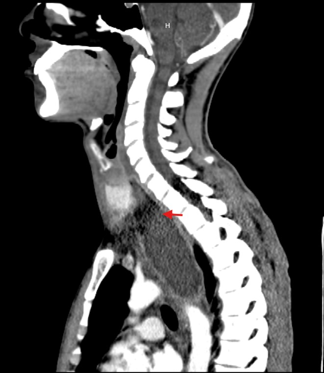
Due to the unclear etiology of the symptoms, combined with unremarkable initial studies and the subsequent development of dyspnea, a CT scan of the neck and thorax was performed. This revealed a large collection centered in the mediastinum, dissecting tissue planes and extending below the carina, reaching into the neck up to the left thyroid lobe. Gas was observed both within the collection and around the thyroid gland and mediastinum, with the esophagus displaced to the right by the mass effect of the abscess (Figure 1-5).

Figure 1 CT: Sagittal view showing an abscess extending from the left thyroid space to the superior mediastinum.
In the setting of a cervicomediastinal collection without a clear etiology, the patient was evaluated by the head and neck surgery team. Given the clinical history, the course of the illness, and the extent of the abscess involving both the thoracic and cervical regions, the team considered a diagnosis of complicated Boerhaave syndrome with abscess formation. Since neither upper endoscopy nor contrast studies of the upper gastrointestinal tract showed any wall defects, spontaneous closure of the lesion was suspected. Due to the persistence of the mediastinal abscess and the patient’s clinical condition, with no possibility of endoscopic resolution, open surgical drainage was indicated.
During surgery, a large left paratracheal abscess was identified, extending from the thyroid space to the superior mediastinum, with no defect in the gastric tube. Approximately 250 cc of hematopurulent material was drained from the abscess. Cultures were taken, followed by extensive saline irrigation and debridement of the affected tissue. The wound was closed in layers, leaving a drainage tube in the left cervicomediastinal space and a nasogastric tube for enteral nutrition for one week. The patient was discharged two days postoperatively with empirical antibiotic therapy, nil per os, and enteral nutrition via nasogastric tube.
At the follow-up appointment, after completing the antibiotic regimen, the nasogastric tube was removed, and the cervical drain, which was inactive, was also removed. Culture results confirmed the presence of oral cavity and gastrointestinal tract organisms in the cervical collection; Schaalia odontolytica and Streptococcus constellatus. Complete resolution of the pathology was considered.
Boerhaave syndrome was first described in 1724 by the German physician Herman Boerhaave. It is a transmural and longitudinal rupture of the esophagus, usually located in its lower third. It is considered a spontaneous rupture, as it occurs in the absence of trauma or iatrogenic interventions. Although rare, it is a clinically serious condition, and its true incidence remains unknown due to underdiagnosis.1,2
This condition usually occurs after intense episodes of vomiting or retching, resulting from a sudden and significant increase in intraluminal esophageal pressure, combined with negative intrathoracic pressure. This mechanism is further intensified by the contraction of the abdominal muscles during vomiting, and it most commonly occurs in an anatomically vulnerable area: the lower third of the esophagus, where the muscle fibers are thinner, especially on the left side, 2–4 cm from the gastroesophageal junction.3
A sudden rise in pressure can cause a full rupture of the esophageal wall, enabling gastrointestinal contents to enter the mediastinum and pleural cavity.1 The communication between the various spaces of the neck and mediastinum allows microorganisms from the esophagus and oral cavity to pass into adjacent tissues, causing infections at this level with dissemination to the thorax. As a result, one of the most severe and inherent complications of this syndrome is mediastinitis. This condition can rapidly progress to septic shock and death if not managed promptly.2,4 This carries a high risk of severe infectious complications such as mediastinitis, empyema, mediastinal abscesses, and septic shock, all of which are associated with high mortality. In the absence of treatment, mortality can reach 100%, while in treated cases, it ranges between 30% and 50%.5,6
The clinical presentation of Boerhaave syndrome is often nonspecific. Classically, Mackler’s triad—vomiting, chest pain, and subcutaneous emphysema—has been described, but it is present in only 15% of cases. Other important symptoms to consider include dyspnea, dysphagia, abdominal or chest pain, hemodynamic instability, and Hamman’s sign (precordial crepitus synchronized with the heartbeat). In some cases, the lesions may be superficial and limited to the mucosa, as in Mallory-Weiss syndrome; however, Boerhaave syndrome involves all layers of the esophageal wall, which leads to a much more severe outcome.7–9
An early diagnosis is essential but remains a clinical challenge. In up to 50% of cases, diagnosis is delayed due to the nonspecific nature of the symptoms. Therefore, a thorough history, appropriate clinical suspicion, and proper use of imaging studies are required. The main diagnostic tools include upper digestive tract endoscopy, chest X-ray, contrast-enhanced computed tomography, water-soluble contrast esophagogram. These allow the identification of findings such as pneumomediastinum, left pleural effusion, or air in the mediastinal soft tissues.10
Although these tests have a sensitivity of 90% to 100%, they are not always conclusive. Contrast-enhanced studies may have a false negative rate of up to 10%, especially in subacute phases or when the perforation is contained or has closed spontaneously. Therefore, the absence of findings does not exclude the diagnosis. Endoscopy is a useful tool in the diagnosis of Boerhaave syndrome, especially when it is performed early.11,12 However, its use is controversial, as it does not always detect the lesion and may worsen the situation in unstable patients. In the cohort described by Schipper et al., endoscopic lesions were found in 90% of cases in the distal portion of the esophagus, on the left side, with an average size of 2.2 cm.5,13
The treatment of Boerhaave syndrome depends on the severity of the clinical presentation and the time since the perforation occurred. Surgical intervention within the first 24 hours is the most recommended strategy, as it significantly improves survival. Surgical options include primary closure of the lesion with mediastinal drainage, esophageal resection with reconstruction in severe cases, while conservative treatments are preferred in stable patients with small perforation.5
Conservative treatment includes broad-spectrum antibiotic therapy (covering both aerobic and anaerobic bacteria), proton pump inhibitors, oral intake restriction with enteral nutritional support if tolerated, and close observation. This approach is reserved for patients without signs of advanced mediastinitis or sepsis, and in whom the perforation is minimal and contained.9,14,15
Considering that mortality associated with Boerhaave syndrome remains high even with early diagnosis, it is essential for healthcare professionals to keep it in mind as a potential diagnosis in any patient presenting with acute chest pain—particularly when preceded by episodes of forceful vomiting. Early identification, combined with an effective multidisciplinary approach, can make a substantial difference in patient outcomes.5,16
Finally, although the diagnosis has traditionally relied on direct visualization of the perforation through imaging or endoscopy, evidence suggests that its absence does not rule out the disease. The case we present exemplifies this, as the patient exhibited symptoms compatible with the condition and radiological findings suggestive of mediastinal collection with gas and cervical extension, without any evident perforation found on initial studies. This suggests a possible contained perforation or spontaneous closure, highlighting the need to consider Boerhaave syndrome even in the presence of negative studies.
We present the case of a 26-year-old previously healthy male who presented with chest pain, dyspnea, and progressive deterioration of his general condition, in the context of recurrent vomiting over several days. Despite endoscopic evaluations and a water-soluble contrast esophagogram showing no evidence of esophageal perforation, imaging revealed a cervicomediastinal collection with the presence of gas. The clinical presentation, sequence of events, and radiologic findings pointed toward a diagnosis of Boerhaave syndrome complicated by a cervicomediastinal abscess, likely with spontaneous sealing of the esophageal perforation. This case underscores the importance of considering this pathology even in the presence of negative studies, relying on clinical impression and a multidisciplinary approach.
None.
The authors declare that there are no conflicts of interest.

©2025 Martinez, et al. This is an open access article distributed under the terms of the, which permits unrestricted use, distribution, and build upon your work non-commercially.