MOJ
eISSN: 2379-6162


Research Article Volume 5 Issue 3
1Dept of Radiodiagnosis, Gajara Raja Medical College, India
2Assistant Professor, School of Pharmacy, ITM University, India
Correspondence: Som Biswas, Dept of Radiodiagnosis, Gajara Raja Medical College, Indian
Received: November 20, 2017 | Published: December 29, 2017
Citation: Biswas S, Biswas S. A study on penile doppler. MOJ Surg. 2017;5(3):196-201. DOI: 10.15406/mojs.2017.05.00110
Erectile dysfunction (ED) or impotence is defined as inability to achieve and/or maintain an erection for satisfactory sexual intercourse. Its prevalence increases with age and affects upto 52% men between age 40-69 years. Although erectile dysfunction(ED) does not affect life expectancy and it is not a life-threatning disorder but is associated with significant morbidity. Sex is important to men and it is important to preserve erection, orgasm and sexual desire. In a society in which sexuality is widely promoted, ED impacts on feeling of self worth and self confidence and may impair the quality of life of affected men and their partners. ED is strongly associated with unsatisfying personal experiences and relationships, highlights the association of ED with emotional and physical satisfaction with sexual partners and with feelings of general happiness.1 Improved erectile function is accomplished by improved quality of life and decrease in general psychiatric symptoms.2
Keywords: dysfunction, decrease, symptoms, doppler sonography, vasculogenic
Study design: Cross sectional study.
Study period: 12months; October 2015-september 2016.
Study group: All those patients which were referred for penile color Doppler study ,in department of Radio diagnosis G.R Medical College Gwalior Madhya Pradesh.
Sample size: 52patients.
Work place: Department of Radiodiagnosis G.R.Medical College and Jayarogya Hospital,Gwalior using ALOKA PROSOUND ALPHA (Aloka Trivilion Pvt.Ltd.Tokyo Japan) USG machine with high(7.5MHz) frequency linear probe.
Inclusion criteria:
Exclusion criteria
Technique: The penile colour duplex examination is typically performed for either;
The examination
Pre Injection: 2.1.1. The examination should be performed in a quiet relaxed environment free of interruption71. The examination should be explained in detail and verbal and written consent obtained. The low risk of priapism should be clearly stated.7 A high-frequency linear transducer is used to perform a B-mode/colour Doppler examination of the penis. There are variable techniques to scanning this region however using a gown or towel to fix the penis with the dorsal aspect accessible can be very useful. The corpus cavernosa, corpus spongiosum and glans are evaluated. The dividing penile septum is a valuable echogenic landmark. Prior to injection, the cavernosal arteries will be seen as fine echogenic parallel lines. Variations in vascular anatomy are common and rarely of clinical significance.
The injection:
Post injection:
E0: No response
E1: Elongation of shaft only
E2: Moderate tumescence, no rigidity
E3: Full tumescence, no rigidity, easily bendable
E4: Full erection, partial rigidity
E5: Full rigidity for at least 20 minutes
These are further divided into four groups:
Penile color doppler
Penile Doppler is a non-invasive, radiation-free and relatively cheap means of screening for penile arterial insufficiency in patients with ED. Patients initially presenting with ED are typically offered pharmacological therapy on the basis of a clinical assessment involving history and physical examination, and basic laboratory tests. Radiological vascular testing is of most use in the evaluation of patients failing to respond to first-line therapy, and in the context of research. Other specific clinical problems are well suited to evaluation with penile Doppler. For example, ‘high flow’ priapism, due to traumatic cavernosal artery tears, gives characteristic appearances of turbulent flow in the arteriovenous fistula (often with a small pseudoaneurysm), high velocity proximal to the arteriovenous fistula and low flow distally (and in the contralateral artery). Color Doppler sonography can be useful in the evaluation of erectile dysfunction, which can result from psychogenic, endocrinologic, neurologic, pharmacologic, and vasogenic causes.3 It is used to determine the integrity of the vascular mechanism. After an intracavernosal injection of a vasodilatory agent, color Doppler sonography is performed to evaluate cavernosal arteries and dorsal vessels. Color flow imaging allows direct visualization of intrapenile anatomy, vascular variants, and disease. It is also helpful in demonstrating transitions in cavernosal and dorsal blood flow. Color Doppler sonography is combined with spectral interrogation of the cavernosal arteries and dorsal veins to help determine peak systolic and end-diastolic velocities. Cavernosal artery size and systolic velocities help diagnose arterial insufficiency. Recent work on cavernosal artery diastolic flow and dorsal vein flow has indicated that color Doppler sonography, when correlated with cavernosographic findings, may be helpful in diagnosing venous incompetence. Temporal variations in transitions in cavernosal artery and dorsal vein flow during various stages of erection are important in the accurate diagnosis of vasogenic impotence.
Interpretation of penile color doppler
We initially scan the flaccid penis to determine the presence of the structural anomalies and plaques. Cavernosal artery diameters can be obtained and are used by some authors in determining arterial integrity. We currently inject 60mg of papaverine intracavernosal near the penile base. Pharmacologic agent used at other institutions to induce erection includes papaverine, phentolamine, prostaglandin E and various combinations. We begin scanning dorsally but ventral transducer placement may be necessary as erection progresses. We alternate scanning of each corpus cavernosum and the dorsum immediately after the injection and at 5minute interval for 20-30minutes.
Temporal response to papaverine
As previously mentioned, we routinely sample both cavernosal arteries at 5minute interval from 1 to 25minutes after papaverine injection or until waveform progression ceases.
Arterial insufficiency
Primary diagnostic criteria for arterial insufficiency include a peak systolic velocity of less than 25cm/sec and waveform dampening. Secondary diagnostic criteria include failure of cavernosal artery dilatation and asymmetry of caversonal flow velocities of greater than 10cm/sec.4,5
Venous insufficiency: Venous incompetence or veno-occlusive failure may represent the most common cause of vasogenicimpotence.6 The principal investigators used in arterial end-diastolic velocity of greater than 5cm/sec to diagnose venous leakage. With increasing intracavernosal pressures, there are specific transitions in spectral waveforms, principally a progressive loss of diastolic flow and ultimately diastolic flow reversal .Therefore, the presence of persistent diastolic flow and elevated end-diastolic velocities are indirect indicators of veno-occlusive failure. It should be emphasized that the diagnosis of venous incompetence is made only if the patient has normal peak systolic velocity (25cm/sec).When end-diastolic velocity is used to diagnose venous incompetence, it should require accurate angle correction. Although transient dorsal vein flow is a normal occurrence, persistent dorsal vein flow may reflect veno-occlusive failure.7–10
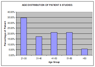
The age distribution of the patients studied was from 21to 66years maximum number of patient (34.6%) was in age group 21-30years (Graphs 1-7).

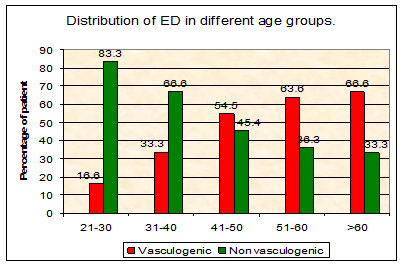
Graph 1 The vasculogenic cause of ED increases with age, percentage distribution in different age group patient was 21-30(16.6%), 31-40(33.3%), 41-50(54.5%), 51-60(63.6%) and >60 years (6.6%).

Graph 2 With increase in age there is increase in arteriogenic cause of ED and venogenic causes of ED are approximately same in different age groups.

Graph 3 Multiple risk factors were found in majority of the patients with vasculogenic causes of ED.

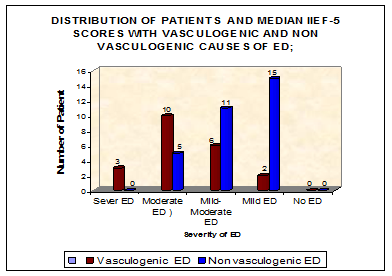
Graph 4 Significant difference was detected in IIEF-5 scores of men with non vasculogenic ED then those with vasculogenic causes of ED.

Graph 5 Medial IIEF-5 score was almost same in arteriogenic and venogenic causes of ED and the difference was not significant statistically.
The age distribution of the patients studied was from 21to 66 years maximum number of patient 18(34.6%) were in age group (21-30)years Table 1 (Graph 1). Penile color Doppler examination of 21(40%)patients showed abnormal color Doppler findings were labeled vasculogenic ED patients and 31(60%) patients revealed normal penile vascular system in patients with ED by history were labeled non vasculogenic ED patients. The vasculogenic cause of ED increases with age, percentage distribution in different age group patient was 21-30(16.6%), 31-40(33.3%), 41-50(54.5%), 51-60(63.6%) and >60years (66.6%) Table 2 (Graph 2). With increase in age there is increase in arteriogenic cause of ED and venogenic causes of ED are approximately same in different age groups Table 3 & (Graph 3). Forty four (84.6%) patients out of a total of 52patients were married and 9(15.4%) were unmarried. In our study 13(25%) patients were smoker, among smokers 8(61%) patients had vasculogenic ED, compare to 5(38%) patients with non vasculogenic ED Table 4 & (Graph 4). Furthermore, our study highlights the potential interaction of buergers disease with ED.11–15 Buerger disease was present in 5(9.6% patients) all of them were smokers and 4 out of 5(80%) were suffering from vasculogenic ED. Total 10(19.2%) patient in our study had history of alcohol intake, out of which 6(60%) were vasculogenic ED patients and 4(40%) were non vasculogenic ED patients. In our study 9(17.3%) patients had Hypertension ,and among these 6(66.6%) patients had vasculogenic ED thus Hypertension is important risk factor for vasculogenic ED. Out of total 8(15.3%) ED patients with diabetes, 5(62.5%) patients had vasculogenic ED and all of them were arteriogenic ED, thus supporting that diabetes is important risk factor of arterial involvement in patients with ED. Peyronies disease with Calcification/fibrosis of tunica albugenia on grey scale ultrasonography was found in 7(13.4%) of the patients with ED out of which 3(43%) were of vasculogenic ED patients,two third patients were of venogenic ED and one third were arteriogenic ED. In our study a significant difference p-value (0.0006) was detected in IIEF-5 scores of men with non vasculogenic ED then those with vasculogenic causes of ED 15.4 verses 11.3 Table 5 & (Graph 5). However medial IIEF-5 score was almost same in arteriogenic, venogenic causes p-value (0.38) of ED Table 6 & (Graph 6). In our study, the erectile response is graded visually from E0–E5 after injection of intracavernosal papaverine .grade E4 and E5 are sufficient for penetration. We divide the patients in 4groups table 7.We compare mean PSV in each group. Mean PSV in Gr-I, II, III and IV is 24.3, 22, 35.9 and 68 respectably PSV in Gr I and II is significantly lower p-value (0.002) then Gr III and IV.15–20
Age in Years |
No of Patients |
Percentage |
21-30 |
18 |
34.60% |
31-40 |
9 |
17.30% |
41-50 |
11 |
21.10% |
51-60 |
11 |
21.10% |
>60 |
3 |
5.70% |
Table 1 Age distribution of patients studied
Causes of ED |
21-30 In Years (%) |
31-40 |
41-50 |
51-60 |
>60 |
Total |
Vasculogenic |
3(16.6) |
3(33.3) |
6(54.5) |
7(63.6) |
2(66.6) |
21 |
Non vasculogenic |
15(83.3) |
6(66.6) |
5(45.4) |
4(36.3) |
1(33.3) |
31 |
Total |
18 |
9 |
11 |
11 |
3 |
52 |
Table 2 Distribution of ED in different age groups
Chi-square: 8.64, DF: 4, p-value : 0.070789
Vascular Cause N(%) |
21-30 |
31-40 |
41-50 |
51-60 |
>60 |
Total |
Arteriogenic |
2(13.3) |
2(15.3) |
5(29) |
6(35) |
2(13.3) |
17 |
Venogenic |
1(25) |
1(25) |
1(25) |
1(25) |
0 |
4 |
Total |
3 |
3 |
6 |
7 |
2 |
Table 3 Age and Specific Causes of Vasculogenic ED
Factors |
With Vasculogenic ED N (%) |
With non Vasculogenic ED N (%) |
P Value |
Smoking |
8(38) |
5(16) |
0.42 |
Alcohol |
6(28) |
4(13) |
0.51 |
HTN |
6(28) |
3(9.6) |
0.58 |
DM |
5(23) |
3(9.6) |
0.61 |
Dyslipidimia (DL) |
3(14) |
1(3) |
0.65 |
Peyronies (PYR) |
4(19) |
3(9.6) |
0.59 |
Burgers disease(B) |
4(19) |
1(3) |
0.63 |
Spinal trauma/infection |
2(9) |
3(9.6) |
0.7 |
Table 4 Clinical Parameter of ED Patients
Specific Causes |
Sever ED (1-7) |
Moderate ED (8-11) |
Mild-Moderate ED (12-16) |
Mild ED (17-21) |
No ED (22-25) |
Vasculogenic ED |
3 |
10 |
6 |
2 |
0 |
Non vasculogenic ED |
0 |
5 |
11 |
15 |
0 |
Total |
3 |
15 |
17 |
17 |
0 |
Table 5 Distribution of patients and median iief-5 scores with vasculogenic and non vasculogenic causes of ED
Chi sq 17.27 , Df 3, P=0.0006
Specific Causes |
Sever ED (1-7) |
Moderate ED (8-11) |
Mild-Moderate ED (12-16) |
Mild ED (17-21) |
No ED (22-25) |
Median IIEF-5 |
Arteriogenic |
2 |
8 |
6 |
1 |
0 |
11.3 |
Venogenic |
1 |
2 |
0 |
1 |
0 |
11.2 |
Table 6 IIEF-5 Score in Specific Causes of Vasculogenic ED
Chi sq 3.05, Df 3, P=0.38
Mean PSV(cm/sec) |
Gr –IV (E4,E5) |
Gr-III (E3) |
Gr-II (E2) |
GrI (E0,E1) |
68 |
35.93 |
22 |
24.3 |
|
Avg RI |
0.99 |
0.92 |
0.88 |
0.83 |
Arteriogenic Ed |
0 |
7 |
8 |
2 |
Venogenic ED |
0 |
3 |
0 |
1 |
Table 7 Penile Rigidity State and Its Hemodynamic Parameters
Chi sq 25.3 , Df 9 , P=0.002
Cases (n=21) |
Mean Resistive Index R.I |
Standard Deviation |
Arteriogenic ED |
0.96 |
0.09 |
Venogenic ED |
0.66 |
0.1 |
Table 8 R.I Value of Cavernosal Artery in Different Causes of Vasculogenic ED
The present study was carried out on 52patients in Department of Radio diagnosis G.R. Medical college Gwalior To assess the role of Penile Color Doppler Sonography in the evaluation of erectile dysfunction. In our study 52 (sample size) patients of erectile dysfunction on the basis of IIEF-5 score were subjected to penile color Doppler and differentiated into vasculogenic and non vasculogenic causes of ED .The age distribution of the patients studied was from 21to 66years maximum number of patient 18(34.6%)were in age group (21-30) years. On penile color Doppler examination of 21(40%)patients showed abnormal color Doppler findings were vasculogenic ED patients and 31(60%) patients revealed normal penile vascular system in patients with ED by history were non vasculogenic ED patients. The vasculogenic cause of ED increases with age. In our study, we used the PSV as the reference standard to diagnose arteriogenic impotence out of 21patients of vasculogenic ED on penile color Doppler , 17(81%) patients with low PSV values (<25cm/s) in the cavernosal artery were considered to have arterial insufficiency and 4(19%)patients with adequate arterial inflow that is, normal PSV, a short duration erection, with the persistent antegrade flow of >5cm/s throughout all phases suggestive of venous incompetence(Venogenic ED). Most common risk factors found were smoking, followed by hypertension, diabetes, alcohol, Peyronies disease and Burgers disease. In our study a significant difference p-value (0.0006) was detected in IIEF-5 scores of men with non vasculogenic ED then those with vasculogenic causes of ED. The IIEF-5 scores of men with nonvasculogenic causes ED are higher than those with vasculogenic ED causes. However medial IIEF-5 score was almost same p-value (0.38) in arteriogenic, venogenic causes of ED. In our study, the erectile response is graded visually from E0–E5 after injection of intracavernosal papaverine. We further divide the patients in 4groups Gr –I to Gr-IV. Group I and II either had no or minimal response to papaverine which is not sufficient for penetration, group III showed moderate and Group IV showed adequate response to papaverine. We compared mean PSV and R.I in each group , none of the patient in group IV, 55% of the patients in group III and all the patients in group I and II were found to have vasculogenic cause of ED on Penile Color Doppler . Patients in group IV had penile erection which is theoretically taken sufficient to result in penetration had mean PSV and RI 68 and 0.99 .Mean PSV and RI in group III was 35.9 and 0.92 ,PSV and R.I in groups I and II was 24.3, 0.83 and 22, 0.88 respectably .The mean PSV and R.I in group I and II were significantly p-value(0.002) below the normal cut off for vasculogenic ED. EDV>5 is associated with venogenic ED but can be found in patients with arteriogenic ED since arterial flow is insufficient to cause enough engorgement of sinusoids that can obstruct the venous outflow. Therefore, such threshold values for EDV can be misleading if arterial insufficiency is present. In our study Average R.I is found to be a significant indicator of differentiating vasculogenic ED avg. R.I (0.88) and non vasculogenic ED avg. R.I (0.98).avg. R.I is found to be helpful in differentiating specific cause of vasculogenic ED ,with venogenic ED patients had significantly lower avg.R.I (0.66) compared to arteriogenic ED avg. R.I (0.96) p-value(0.002). Thus the present study provides information as to confirm that an endothelial dysfunction can manifest itself as ED, namely (Arteriogenic or Venogenic) based upon the data provided by PSV and RI levels measurements.20–23
Evaluation of patients based on the International Index of Erectile Function (IIEF-5) is a useful tool to screen a patient with erectile Dysfunction for the etiology and level of dysfunction. However, it is also limited in its usefulness because of the inaccurate and reserved history provided by the patients. A positive response to the pharmacoerection test in terms of rigidity and duration implies normal veno occlusive mechanisms. A duplex ultrasound of the penile arteries should be performed to provide a better selection of therapeutic options.
Limitations of penile Doppler include diagnostic misinterpretation as a consequence of the high prevalence of arterial anatomical variants in the penile circulation. For example, distal perforator branches may lead to the spuriously low flow recorded in the proximal cavernosal artery at the base of the penis.
None.
The author declares no conflict of interest.

©2017 Biswas, et al. This is an open access article distributed under the terms of the, which permits unrestricted use, distribution, and build upon your work non-commercially.