MOJ
eISSN: 2379-6162


Background: The senior authors previously published a paper on abdominal wall reconstruction using Strattice acellular dermal matrix demonstrating effective aesthetic and functional results. Despite the common and effective use of Strattice acellular dermal matrix for abdominal wall reconstruction, insurance companies do not universally approve its usage. Critics of its usage state that its intra-abdominal effects are unknown.
Purpose: To further demonstrate that Strattice acellular dermal matrix is a safe and durable method to reconstruct the abdominal wall with maximal outcomes, minimal intra-abdominal morbidity, and adhesion formation.
Methods: A retrospective chart review was done on our patient who underwent abdominal wall reconstruction of a ventral hernia with component separation and placement of Strattice acellular dermal matrix several years prior. The patient happened to develop appendicitis several years post abdominal wall reconstruction, requiring laparoscopic appendectomy. Photographic analysis was used to document the Strattice and absence of adhesions.
Results: Strattice acellular dermal matrix was successfully used in the underlay intra-abdominal position to reinforce a midline hernia repair and external oblique component separation. At three years post hernia repair, the patient had no evidence of hernia. CT-scan demonstrated well opposed rectus abdominus muscles. On laparoscopic examination of the intraperitoneal cavity during appendectomy, there was no evidence of bowel adhesions to the abdominal wall or Strattice acellular dermal matrix. The Strattice was lined with peritoneum.
Conclusion: Successful repair of primary and recurrent abdominal hernia with Strattice acellular dermal matrix is effective. In our experience, the production of adhesions between the bowl and the intraperitoneal placed Strattice is low.
Keywords: ventral hernia, recurrent hernia, abdominal wall reconstruction, component separation, acellular dermal matrix, Strattice, adhesions
IV, intravenous; JP, Jackson-Pratt
The senior authors on the manuscript previously published on abdominal wall reconstruction using Strattice acellular dermal matrix with acceptable aesthetic and functional results.1 Our premise was to repair and restore the abdominal wall to minimize recurrences and complications. Hernias are prevalent in the population and hernia repair has become one of the most common conditions that general surgeons encounter.2–4 As the population ages and as comorbidities such as obesity, pulmonary disease, DVT, cardiac disease, and recurrent hernias become more prevalent, the complexity of hernia repairs and their post operative management increases.
Abdominal wall reconstruction is challenging and humbling for both patients and surgeons. Complications can occur with both prosthetic and biologic mesh.5–10 Our impetus for using biologic mesh or Strattice acellular dermal matrix was to decrease the risk of prosthetic mesh complications, such as infection and bowel adhesions in those patients with multiple previous operations or patients with significant comorbidities. The load sharing approach of bony fracture repair was applied to abdominal wall hernia repair. In mandible fractures, the plate can “bear the load” of the forces on the mandible, but an alternative construct often needed for larger bones is the “load sharing” principle that allows the bone to begin to withstand the more of the compressive forces. We tried to learn, study, and adapt this “load sharing principle” to large abdominal wall reconstructions. The only difference is the vector of the forces. The predominant load sharing materials in our experience of abdominal wall reconstruction is Strattice acellular dermal matrix. This was usually chosen given the thickness and tensile strength that was greater than human matrices. Strattice was also generally chosen because of strength and biologic nature and safety with a lower risk of infection and more easily to manage complications in a higher risk population of patients who may be at higher risk given their previous cancer, infectious abdominal processes, or additional comorbidities in our abdominal wall reconstruction population.5–10
A retrospective chart review was conducted on a patient who underwent abdominal wall reconstruction with component separation and Strattice acellular dermal matrix repair of a ventral hernia placed in the intraperitoneal spaces by the authors. The chart was reviewed. Pre-operative CT-scan prior to the hernia repair and hernia repair post-operative CT-scan and at the time of presenting appendicitis was compared for functional hernia repair assessment. Before and after photos were examined and charts were reviewed for complications.
All participants provided written consent for their use of photographs for presentation and publication.
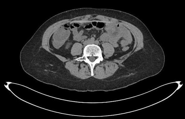
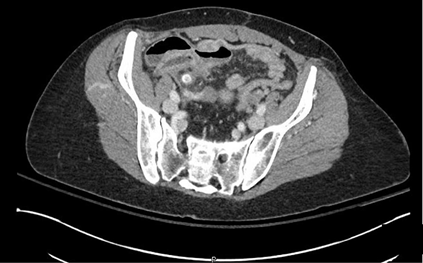
A 48 year old female with a history of a previous cesarean section and lower abdominal ventral hernia (Figure 1) underwent abdominal wall reconstruction with component separation including external oblique release, intra-abdominal placement of Strattice acellular dermal matrix with Prolene transfascial sutures, and direct midline closure with permanent sutures of prolene and rectus muscle plication with permanent prolene sutures. The patient had an uneventful postoperative course and at six weeks returned to unrestricted physical activity without limitations. A postoperative CT-scan obtained several months later demonstrated a well repaired ventral hernia with approximation of the rectus muscles as well as the visible presence of the Strattice acellular dermal matrix in the underlay position (Figure 2).

Figure 1 Pre-operative CT scan showing midline umbilical and ventral hernia as well as rectus diastasis.

Figure 2 Post-operative CT scan at a level just below the umbilicus showing reapproximation of the rectus abdominis muscles. The Strattice underlay mesh is visible and in position intraperitoneally. A functional abdominal wall is restored.
Three years after successful abdominal wall reconstruction at age 51, the patient developed abdominal discomfort that she normally associated with her menstrual cycle and dismissed the pain. The pain eventually progressed to consistent right sided lower abdominal pain and she presented to our hospital with leukocytosis of (13.7) and a CT-scan of the abdomen that demonstrated appendicitis with no evidence of rupture or abscess (Figure 3).

Figure 3 CT scan of the abdomen at the time of acute appendicitis at a level below the umbilicus at the level of the appendix showing reapproximation of the rectus abdominis muscles. The Strattice underlay mesh is visible and in position intraperitoneally. A functional abdominal wall is restored.
The patient was brought to the operating room where she underwent laparoscopic appendectomy. During the laparoscopy it was observed that the Strattice acellular dermal matrix was covered with peritoneum and there were no adhesions of the bowel to the abdominal wall.
The appendix was removed in a straightforward manner without additional adhesiolysis of adjacent bowel and no removal of bowel attached to the acellular dermal matrix (Figure 4) (Figure 5).

Figure 4 Laparoscopic view at the time of appendicitis showing Strattice acelluar dermal matrix overed with vascularied peritoneam located at the top of the image. The inflammed appendix is present at the bottom of the screen. There were no adhesions between the bowel and the anterior abdominal wall and Strattice acellular dermal matrix.
Abdominal wall reconstruction is a challenging aspect of General Surgery and Plastic & Reconstructive Surgery. Not only are the anatomical problems challenging, but so are the comorbidities of the patients themselves. Given the frequency of ventral and incisional hernias and the challenging patient population, the likelihood of having to return intra-abdominally to address an infectious problem such as appendicitis or colitis. Furthermore the possibility of colon cancer or some other enteric cancer is high in the aging population. For these reasons we often choose as our preferred load sharing mesh, extra thick Strattcie acellular dermal matrix. The Strattice acellular dermal matrix is sturdy, holds sutures, and has a high tensile strength.
Our case report of appendicitis after successful repair of an abdominal wall hernia highlights several important components that can be approached in the post abdominal wall reconstruction patient. The first is that the Strattice acellular dermal matrix can often be identified on CT-scan imaging. Knowing the location of the Strattice acellular dermal matrix can be helpful when planning placement of laparoscopic ports for diagnostic or therapeutic reasons. Secondly, when a therapeutic intervention is necessary, performing the procedure laparoscopically when possible, can prevent having to undo and then reconstruct the abdominal wall repair. Thirdly, in our experience we have found and have demonstrated photographically, that no minimal adhesions occur between the enteric contents and the Strattice acellular dermal matrix. When we have encountered Strattice placed by other surgeons and have had to enter the abdomen for oncologic resections, we have found that any adhesions between the Strattice and the enteric contents can be safely mobilized away from the abdominal wall without the inadvertent creation of enterotomies.
Strattice acellular dermal matrix has been effective in successfully repairing ventral, incisional, and recurrent hernias of the abdominal wall. In our experience adhesion formation is minimal to the abdominal wall and re-operative surgery can be performed safely in patients who have had Strattice placed in the intraperitoneal position.
The authors would like to thank Sundae Zehner and Heather Peffley for her help in these complex reconstructive cases. Sundae is reliable, dependable, and makes these complex reconstructive cases possible.
Ethics approval and Consent to Participate. The data collection for the publication was completed via chart review and a case report format. Informed consent, obtained from all patients, for photographic consent was obtained in written form for all patients.
Consent for publication. All participants provided written consent for their use of photographs for presentation and publication. Patients are not identified by name in any photographs or text. Patients are aware that in some circumstances, the photographs may make their identity recognizable. In addition, patient identifying information has been removed from images and faces are omitted from photographs.
Availability of data and material: The chart and photographic review material are available from chart review from the office of Brian P. Dickinson, M.D., Inc.
Consent to participate. Photographic consent was obtained from the patients in the chart review for presentation and publication.
Authors' contributions: The manuscript work presented has not been published before and it is not under consideration for publication anywhere else. The publication has been approved by all co-authors, as well as by the institute where the work has been carried out.
There was no funding for the completion of the chart review or preparation of the manuscript.
The authors have no conflicts of interest and no competing interests.

© . This is an open access article distributed under the terms of the, which permits unrestricted use, distribution, and build upon your work non-commercially.