MOJ
eISSN: 2379-6162


The presence of portal venous gas (PVG) and pneumatosis intestinalis (PI) is often considered an ominous radiological sign. We present a case of diffuse PI with extensive PVG seen in a patient two days after an incisional hernia repair. The patient underwent a laparotomy primarily on the basis of these extensive radiological signs, however with no discernable findings. To our knowledge, this is the first reported case which demonstrated extensive PVG and PI in a patient that subsequently underwent a negative laparotomy and had complete resolution of her PI and PVG after 24hours of repeat imaging. We discuss the causes of PVG/PI, its clinical significance, and whether surgical intervention is always needed in these situations.
Keywords: radiological findings, clinical picture, gastrointestinal ischemia, peritoneal signs, leukocytosis
PVG, portal venous gas; PI, pneumatics intestinalis; CT, computed tomography
The presence of portal venous gas (PVG) and pneumatosis intestinalis (PI) on computed tomography (CT) scan is a potentially ominous sign that presents a diagnostic challenge for surgeons. Early studies demonstrated mesenteric ischemia as the most common cause with an associated mortality of greater than 75%-90%.1Conversely, these studies generally used plain abdominal films for diagnosis. The increase of reported cases of PVG and PI is likely due to the advancements of CT over the past two decades; with more recent literature demonstrating a lower mortality rate.2 we present a case of PVG and PI in a post-operative patient, which is unusual in two respects. First, the extensive amount of PI with PVG and complete resolution after 48 hours, providing remarkable radiological images to review. And secondly, the disparity between the radiological findings and operative findings.
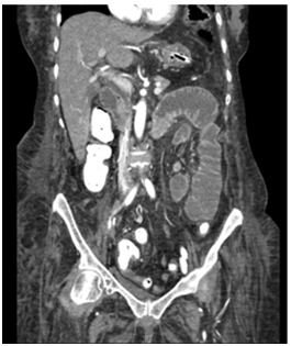
Here we have a 60year old female who presented to our service with features of a partial small bowl obstruction related to a hernia from a previous transplant nephrectomy. Her incisional hernia was repaired primarily with lysis of adhesions without any complications. Her post-operative course was unremarkable, and had return of bowel function while tolerating a diet on post-operative day two. Prior to her discharge on post-operative day two, she began having diffuse abdominal pain with nausea and retching. Her labs were unremarkable with no leukocytosis and normal lactate. She was tachycardic but remained normotensive. On examination her abdomen was soft, slightly distended, with tenderness but no peritoneal signs. An abdominal CT was obtained and revealed PI in the small bowel as well as PVG, concerning for extensive small bowel ischemia (Figure 1).

The patient had a nasogastrictube placed for decompression, and broad-spectrum antibiotics were started, prior to being taken back for a laparotomy. Findings during the laparotomy were unexpected; no evidence of gastrointestinal ischemia, perforation, necrotic tissue, or mechanical obstruction. Pneumatosis was noted on the bowel wall, once again, with no evidence of perforation. She was closed primarily. The patient remained stable throughout the procedure and was extubated post-operatively and taken back to unit for observation. Her labs and vitals continued to be unremarkable and her abdominal exam was not peritonitic. Given the negative findings of the laparotomy and the concern for possible mesenteric ischemia, a CT angiogram of abdomen and pelvis was done. Interestingly, her PI and PVG had completely resolved (Figure 2). The patient had an unremarkable recovery from this, her nasogastric tube was removed on post-operative day five and she was started on a diet which she tolerated. She had return of bowel function on post operative day seven and was ultimately discharged with a complete recovery.
Concomitant PI with PVG is an alarming finding has traditionally been associated bowel ischemia and necrosis with requiring emergency surgical management.2However, with increased availability to imaging studies and a better understanding of the variety of etiologies, this may not always be necessary.2,3 Gastric dilatation has been suggested as a possible cause of PVG4 which was present in our case, however, this would not explain the presence of air in the superior mesenteric vein and PI throughout the small bowel. She had undergone a hernia repair with lysis of adhesions for a partial small bowel obstruction 3 days prior, so the consideration of a bowel injury would be high, although we did not find one Wayne et al.3 have developed a management algorithm for patients with PI and/or PVG. The algorithm includes mechanical disease, trauma, and a cardiovascular disease score to determine risk of acute mesenteric ischemia. They identify three major clinical subgroups: mechanical causes, acute mesenteric ischemia, and benign idiopathic. Analysis of 88 patients managed with their algorithm found that patients with acute mesenteric ischemia were associated with abdominal pain (p=0.01), elevated lactate (≥3.0mg/dL) (p<0.01), small bowel PI (p=0.04), and calculated vascular disease score (p<0.01). The management algorithm was able to distinguish the three subgroups with a sensitivity of 89%, specificity of 100% and positive predictive value of 100%.3 However, reported cases where PVG was managed conservatively have been those where the radiological feature are more subtle.2–4 Our case is particularly unusual in terms of the extent of the PVG and PI, the absence of any identifiable cause, and the complete resolution of PVG and PI within 48 hours of initial imaging.
Our patient underwent an exploratory laparotomy primarily on the basis of the extensive radiological signs. Despite the fact that her laboratory results and clinical features were normal and not consistent with extensive bowel ischemia, she had undergone a laparotomy three days prior thus a surgical complication needed to be ruled out. A conservative approach has been proposed where there is a discord between the radiological signs and the overall clinical picture.3 However, in our patient it is hard to say if we would have done anything different in the management. In conclusion, this case demonstrates that extensive PI with PVG is not always anominous sign in select patients. A definite etiology for PI with PVG is not always identified which makes it challenging to define the most appropriate management.
None.
The authors declared that there are no conflicts of interest.

© . This is an open access article distributed under the terms of the, which permits unrestricted use, distribution, and build upon your work non-commercially.