MOJ
eISSN: 2379-6383


Research Article Volume 11 Issue 2
1Postgraduate Student of Department of Stomatology, School of Dentistry, University of São Paulo, Brazil
2Dentist of the Department of Stomatology, Public Oral Healthy and Forensic Dentistry, Ribeirão Preto School of Dentistry, University of São Paulo, Brazil
3Full Professor of Department of Stomatology, Public Oral Healthy and Forensic Dentistry, Ribeirão Preto School of Dentistry, University of São Paulo, Brazil
Correspondence: Plauto Christopher Aranha Watanabe, Ribeirão Preto School of Dentistry, Department of Stomatology, Public Health and Forensic Dentistry, University of São Paulo, Av. do Café, s/n, CEP: 14040-904, Ribeirão Preto, SP, Brazil, Tel +551633153993
Received: August 03, 2021 | Published: May 3, 2022
Citation: Rodrigues GA, Martins FB, Bottacin FS, et al. Iatrogenics in dentistry: importance of radiographic examinations in identifying dental treatment failures, study by use trough the analysis of panoramic radiographs. MOJ Public Health. 2022;11(2):58-62. DOI: 10.15406/mojph.2022.11.00376
Background: Technological advancement has also allowed dentistry to obtain new materials and techniques, as well as more predictable results in dental treatment. However, there are still many flaws related to the technique and choice of materials by dentists. Thus, the aim of this study was to evaluate through panoramic radiographs failures in dental treatment.
Methods: 1250 panoramic radiographs were selected and evaluated by an experienced specialist, 609 of which had one or more iatrogenic
Results: 48.72% of the radiographs analyzed showed some failure in dental treatment that focused on the age group of 31-50 years.
Conclusion: Thus, it is necessary that dentists are attentive to the detection and correction of iatrogenesis, seeking a better quality of life for patients.
Keywords: iatrogenic, panoramic radiographs, dental treatment failures
Technological advances have allowed dentistry to develop new materials and forms of treatment aimed at greater biocompatibility with oral tissues, as well as better clinical planning, thus aiming at the prevention and recovery of the structures that make up the stomatognathic system. In Brazil, the population's access to dental treatment has increased, but in regions with lower socioeconomic indices, the search for dental surgeons only occurs in times of urgency such as trauma and diseases related to dental pulp.
In recent years, with the increase in the supply of dentistry courses across the country, the number of dental professionals and the ease of access to dental treatment have increased, however, there has also been an increase in failures in the treatments performed that may be related to low experience of newly graduated dentists or the decrease in the quality of dental materials due to the demand for inputs and instruments of lower economic value.
The most common failures committed by Brazilian dentists are failures in dental restorations (excess or lack of restorative material), root perforation during endodontic treatment, fractures of instruments inside root canals, poorly adapted dental prostheses and failures in filling root canals.
Failures during dental treatment can occur and dentists must be able to resolve them in the best possible way, as well as adopt clinical protocols that increase the predictability of their treatments and reduce the chances of errors or iatrogenic. Thus, radiographic exams are allied to the professional, as they allow for better clinical planning and detection of possible failures.
Thus, this study aimed to evaluate panoramic radiographs of patients treated at the oral medicine service of the Ribeirão School of Dentistry in order to detect images compatible with failures or iatrogenic in patients from the Brazilian Unified Health System and admitted for dental treatment in the institution.
Digital panoramic radiographs from the oral medicine service of the Ribeirão Preto School of Dentistry were analyzed by a single experienced observer to catalog dental treatment failures. All radiographs are from patients referred to the oral diagnostic service and were performed by a single operator on the Vera view epochs J.Morita Co device. After radiographs were selected, patients' clinical and demographic data were collected and images separated according to age and type of treatment failure. For this, we considered iatrogenic radiographic images compatible with excess or lack of restorative or obturator material, root perforation during end odontic treatment, fractures of instruments inside the root canals and poorly fitted dental prostheses. During the preparation of the patient for the panoramic radiography, the Frankfurt plane washigher, thus producing a radiographic image with the most distant interproximal spaces in the posterior teeth, which facilitates the analysis of the contours of restorations and does not impact the interpretation of the teeth of the anterior region of the maxilla and mandible.
1250 panoramic radiographs of the image archive of the Ribeirão Preto School of Dentistry were selected for this study. An experienced observer (dental radiologist) evaluated the images and excluded 641 (51.28%) of the radiographs because they showed no images suggestive of iatrogenesis or dental treatment failures. Thus, 609 (48.72%) panoramic radiographs were reanalyzed by the same observer who divided them according to age and type of iatrogenesis found Table 1 & 2.
Variables |
Patients |
|
Iatrogenics |
No iatrogenics |
|
N (%) |
N (%) |
|
Age |
||
0-10 |
2 (0,33) |
24 (3,74) |
20-Nov |
17 (2,79) |
169 (26,37) |
21-30 |
110 (18,06) |
219 (34,17) |
31-40 |
178 (29,23) |
123 (19,19) |
41-50 |
169 (27,75) |
64 (9,98) |
51-60 |
92 (15,11) |
34 (5,30) |
61-80 |
41 (6,73) |
8 (1,25) |
Table 1 Clinical and demographic characteristics of patients with iatrogenics (N=609) and patients without iatrogenics (N=641)
Iatrogenics N (%) |
Patients |
Over-restoration |
375 (61,57) |
Failures in endodontic treatment |
160 (26,27) |
Both |
74 (12,16) |
Sample total |
609 (100) |
Table 2 Distribution of patients analyzed with the presence of iatrogenesis (N = 609) according to the type
Radiographic images compatible with excess or lack of restorative material was the most common iatrogenesis in the analyzed images (61.57%), followed by insufficient endodontic treatment (26.27%) where findings of lack of obturator material, apical obturation below were found beyond the radiographic apex. Also, 74 (12.16%) radiographs showed two or more types of iatrogenesis Figure 1 & 2.

Figure 1 Interproximal radiography of the Molares D region, diagnosed on panoramic radiography. The arrow points to the distal of tooth 16, showing excess of the prosthetic crown, that causes iatrogeny.

Figure 2Periapical radiography of the lower molars D region, diagnosed on panoramic radiography. The yellow arrows point to the distal of tooth 45 and mesial of tooth 47, showing a gross excess of amalgam restorations, which characterize iatrogeny. The red arrows point to the resorption of the alveolar bone crest, due to iatrogenesis. Note that tooth 46 is absent, with teeth 45 and 47 tilted in that space.
Radiographic examinations are part of the routine of health professionals and in dentistry the use of X-rays is essential for a correct diagnosis as well as for the clinical planning of the treatment to be offered to patients. As pointed out by Watanabe et al., 1999, current dentistry, especially dental radiology, are working with digital images that are analyzed on the computer, which may have contributed to the increased detection of failures in dental treatments because resources such as image magnification, brightness and contrast are manipulated through specific software and thus facilitate the diagnosis of dental fractures, excess materials, poor prosthetic positioning and the quality of dental treatment.
This study used digital panoramic images from patients seen at a center of excellence in dentistry in the state of São Paulo - Brazil and an academic reference throughout Brazil and Latin America. With this, the researchers point out that panoramic radiography is not the most used technique for the evaluation of restorations, cemented prostheses or endodontic treatment, but in order to avoid excessive patient exposure to X-rays, panoramic radiography becomes an important examination to be considered in the clinical practice of dentists.1,2,4,6-10
According to Healey & Phillips, most failures of amalgam restorations occurred due to technical errors by the operator (56% due to incorrect cavity preparation and 40% due to improper material handling), and only 4% occurred due to several causes, which can also be validated with the data produced by this study, where most failures occurred in restorations with silver amalgam, and it is possible to note an excess of this restorative material and failures in its adaptation to the tooth.12–14,16,18,20
Gingivitis is the most common form of periodontal disease, and its etiology is related to the accumulation of biofilm. Excessive restorations favor this accumulation, which consequently leads to the development of periodontal disease.11,6,15,17,19 In this study, clinical periodontal parameters of the patients were not evaluated, as only radiographic images were analyzed, therefore, other studies need to be carried out in order to evaluate radiographic examinations correlating them with a clinical examination of the patient to demonstrate the impact of the failures of the treatment. Gilmore et al.11 analyzed 1763 panoramic radiographs, and the result was that 32% of the patients had one or more over-restored teeth. Hakkarainen et al.15 observed in their study a prevalence of about 50% of excess restorations, found mainly in men, and that the excess restoration is a more retentive site for plaque than on a free dental face, causing gingivitis and subsequent bone loss.
This number is close to that observed in the study carried out, where 61.57% of patients had an excess of restorative material, although the studies by,31,24 have not found a direct relationship between the quality of restorations and greater periodontal attachment loss, the studies by 31,24,3 reported excess restorative material in an average of 69.8% of the restorations, numbers very close to those observed in our study.
In the study by Brunsvold & Lane,5 33% of adult patients had excess restorations and they found that these restorations are related to the progression of periodontal disease, as in addition to promoting the accumulation of biofilm, they contribute to an alteration in the subgingival microbiota. Waerhaug reports that alveolar bone loss in regions with excess restorations is more associated with biofilm accumulation, with a percentage of 32% of proximal excess in the cases analyzed by him and his team, which was corroborated by the study by,25 who found the presence of 40.69% of teeth restored with proximal excess Figure 3–12.
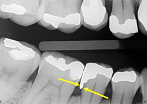
Figure 3Interproximal radiography of the molars and premolars D region, diagnosed on panoramic radiography. The arrows point to the distal of tooth 45, showing excess of amalgam restoration, in the distal box, which characterize iatrogeny. Note that the alveolar bone crest is already in the process of resorption.
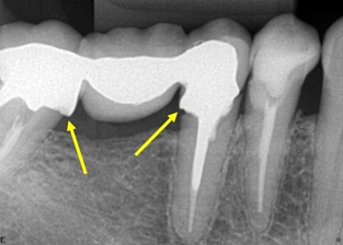
Figure 4 Periapical radiography of the molars and lower premolars region D. There is a fixed prosthesis with three elements, teeth 45 and 47 being pillars. Suspended element 46. Note the lack of material in the distal of tooth 45 and the excess in the mesial of the tooth 47, which characterize iatrogeny. Both teeth are treated endodontically. The endodontic treatment of teeth 44 and 45 is correct, apparently.

Figure 5 Periapical radiography of the molars and lower premolars region D. The dotted oval areas identify endodontic treatments for tooth roots 46. There is a prosthetic fused metal core to support the metallic crown on that same tooth. See that the root fillings are below the apical limit, and this characterizes iatrogeny. There is an apical radiolucent image, which may mean repair.
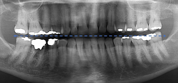
Figure 6 Panoramic radiographic technique performed with the Frankfurt Plan slightly higher than the standard position, to "open" the interproximal spaces in the posterior teeth, making the smile line straighter .
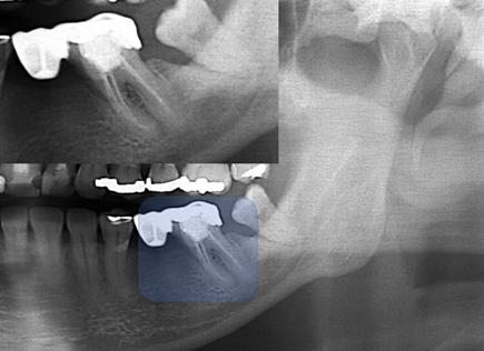
Figure 7 Panoramic X-ray cutout showing highlighted tooth 36, mesiangulated, with a coarse excess on its distal face, that causes iatrogeny, even though it has free access due to the absence of tooth 37. This tooth was treated endodontically, at first, in a correct way.

Figure 8 Panoramic X-ray cutout showing highlighted teeth 36 and 38, with a coarse excess on its distal face of tooth 36. In fact, there is an entire contact face, instead of a contact point, too, due to mesiangulation of tooth 38 that causes iatrogeny .

Figure 9 Panoramic X-ray cutout showing highlighted tooth 48, with some mesiangulation. Note that in iatrogenic endodontic treatment of the mesial root, one of the root canals is partially filled. This may be causing the space in the apical periodontal ligament to increase in that root .
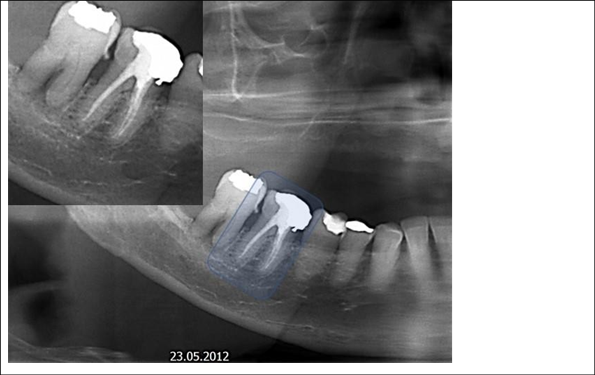
Figure 10 Panoramic X-ray cutout showing highlighted tooth 46, with correct endodontic treatment and with a coarse excess on its mesial surface, that causes iatrogeny. As the tooth is dead, the patient will be slow to feel the symptoms of this iatrogeny, excess restoration, which will possibly destroy the tooth. Note that there is already a caries infiltration.
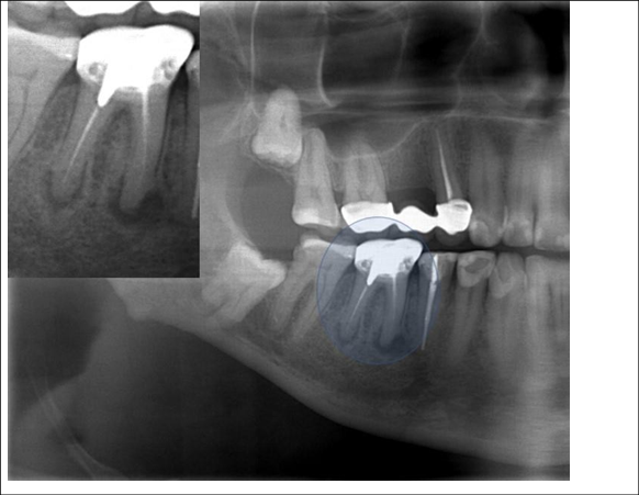
Figure 11 Panoramic X-ray cutout showing highlighted tooth 46, with iatrogenic endodontic treatment. There is a prosthetic crown with a fused metallic core with a larger support at the distal root. Note that in endodontic treatment there is a lack of material in both roots, and apical radiolucent images of these, partially diffuse, greater in the mesial root, compatible with apical lesion such as chronic abscess or granuloma. Both have a slow characteristic, due to the halo, or condensing osteitis that has formed around the lesions. Tooth 45 also has an endodontic treatment, but it is correct.
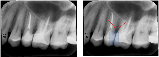
Figure 12 Periapical radiographic images of the premolars and left molars, where we point with the red arrows, the resorption of the alveolar bone crest between teeth 25-26, due to the lack of contact point between these teeth, exacerbated by the lack of contour of the teeth. restorations in the respective proximal boxes. This iatrogenesis is causing the periodontal problem.
Torsional and bending forces to which endodontic files are subjected during the chemical-mechanical preparation of the root canal in endodontic treatment cause tensions that can lead to fracture. These fractures within the root canals occur due to the forces that these instruments receive, the movements used, the anatomical situation of the dental root, the action of auxiliary chemicals during the canal preparation and the sterilization cycles, which can affect the metal structure. from which instruments are made.30
In addition to instrument fractures, anatomical deformations of the canal can occur, such as the formation of steps and root perforations,26 our data show that 26.27% of the analyzed patients have some failure of the endodontic treatment. Thus, some factors can be raised in order to explain the data found in this work.21-23,27–29,32
With the popularization of dentistry courses in Brazil, more professionals were inserted in the labor market and in order to offer a more accessible training, the courses no longer promote intensive practical training and adequate training and development of manual skills, which are essential for profession. Thus, with professionals trained in less time and with less practical training, combined with the need to reduce the prices of dental materials, it was possible to notice a greater number of failures in the treatments performed.
In endodontics, the use of digital image receptors, as well as digital root apical foramen locators are instruments that can avoid the inherent failures of endodontic treatment and oral scanning technologies (CAD/CAM) and impression of dental prostheses using the 3D technology allows the dentist to offer greater predictability of results to the treatments developed.33–44
Thus, the objective of this work was to demonstrate that there are many failures in dental treatment and draws attention to the fact that dental professionals can select the best technique and material needed to have predictability in their treatments, contributing to the evolution of dentistry that reestablishes the masticatory, aesthetic and functional functions of patients.
None.

©2022 Rodrigues, et al. This is an open access article distributed under the terms of the, which permits unrestricted use, distribution, and build upon your work non-commercially.