MOJ
eISSN: 2374-6939


Research Article Volume 17 Issue 4
 Mohamed Nasr Akl,2 Elsherbiny Ali Elsherbiny,2 Mahmoud gaber abdelazim,2 Abdelhameed Elsaid Abdelhameed Hendy,2 Ahmed M Khairy,2 Abdulhamid Elzoghby Abdulhamid Ebeed,2 Mohammed Ahmed Ismail Ismail2
1Professor of Orthopedics, El Hussein University Hospital, Egypt
2Lecture of Orthopedic, El Hussein University Hospital, Egypt
Correspondence: Sameh Elsied Anwar, Lecture of Orthopedic, El Hussein University Hospital, Egypt
Received: July 14, 2025 | Published: August 5, 2025
Citation: Kornah B, Anwar SE, Akl MN, et al. Value of procalcitonin in two- stage revision of periprosthetic hip infection. MOJ Orthop Rheumatol. 2025;17(4):98-102. DOI: 10.15406/mojor.2025.17.00708
Background: To this day, periprosthetic joint infections (PJIs) continue to rank among orthopedic surgery's most difficult consequence.
Aim: to learn more about pro-calcitonin (PCT) and its role in PJI diagnosis and treatment.
Patients and methods: Thirty patients at El-Hussien University Hospital were monitored prospectively for two years after a PJI diagnosis.
Results: All cases examined showed elevated levels of C-reactive protein (CRP), with a mean value of 51.8 (±23.64 SD) and a range from 12 to 102. Similarly, all cases showed increased erythrocyte sedimentation rate (ESR), with a mean of 69.8 (±24.59 SD) and a range of 18 to 114. Additionally, 24 cases presented elevated pro-calcitonin levels, while 6 cases were within normal limits, yielding a median of 0.35 (IQR: 0.24-0.43) and a range of 0.01 to 0.49. Among the cases studied, 3 (10%) had negative cultures, whereas 27 (90%) had positive cultures, with Staphylococcus aureus being the most frequently identified organism in 12 (40%) of the cases. At the follow-up stage, all cases demonstrated normal levels of CRP and ESR, but 3 cases showed elevated pro-calcitonin.
Conclusion: Procalcitonin (PCT) can detect infection control and patient outcomes evaluating infection state in a two-phase revision procedure for periprosthetic hip infections and determining optimal reimplantation timing, in combination with other strong inflammatory marker.
Keywords: preprothtic Joint infection, total Joint infection, pro- calcitonin
The gold standard for treating severe arthritis is a total replacement of the joint (TJA), but a common cause for postoperative revision is periprosthetic infection of the joint (PJI), a dangerous consequence.1 After arthroplasty, PJI occurred in 0.7 percent of cases of total hip arthroplasty which is the incidence of PJI.2 A patient's quality of life and death rate may be significantly impacted by PJI, even when they get the most effective therapy.3 Kurtz et al. found that the risk of PJI is 0.7% in the first year after total hip arthroplasty (THA) and 1.1% in the fifth year. There was an 88.7 percent one-year survival rate and a 67.2 percent five-year survival rate for THA after a PJI diagnosis. Addressing PJI is the primary goal of revision TJA procedures, which constitute 25% of all cases.4 Patients might be significantly impacted by the wide variation in the treatment of infected and non-infected failures after TJA. Because of the potential for increased rates of sickness, needless expenditures, and avoidable surgical operations, it is essential to avoid confusing a healthy joint for an infected one. In order to avoid these unfavorable outcomes and guarantee appropriate treatment, accurate and trustworthy diagnoses are essential in clinical practice. In certain cases, definite diagnostic criteria for PJI include the presence of a sinus tract or pus around the prosthesis, which may make the diagnosis easy.5 A peptide that develops into the hormone calcitonin is known as procalcitonin. It has very low concentrations in the bloodstream when everything is working properly. A systemic bacterial infection, on the other hand, causes a dramatic rise in PCT production. An important host-response biomarker, procalcitonin (PCT) may help doctors determine if a patient has an infection as well as how to best treat them with antibiotics.6 Assicot et al. found in 1993 that patients with positive results for bacterial infection and sepsis were more likely to have high blood levels of PCT. In addition, they proved that PCT levels in the blood would drop if the right antibiotic treatments were administered, and that PCT levels in the blood would not rise during viral infections.7 Cells in the parafollicular thyroid gland and neuroendocrine cells assemble to form PCT, a protein of 116 amino acids.8 Elevated serum calcitonin is often seen in cases of aseptic infections, which may manifest as severe burns or the failure of numerous organs. Early infection with PJI occurs in fewer than 1% of cases, and the illness is often progressed. A local joint infection will not lead to an increase in PCT until the illness spreads throughout the body, since PCT reacts immediately to bacterial endotoxin. In most situations, PJI cannot create considerable levels of PCT since it is a limited low-toxicity infection.9 In individuals, a PCT level below 0.1 ng/mL is considered normal. PCT levels slightly above normal, ranging from 0.15 to 2 ng/mL, are indicative of noninfectious systemic inflammation, untreated end-stage renal illness, or a mild to severe bacterial infection in a specific area.4 It has also been shown to be higher in individuals with autoimmune diseases and trauma.5,10 Serious bacterial infections, medullary thyroid cancer, or bacterial sepsis are all linked to PCT levels above 2 ng/mL. A frequent and successful method for treating persistent periprosthetic hip infections is a two-stage revision procedure. Here, precise biomarkers reflecting the infection's activity and resolution are essential for directing when reimplantation should be done. One possible biomarker in this context is procalcitonin (PCT).5,10 Due to its ability to forecast the severity of bacterial infections and its effectiveness in decreasing the inappropriate use of antibiotics, PCT has become a crucial laboratory test, particularly in the in-patient hospital environment. This has led to a drop in antibiotic resistance and costs.11 Examining the role of pro-calcitonin (PCT) in PJI diagnosis and therapy decision-making was the primary objective of this research.
Thirty patients with PJI were the subjects of this prospective trial, which ran from 2018 to 2020 at a single site. We conducted a study to assess the diagnostic utility of PCT in the diagnosis of PJI in patients who had undergone revision hip arthroplasties at our institution.
Inclusion criteria: Over an extended two-year monitoring period, patients who had a history of periprosthetic hip infection of the joint and had undergone a scheduled two-phase revision and reimplantation procedure were assessed according to the recommendations of the Musculoskeletal Infection Society (MSIS). In particular, we looked through medical records for cases of infection that met one of the following criteria: a connecting sinus tract with the prosthesis; a pathogen identified by culture from more than one joint tissue or fluid samples; or 4 out of 6: Symptoms may include a boost in ESR and CRP levels, an increase in synovial white blood cell count and synovial polymorphonuclear percentage terms, purulence in the affected joint, culture isolation of a microbes from a tissue or fluid sample, and a count of over five neutrophils each high power field in five fields with a magnification of ×400 during a histological study.
Exclusion criteria: Patients with aseptic periprosthetic joint, individuals with arthritis that is inflammatory, bone malignancies, or a history of TB.
Surgical technique
First stage
All patients underwent a lateral approach to the affected hip. Comprehensive debridement and excision of all necrotic tissue. A total of 5 samples of soft tissue were obtained for in-depth cytological and culture analysis. Polymethyl methacrylate (PMMA) impregnated with gentamycin and vancomycin (4gm for every PMMA packet) was hand-made molded as articulating spacer or fabricated cement spacer with different sizes and lengths of the femoral head and stem.
Interval period
We recommend weight-bearing activities for all patients to reduce mechanical complications related to spacers. Antibiotics are given according to the organisms found in preoperative cultures. If no growth is detected, we choose broad-spectrum antibiotics. We suggest an injection for two weeks, followed by ongoing oral medication. Inflammatory lab marker and surgical wound were the indicators for 2nd stage, otherwise, debridement was performed, and the spacer was replaced.
Second stage
After removing the spacer using a lateral technique, we debrided the area using irrigation. The reconstruction of the acetabular defect was tailored to its specific type, utilizing allografts, augmentations, and, in some cases, reconstruction rings along with femoral stem reconstruction. The wound was closed with effective hemostasis, and drains were placed as necessary.
Consideration of ethics
The hospital administrators oversaw the trial and gave their stamp of approval to the procedure. Every individual who took part in the research gave their written, informed permission. At every stage of the research, participants' right to privacy and confidentiality was honored. No further use will be made of the collected data.
The mean age of the examined cases was 63.5 (±6.97 SD) with 50 to 75 years, among the examined cases there were 15 (50%) females and 15 (50%) males, From one to seven years passed between initial arthroplasty and the initial reoperation, with a median of three years (range: 0-8 months). The average period from the beginning of symptoms to the initial reoperation was three months (range: 2–4 months). Among the patients studied, 9 had diabetes and 12 had hypertension, accounting for 30% and 40% of the total, respectively as shown in Table 1.
|
Subjects (n = 30) |
||
|
Age |
63.5± 6.97 |
|
|
Mean±SD. |
||
|
Range (min-max) |
50 – 75 |
|
|
Median (IQR) |
64 (58-70) |
|
|
Sex |
No. |
% |
|
Female |
15 |
50 |
|
Male |
15 |
50 |
|
Reoperation time after primary arthroplasty (years) |
3.4±1.92 |
|
|
Range (min-max) |
1 – 7 |
|
|
Median (IQR) |
3 (2 – 5) |
|
|
Temporary symptomatology before first surgical intervention (months) |
3.13±1.97 |
|
|
Range (min-max) |
0 – 8 |
|
|
Median (IQR) |
3 (2 – 4) |
|
|
No. |
% |
|
|
Diabetes mellitus |
9 |
30 |
|
Hypertension |
12 |
40 |
Table 1 Distribution of the examined cases according to demographic data
All the examined cases had elevated CRP with a mean of 51.8 (±23.64 SD) with a range of (12-102 mg/L), All the examined cases had elevated ESR with a mean of 69.8 (±24.59SD) and a range of (18-114 mm/h), the mean platelet was 306.45 (±83.45 SD) with a range of (201-438) and only 24 the examined cases had elevated pro- calcitonin with a median 0.35 (0.24 – 0.43 IQR), from 0.01-0.49 ng/L as shown in the Table 2.
|
Subjects (n = 30) |
||
|
CRP (mg/L) |
51.8±23.64 |
|
|
Range (min-max) |
12 – 102 |
|
|
Median (IQR) |
46 (36 - 66) |
|
|
Normal (0-8) |
0 |
0 |
|
Abnormal |
30 |
100 |
|
ESR (mm/h) |
69.8±24.59 |
|
|
Range (min-max) |
18 – 114 |
|
|
Median (IQR) |
69.5 (53 - 85) |
|
|
Normal (0-15 male, 0-25 female) |
0 |
0 |
|
Abnormal |
30 |
100 |
|
PCT (ng/ml) |
0.32±0.13 |
|
|
Range (min-max) |
0.01 – 0.49 |
|
|
Median (IQR) |
0.35 (0.24 – 0.43) |
|
|
Normal (0-0.05) |
6 |
20 |
|
Abnormal |
24 |
80 |
Table 2 Distribution of the examined cases according to baseline Lab investigation
Among the examined cases there were 6 (20%) with negative culture and 24 (80%) with positive culture, and According to Table 3, Staphylococcus aureus was the most prevalent culture in 12 instances, accounting for 40% of the total.
|
Culture organisms |
Subjects (n = 30) |
|
|
|
No. |
% |
|
Positive culture |
27 |
90.0 |
|
Candida tropicalis |
1 |
3.3 |
|
E. coli |
5 |
16.7 |
|
Klebsiella pneumoniae |
5 |
16.7 |
|
Staphylococcus aureus |
12 |
40.0 |
|
Staphylococcus epidermidis |
1 |
3.3 |
|
Staphylococcus Haemolyticus |
1 |
3.3 |
|
Streptococcus agalactiae |
2 |
6.7 |
|
Negative culture |
3 |
10.0 |
Table 3 Distribution of the examined cases according to culture organisms
Data are presented as frequency (percentages)
All the examined cases had normal CRP, but 7 cases with elevated ESR, and also there were 3 cases of positive PCT at the follow-up stage as shown in table 4.
|
|
Subjects (n = 30) |
|
|
CRP (mg/L) |
4.33±2.03 |
|
|
Range (min-max) |
0.3– 7.8 |
|
|
Median (IQR) |
3.9 (2.6-5.9) |
|
|
Normal (0-8) |
30 |
100.0 |
|
Abnormal |
0 |
0.0 |
|
ESR (mm/h) |
15.36±11.67 |
|
|
Range (min-max) |
0 – 44 |
|
|
Median (IQR) |
13.5 (7 - 24) |
|
|
Normal (0-15 male, 0-25 female) |
23 |
69.6 |
|
Abnormal |
7 |
30.4 |
|
PCT (ng/ml) |
0.027±0.016 |
|
|
Range (min-max) |
0 – 0.06 |
|
|
Median (IQR) |
0.03 (0.01-.04) |
|
|
Normal (0-0.05) |
27 |
90.0 |
|
Abnormal |
3 |
10.0 |
Table 4 Distribution of the examined cases according to follow-up Lab investigation
Complications
Two patients showed postoperative signs of recurrent infection 1 month and 3 weeks after cement spacer with elevated CRP by 96 and 48 and PCT by 0.2 and 0.28, respectively. They are treated with debridment with new culture taken and follow up by new laboratory marker.
One case of a traumatic broken and dislocated spacer occurring two weeks post-implantation is presented, which was revised with a new spacer. One case developed preprosthetic fracture after 7 months of reimplantation with normal inflammatory marker, treated by preprosthetic plate fixation.
One case of dislocation was treated using a closed reduction technique along with an internal rotation splint. Wound dehiscence was observed in one patient and was effectively treated with oral antibiotic therapy.
Case Title: “Two-Stage Revision of an Infected THA”
Demographics
Age: 54 years old
Sex: Female
BMI: 29.92
Relevant past medical history
Principal pathologies: HCV+
Medication: nothing
History of Presenting Complaint: In 2015, patient complained from pain in Rt hip diagnosed as arthritic hip treated by Rt THA. IN 2020, patient complained from painful hip and sinus discharging pus since the X-rays clearly show that the stem is becoming loose (Figure 1).
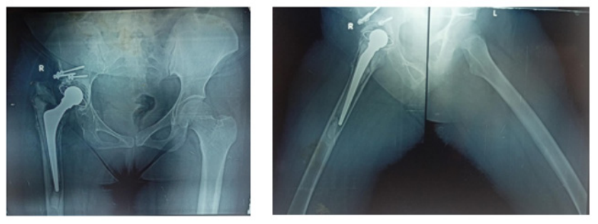
Figure 1 Preoperative X-rays of the pelvis show the loosening of the stem.
Clinical examination
The patient complained of hip pain, a stuffy nasal, and the inability to walk without the use of crutches.
Range of motion: Pain severely limits ROM.
Clinical finding: Symptoms of a swollen thigh (after a few months of sinus closure) with antalgic gait.
Main disability: Pain and severe walking limitation
Neurovascular evaluation: No neurovascular lesions detected.
LAB before the first stage: CRP 12 mg/dl, ESR 44, PCT 0.48, culture ia staph aureus
LAB before the second stage: CRP 6.9 mg/dl, ESR 14, PCT 0.01.
Preoperative radiological imaging
Figure 1
Preoperative planning
Diagnosis: late infected THA.
Alternative methods of treatment: Resection arthroplasty, one-stage revision, two-phase revision with a prefabricated antibiotic-loaded cement spacer, or retention of the prosthetic with debridement are some of the curative possibilities.
Treatment approach selected: A two-stage procedure using a cement spacer laced with antibiotics and crafted by hand
Expected difficulties
In the first step (Figure 2), the stem and cup are removed. The femur and thigh must be thoroughly debrided of any infected tissues that extend distally. Step two involves removing the spacer, getting ready for the new implant, and selecting a stable implant.
Approaches to conquer challenges: First step: universal instruments for cup removal are available. An option to use antibiotic-infused cement with a lengthy spacer was available.
Stage two (Figure 3), the implantation of revision implant. Intraoperative histology and the leukocytes esterase with absence of infection with normal lab marker.
Post-spacer radiographs after the first stage
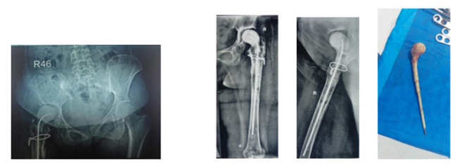
Figure 2 X-rays of the pelvis showing the spacer.
Radiographs after the second stage
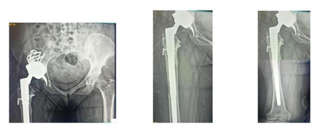
Figure 3 X-rays of the pelvis showing the revision prosthesis.
Postoperative management: Antimicrobial and anticoagulant treatment period: for five weeks from the surgery as protocol. Based on the results of the intraoperative cultures for the bacteria Staphylococcus aureus, the first stage of treatment consists of two weeks of vancomycin with piperacillin/tazobactam, followed by a single month of cefazolin, and an additional two months of levofloxacin. Next, until the intraoperative cultures come back negative, the patient will be administered doxycycline for two weeks and vancomycin for three days as a prophylactic measure. The patient used two crutches and only put some of her weight on her feet up to the second stage of operation, during which she was fully loaded. Three months after the second stage, you must use crutches to support some of your body weight.
With a range of 50-78 years, our research found that the average age of the patients was 64.65 (±8.91 SD). Of the cases we looked at, 9 were male and 11 were female. The median period from initial arthroplasty to the first reoperation was 5 years, with a range of 1-7 years. Among the patients studied, 4 (20%) had diabetes and 7 (35%) had hypertension; the mean period from the beginning of symptoms to the first reoperation was 3.5 months ((1-9) IQR) with a range of 0–15 months. Among the 30 infected patients before the first stage, only 24 had a high PCT, indicating a sensitivity of 80%. A specificity of 90% was achieved in the third out of thirty instances when the PCT came back false positive before phase two.
In their diagnostic meta-analysis, Sun et al.9 included 621 patients from nine different trials. The combined sensitivity and specificity of serum PCT in diagnosing PJI were 44.1% and 85.2%, respectively. The results of this investigation demonstrated that the diagnostic accuracy of PCT detection of PJI was low. Therefore, PCT in the blood is not a good indicator of PJI. From January 2013 through July 2020, 77 patients who had revision arthroplasties were retrospectively reviewed by Sun et al.12 The aseptic loosening (AL) group included of 36 patients, whereas the PJI group had 41 individuals. The specificity for the diagnosis of PJI was 83.33% and the sensitivity was 19.51% according to the serum PCT. The positive probability ratio for PCT was 57.1% and the negative likelihood ratio was 47.6% when the best cut-off value was established at 0.05 ng/ml. Prior to revision arthroplasties, they advised against using serum PCT as a biomarker since it did not seem to be a reliable indicator of PJI or aseptic loosening. Nevertheless, the diagnosis accuracy is further enhanced when PCT is coupled with additional biomarkers.
In their study, Zhang et al.13 monitored 150 patients who had primary hip arthroplasty for serum PCT and white blood cell (WBC) levels. One set of patients, 34, had postoperative infection, whereas the other group, 116, did not. The specificity and sensitivity of PCT diagnosis were 100% and 36%, respectively. The specificity of WBC was 44% and the sensitivity was 64%. As a potential diagnostic measure for infection, they suggested that PCT offers a high specificity for perioperative infection in patients undergoing primary hip replacement.
According to Busch et al.,14 47 patients were categorized as aseptic, whereas 23 were classified as PJI. The PJI group had much greater mean Serum-PCT levels than the aseptic group. The aseptic group had significantly higher mean PCT levels in synovial fluid compared to the PJI group. The specificity of S-PCT was 91.0 percent, while its sensitivity was 13.1 percent. The sensitivity and specificity of SF-PCT were 13.0% and 52.0%, respectively. When it came time to differentiate between aseptic loosening in total joint arthroplasty and PJI, they found that S-PCT and SF-PCT did not seem to be valid biomarkers. To determine if PCT is effective in diagnosing PJI, Saleh et al..15 conducted a meta-analysis of six trials. The pooled sensitivity, specificity, DOR, and AUC for PCT were determined to be 53%, 92%, 13, and 0.76, correspondingly. These findings suggest that the limited sensitivity of serum PCT makes it an unsuitable biomarker for the diagnosis of PJI.
Xie et al.16 conducted a meta-analysis and systematic review using 12 publications, 6 pertaining to PCT and 6 to α-defensin. The results of this meta-analysis demonstrated that α-defensin is very useful as a diagnostic tool for PJI, with a specificity of 0.95 and a sensitivity of 0.96. The poor sensitivity of serum PCT renders it of little use in clinical practice. Out of the 18 studies found by Yoon et al.,17 16 focused on IL-6 and 6 on PCT, with a total of 1,835 individuals. On the one hand, IL-6 had a sensitivity of 0.83 (95% CI, 0.74-0.89) and PCT of 0.58 (95% CI, 0.31-0.81). With a 95% confidence interval of 0.84-0.95 for IL-6 and 0.95 (95% CI, 0.63-1.00) for PCT, the combined specificity was 0.91. In this meta-analysis, we found that IL-6 is more useful than PCT for diagnosing PJI. In addition, the IL-6 test has a greater specificity than sensitivity. On the other hand, PCT should not be used to rule out potential diagnoses.
An observational prospective analysis of seventy-one patients (74 hips) receiving revision total hip arthroplasty was carried out by Yuan et al.18 Statistically, the levels of procalcitonin and CRP serum as well as white blood cell count were greater in the PJI group compared to the no PJI group (p<0.05). The area under the curve (AUC) for PCT was 0.851 (95% CI 0.773-0.929), for CRP it was 0.830 (95% CI 0.751-0.910), and for white blood cell count it was 0.633 (95% CI 0.518-0.747). There was a statistically significant difference between the diagnostic abilities of serum PCT and CRP and WBC count (p<0.05). The researchers Bottner et al.19 conducted a prospective trial to determine the effectiveness of novel laboratory markers in identifying deep implant infections in 78 patients who had undergone revision total hip or knee replacements. They discovered no statistically significant difference between serum PCT and CRP (p=0.367). A total of 57 patients underwent an aseptic total joint replacement, whereas 21 individuals developed septic infections detected by intra-operative cultures. The levels of C-reactive protein, interleukin-6, procalcitonin, and tumor necrosis factor (TNF)-α were determined in blood samples taken before to the procedure, along with the white blood cell count and erythrocyte sedimentation rate. The most sensitive markers, according to this research, are C-reactive protein and interleukin-6 (0.95). Compared to C-reactive protein, interleukin-6 lacks specificity. If you combine C-reactive protein with interleukin-6, you can find every patient who has a deep implant infection. Although procalcitonin and TNF-α are highly specific, they are not extremely sensitive. They both thought that measuring interleukin-6 and C-reactive protein together are great screening assays for deep implant infections. Patients with actual positive levels of C-reactive protein and/or interleukin-6 might be identified using a very specific marker like procalcitonin in conjunction with pre-operative joint aspiration.
Additional large-scale, future research efforts are necessary to completely verify procalcitonin's function in the two-stage revision process, however some studies have shown encouraging findings about its efficacy in guiding treatment choices in PJIs.
None.
The authors declare that there are no conflicts of interest.

©2025 Kornah, et al. This is an open access article distributed under the terms of the, which permits unrestricted use, distribution, and build upon your work non-commercially.
