MOJ
eISSN: 2374-6939


Review Article Volume 15 Issue 4
1Specialist in Orthopedics and Traumatology, Universidad Central del Ecuador, Sub specialist in Pediatric Orthopedics UNAM, Ecuador
2Rotational Internship in Medicine. University of the Americas Quito- Ecuador, Ecuador
Correspondence: Guerra L, Specialist in Orthopedics and Traumatology, Universidad Central del Ecuador, Sub specialist in Pediatric Orthopedics UNAM, Ecuador
Received: July 15, 2023 | Published: July 31, 2023
Citation: Guerra SA, Llerena R, Vaca V. Ultrasound diagnosis in developmental dysplasia of the hip: literature review. MOJ Orthop Rheumatol. 2023;15(4):141-143. DOI: 10.15406/mojor.2023.15.00634
Developmental dysplasia of the hip (DDH) covers a wide range of abnormalities from dysplasia to dislocation of the femoral head in relation to the acetabulum. It is currently known that there are risk factors that tend to this pathology, among them, family history of DDH, female sex, poor position during pregnancy, multiple pregnancy, oligohydramnios, and fetal macrosomia. The clinical examination in the first months of life, especially the Barlow and Ortolani techniques, together with the prenatal history already mentioned, are the indications to perform imaging screening in the subgroup of the population at risk. From the age of 4 8months, it is performed with AP radiographs of the pelvis, when the hip bones begin their ossification and allow a correct assessment. Today we know that if the diagnosis is made at 4 months, we are talking about a late diagnosis, since the hip joint in the first months of life has a very important remodeling capacity, so an early diagnosis will allow us to offer a timely treatment and avoid the damage in this joint in long term. Dr. Graf designed an ultrasound method, in which both static and dynamic measurements are taken, and can be performed from the 2nd week of life, when performed correctly by trained personnel to interpret the images, the treatment of developmental dysplasia of the hip, would be performed in a timely manner which in the long term means less morbidity in the hip of the newborn, less need for surgical resolution at an early age and less arthroplasty in adulthood.
Objective: to compile the most current literature to assess the results of ultrasound screening of newborns for developmental dysplasia of the hip, with ultrasound using the Graf method.
Results: A total of 21 articles were included for review. The search was performed using the following databases: Cochrane Library, PubMed, Springer, Tripdatabase, evidence was analyzed for screening children with risk factors for CDD or who on initial physical examination show signs of joint instability, is it cost effective to perform hip ultrasound
Conclusions: CDD, the most common pediatric hip condition, when detected in early stages, can be treated with orthopedic methods for this reason it is important to implement guidelines to screen children from the 2nd week of life, which is
Keywords: Developmental dysplasia of the hip, ultrasound, imaging measurements in developmental dysplasia of the hip
Developmental dysplasia of the hip (DDH) is a clinical entity that encompasses a wide spectrum of presentations, from acetabular dysplasia to dislocation of the femoral head of the acetabulum. With an incidence of 1 to 2.5 per 1000 live births. This represents a global health problem.1,2 For this reason, screening programs have been developed to obtain an early diagnosis.2 Early diagnosis of CDD has the ultimate goal of preventing morphological changes of the hip,3 which would lead to increased costs and complex treatments.4 Universal screening includes a clinical examination with Barlow and Ortolani maneuvers plus imaging studies such as hip ultrasound,5 this method is used in infants up to 4 or 6 months of age, due to the lack of ossification of the hip, which does not allow it to be reliably assessed by radiography.6,7 History of treatment of developmental dysplasia of the hip.8
During the last century, there were advances in the treatment of hip dysplasia, previously this was diagnosed when the child walked, Lorenz was the first to propose the realization of a closed reduction in forced abduction, at the beginning of 1900 Ortolani,9 was who proposed the realization of physical examination to babies before 12 months of age which improved the clinical diagnosis.7 In 1950 Arnold Pavlik introduced the concept of dynamic immobilization, with the use of a harness with straps, thus reducing the risk of avascular necrosis of the femoral head, by introducing the concept of "Ramsey's safety zone".2,10,11
In the 1980s, orthopedist Dr. Graf was the pioneer in the ultrasound technique, who emphasized a morphological approach to ultrasound examination, based on a coronal image obtained through a transducer placed on the lateral aspect of the limb, with the infant in the lateral decubitus position, Novick and subsequently Harcke and Clarke, developed a technique based on dynamic multiplanar scanning that assesses the hip in the positions produced by the already known Ortolani and Barlow maneuvers.12
Screening for developmental dysplasia of the hip
There are several programs worldwide for the detection of hip dysplasia,13,14 ranging from clinical examination without imaging studies, ultrasound in patients with risk factors, ultrasound in patients with positive clinical examination for hip instability, and universal screening.1,3
Early detection of CDD can be based on clinical criteria, it has been suggested that clinical examination for CDD should be performed at birth and after the neonatal period, due to the high rate of spontaneous stabilization in the first 28 days of life.2 The prevalence of clinical instability is known to be age-dependent, due to increased muscle tone and hormonal changes.15 Several studies have suggested that dislocatable hips at birth could be safely treated with ultrasound monitoring at 2 weeks after birth and for 2 weeks thereafter, before determining the course of treatment, reducing the number of infants with false positive diagnoses.2,3,16
Graf's method
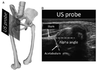
The Graf ultrasound method is characterized by measurements, the most important of which is the alpha angle, which is the measure of the depth of the bony acetabulum, formed between the acetabular roof and the vertical cortex of the ilium, measured in a two-dimensional (2D) coronal image.3 The beta angle is the angle formed between the vertical cortex of the ilium and the triangular fibrocartilage of the labrum (Figure 1, 2).17,18

Figure 1 Ultrasound examination of the hip of the newborn according to Graf. The newborn is placed in the positioning device (1), the ultrasound probe (2) is fixed in the guidance system (3). The examiner operates the ultrasound. probe with the left hand (4) and additionally guides it with the right hand (5) while the mother of the newborn or the nurse additionally fixates the newborn with both hands (6).12

Figure 2 (A) in a coronal plane. (B) an example measurement of the alpha angle in a US image collected in a coronal plane. US indicates ultrasound.4
The main measurements performed are:
The roof of the acetabulum, as it articulates with the femoral head, appears concave or flat with an angulated lateral border. In the presence of hip dysplasia, the acetabulum acquires a convex shape with a rounded lateral border. The femoral head is central within the acetabular cavity, and the acetabulum covers it by 50%. The fibrocartilage of the acetabular labrum, a hyperechoic structure, appears from the lateral edge of the acetabulum, is triangular in shape and covers the lateral part of the femoral head. In cases of dislocation or subluxation of the hip, the labrum can become deformed and interpose between the femoral head and the acetabulum, preventing its reduction (Figure 3).4

Figure 3 1. Hyaline cartilage of the proximal epiphysis of the femur (femoral head). 2. Lateral border of the ilium. 3. Acetabular roof. 4. Fibrocartilage of the acetabular labrum. 5. Gluteus minimus muscle. 6. Gluteus maximus muscle.
Graf's classification of CDD is divided into four groups:
Group I: mature hip, alpha angle is greater than 60º and beta angle is less than 55º.
Group II: delayed ossification. Acetabular rim elevated due to increased hyaline cartilage, alpha angle between 44-60º and beta between 55-77º.
Group II-A, in which there is physiological immaturity.
Group II-B, which is from three months of age.
Group III: significant delay of ossification, presenting an alpha angle less than 43º and beta greater than 77º.
Group IV: Presence of the head is dislocated, with alpha angle less than 37º.
A systematized search was carried out in the main databases, using MESH terms to formulate a search strategy, which will be obtained from PICOT questions, will be subjected to critical reading to determine the most relevant articles.
Eleven articles related to ultrasound diagnosis of developmental dysplasia of the hip were selected. Systematic review articles were included, meeting PRISMA criteria, and review articles, clinical practice guidelines, retrospective and prospective observational studies, case report articles were excluded.19
Universal ultrasound, all newborns, with or without risk factors or with normal physical examination, there is no evidence to support its performance (20) On the other hand, a large number of studies show strong evidence for screening children with risk factors for CDD or who show signs of joint instability at the initial physical examination, it is cost effective to perform hip ultrasound,1 at 2 weeks of life and continue with ultrasound scans after 2 weeks of the first ultrasound.20
CDD, the most common pediatric hip condition, when detected at early stages, it can be treated with orthopedic methods and decrease in the long term the need for surgical resolution in childhood and the need for arthroplasty in early adulthood.
For this reason, several European countries have implemented guidelines for screening children from the 2nd week of life, which has been cost-effective, and this measure is being implemented in several countries.2 Although clinical examination of all newborn hips for instability is universal, imaging screening, either with ultrasound or radiography, has not reached a consensus to become an important component of universal or selective screening. The limitations in the case of ultrasound there is an interevaluator variation so many studies have been generated to validate the reliability of such measures.4,21 So in recent years many studies have been conducted, among them Chavoshi et al. have indicated that the combined sensitivity rate of 93% and specificity of 98%, which showed that ultrasound is an acceptable modality for both confirmation and detection of CDD.4
CDD covers a wide spectrum of presentations. Early detection programs for this pathology vary around the world, and more studies are needed to standardize its management,2 with timely detection and early treatment and thus avoid the appearance of sequelae.4
Careful clinical evaluation is the primary and most important way to diagnose hip dysplasia in newborns. Ultrasound represents the safest, cheapest and easiest imaging technique for the evaluation of the main hip problems in infant age patients.4 Hip ultrasound using the Graf method, allows an early diagnosis of this pathology, with the limitations of being observer dependent.2 Therefore, implementing training strategies would reduce the operator-dependent errors.
Free and limited bibliographic resources were used. The information used is available upon request to the main author.
None.
The author declares that she has no conflicts of interest in the preparation of this article and that she has complied with all the ethical and legal requirements necessary for its publication.
The authors' own resources were used.

©2023 Guerra, et al. This is an open access article distributed under the terms of the, which permits unrestricted use, distribution, and build upon your work non-commercially.