MOJ
eISSN: 2374-6939


Case Report Volume 14 Issue 5
Rheumatology Service, Naval Hospital, Buenos Aires, Argentine
Correspondence: Baied Carmen M, Rheumatology Service, Naval Hospital. Buenos Aires, Argentine
Received: September 10, 2022 | Published: September 21, 2022
Citation: Baied CM, Gutfraind E. Ulcerating variety of carpal tunnel syndrome mimicking scleroderma. MOJ Orthop Rheumatol. 2022;14(5):135-137. DOI: 10.15406/mojor.2022.14.00594
Carpal tunnel syndrome is the most common entrapment neuropathy, affecting 1 to 3% of the population. Digital ulcers and skin lesions are unusually related to this neuropathy. We present the case of a patient with digital ulcers secondary to carpal tunnel syndrome.
Keywords: digital ulcers, carpal tunnel syndrome
Digital ulcers and skin lesions are exceptional in carpal tunnel syndrome (CTS) and there are few reports in literature. Abnormalities include painless digital ulcers, blisters, nail discoloration, onycholysis, gangrene, self-amputation, and acroosteolysis.1–4 Vasomotor dysfunction can lead to Raynaud's phenomenon. Moreover, skin necrosis and sensory involvement can result in mechanical or thermal injury that can exacerbate digital necrosis.5
A 68-year-old male patient with a history of insulin-dependent diabetes 15 years before. Eight years prior to this consultation, a carpal tunnel syndrome (CTS) surgery was performed in the patient’s left hand. He had arterial hypertension and hypothyroidism. He presented ulcerated lesions on the right hand, without pain for 3 months. He had no history of trauma. Occupation: he was a baker.
On examination the patient had sclerodactyly, edema in the index and middle finger of the right hand (dominant hand), digital ulcers in four fingers and onychodystrophy. He had unilateral Raynaud's phenomenon in his right hand. At the examination we thought he was a patient with scleroderma. The hand was swollen, with erythema and edema in phalanges associated with Raynaud's phenomenon.
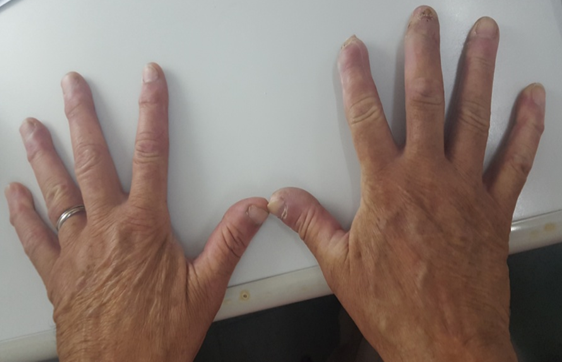
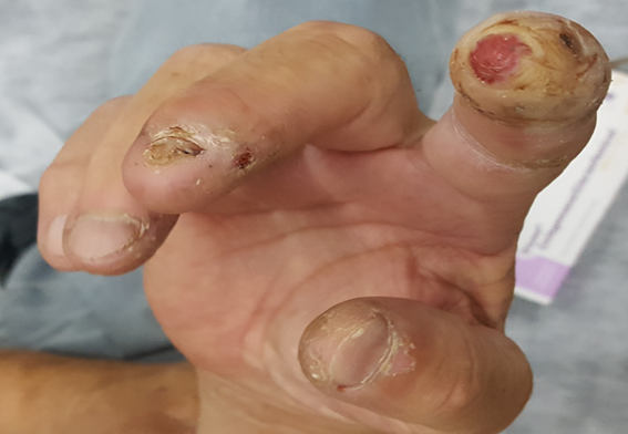
He presented sclerodactyly with moderately exudative ulcers, in the distal phalanges of the 1st, 2nd, 3rd and 4th fingers of the right hand, with loss of the almost total nail plate of the 2nd and 3rd finger and partial of the 1st finger of the right hand (Figure 1 & 2).

Figure 1 Photo of both hands where the right hand involvement with digital ulcers on 1,2 and 3rd fingers is highlighted.

Figure 2 Ulcers in the granular stage of minimally exudative healing in distal regions of the phalanges of fingers 1 to 4 of the right hand. With total loss of the nail plate of the 2nd finger and partially of the 3rd finger.
We noticed that the injuries occurred only in the right hand, without injuries in the other hand. However, the following differential diagnoses were investigated:
At laboratory tests, complete blood count, renal function and complete urine were within normal range, except glycemia 182, the ESR was 7mm/h. The laboratory included a screening for autoimmunity: anti-nuclear antibodies, anti -La antibody, anti-Ro antibody, rheumatoid factor, anti- DNA antibody, anti- centromere antibody, Anti -Scl 70 antibody, Anti RNP antibody, Anti-neutrophil cytoplasmic antibodies, cryoglobulins, anticardiolipin IgM and IgG antibodies, lupus anticoagulant IgM and IgG antibodies, Beta 2 glycoprotein antibody were all negative.
Capillaroscopy could not be performed due to the digital ulcers he presented. With regard to the treatment, the use of emollients, cleaning with white codex water, fusidic acid to be placed on ulcerated lesions, nifedipine 30 mg per day was indicated. He was evaluated by otorhinolaryngology to see if he was in condition to perform hyperbaric chamber. The chest x-ray was normal and an electrocardiogram had no abnormalities. He was given 10 sessions of hyperbaric chamber 9 (Figure 3 & 4).6,7

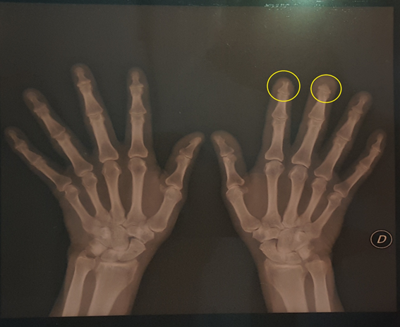
Figure 3 X-ray of the right hand where the acroosteolysis of the 2nd and third fingers is observed more closely.

Figure 4 Panoramic view of both hands where the involvement in the right hand is observed with bone resorption and loss of the distal tuft structures corresponding to the second and third fingers. There are lesions compatible with osteoarthritis and without particularities in the left hand.
The patient presented partial improvement of the Raynaud's phenomenon, with a decrease in erythema, in distal phalanges and a slight decrease in edema, healing in the remodeling stage of the ulcers in the distal phalanges of the 1st, 2nd, and 4th fingers of the right hand.
The patient was questioned about his symptoms of sensitivity: he refers paresthesias in terms of neurological semiology, the Phallen maneuvers and Tinnel sign were positive for the right hand. This case made us suspect the presumptive diagnosis of carpal tunnel syndrome as the etiology of ulcer digitals. The electromyogram with conduction velocity confirmed severe carpal tunnel syndrome in the right hand. The patient had as predisposing factors to CTS diabetes, hypothyroidism and his job as a baker (due to repetitive movements).
A relative improvement of Raynaud's phenomenon was observed. The ulcer in the distal phalanx of the 2nd finger of the right hand and dystrophy with nail thickening of the 3rd homolateral finger of the hand continued. A referral was made to the traumatology service to schedule the patient's surgery.
CTS is the most common compressive neuropathy of the upper limbs. It is caused by compression of the median nerve in the tunnel formed by the bones of the wrist and the annular ligament of the carpus. This condition affects between 1% to 3% of the population, and is four times more common in women than in men. The peak incidence is between 40 and 60 years, and more than half of the cases are unilateral. CTS presents as a triad of night pain, hypoesthesia, and tenar atrophy.8
Digital ulcers on CTS are very rare. This variety has been reported in patients with severe chronic diseases, including hypertension, cardiac arrhythmias, amyloidosis and in some patients with diabetes.9
Dr. Bouvier in 1979 was the first to describe the ulcerated and mutilating variety of this clinical-pathological entity. It is a spectrum of destructive dystrophic changes in the skin, nails, and finally bones.5
Skin manifestations include skin atrophy, anhidrosis, erythema, edema, bullas, sclerodactyly, painless fingertip ulcers, and acro-osteolysis in the area innervated by the median nerve. In this variety nail injuries occur frequently. Hyperkeratosis of the cuticle, and thickening of the nail bed, distortion and brownish discoloration are consequences of insufficient blood supply. Beau's lines are also seen on the affected nails and onychomadesis1 can also be observed. Acral ulcers and osteolysis are often unilateral, although some cases of bilateral involvement have been reported.5,8
Skin involvement is observed in 20% of cases and is caused by compression of the autonomic fibers of the affected nerve. Vasomotor dysfunction can lead to Raynaud's phenomenon and skin necrosis.1 Acral osteolysis typically begins in bone resorption in these tufts. Both mechanical compression of the autonomic fibers of the median nerve and altered distal vascularization are involved. Autonomic dysfunction is a common component of the syndrome because autonomic fibers carried by the median nerve pass through the carpal tunnel before the nerve divides. Decreased or absent sweating, edema of the hand, increased temperature due to vasomotor abnormalities are traits of autonomic neuropathy. Surgical decompression is the definitive treatment for this severe form of carpal tunnel syndrome. Skin and nail changes can show considerable improvement after the surgery.1
There are few reports of digital ulcers on CTS. The autonomic dysfunction in this pathology as a cause of digital ulcers is usually underestimated. Acral ulcers and osteolysis are rare in CTS. Most are unilateral and respond to surgery.8 It is important to consider the etiology of carpal tunnel syndrome in patients with digital ulcers mimicking scleroderma, given that the ulcerative mutilating variety is rare and not widely known in the medical population.
One of the relevant keys in our patient was unilateral involvement, the appearance of blisters affecting the index finger of the right hand, the sclerodactyly and the loss of nail plates. On the x-ray of the hands, the acro-osteolysis of the distal phalanx of the second and third fingers of the right hand is notorious. The objective of this presentation is to highlight the importance of the suspicion of this etiology as a cause of digital ulcers; the interdisciplinary work to optimize timely treatment (which is surgery) in this type of serious and disabling pathologies.
None.
The authors declare no conflicts of interest.

©2022 Baied, et al. This is an open access article distributed under the terms of the, which permits unrestricted use, distribution, and build upon your work non-commercially.