MOJ
eISSN: 2374-6939


Case Report Volume 4 Issue 2
Burjeel Hospital for Advanced Surgery Dubai, UAE
Correspondence: Lucia Heras-Garcia, Consultant Hand & Orthopedic Surgeon, Chairman of the Hand Unit, Burjeel Hospital for Advanced Surgery, Dubai, PO Box 42683, Abu Dhabi, Tel 972000000000
Received: December 03, 2015 | Published: January 25, 2016
Citation: Heras-Garcia L (2016) Traumatic Metacarpal Synostosis. MOJ Orthop Rheumatol 4(2): 00132. DOI: 10.15406/mojor.2016.04.00132
Fixation, Trauma, Achieve, Synostosis, Syndromes
We present an unusual case of cross union or synostosis involving the third, fourth and fifth metacarpal bones in a little Palestinian girl following a trauma in the hand that produced fractures of those metacarpals, a none adequate treatment left her with a serious complication, leading to a severe hand deformity with reduction of motion and function of her dominant hand.
Our treatment was done in a deprived hospital in the Gaza Strip during a humanitarian mission with the help of a motivated team, but with lack of proper materials. The surgical plan was to reproduce the fracture patterns, aligning the fractured metacarpals and fixing them with the small hand plates available.
Metacarpal synostosis following trauma is a very rare condition which has been reported in the medical literature only once before. Congenital metacarpal synostosis is a well-known identity, also present with a low incidence; and appearing more often associated to different syndromes. Multiple fractures in the hand are the result of high energy trauma; to achieve a good and prompt recovery is needed an adequate fixation, stable enough to start an early rehabilitation to be able to recover a full function. Under developed countries have a difficult situation of lack of medical suppliers that may be responsible of sometimes under standards treatments, this situation is even worse when those countries are involved in a war.
A 12 years old Palestinian girl was seen in consultation in a hospital in her own country, complaining of having a deformed hand with which she was unable to write in the school, since the time of her accident. Her mother referred a history of a trauma occurred in the hand at the age of six, when her hand was crushed in a heavy door. According to her mother the child was treated with an open reduction and fixation with Kirschner wires. Not written information or radiographies of that period were provided by the family.
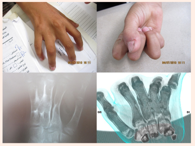
On clinical examination she presented with an obvious functional and cosmetic impairment of the hand, with disability on grasping and also she was unable to make a strong grip because the fixed position of the ring and little finger and the inability of close them together (Figure 1A & 1B). Two old scars were clearly seen in the dorsum of her hand. The third web space was wider that the others web spaces. The ulnar two metacarpals were shorter and ulnarly angulated with not adduction/abduction motion between them. The physiological arch of the hand was lost as well as the normal position of the knuckles, but no rotation deformity was appreciated.
No neurovascular deficit were associated, The radiographies and CT scan revealed and confirmed an old healed fracture of the second metacarpal less than 20 degrees angulate radically, healed fractures producing complete post-traumatic fusion of the third, fourth and fifth metacarpal at mid shaft level (Figure 1C & 1D) with 30 degrees of ulnar, palmar angulation and associated shortening of the fourth and fifth metacarpals. After careful study of the CT scans (Figure 1D), a detailed surgical plan was decided with the idea of reproduce the previous fracture pattern, doing the corrective osteotomies at the level of the intra callus area. Under general anaesthesia and tourniquet application, a single incision was done across the dorsum of the hand between the fourth and fifth metacarpal, the post-traumatic metacarpal synostosis area was exposed after a careful separation of the extensor tendons.

Figure 1A Shows hand deformity. Two old scars appear in the dorsum of the hand.
Figure 1B Third space is wider with not adduction/abduction between the 3rd and 4th. Unable to make a strong grip.
Figure 1C Radiography, oblique view.
Figure 1D CT scan reveals an old healed fracture of the second metacarpal less than 20 degrees angulated radially, complete post-traumatic fusion of the third, fourth and fifth metacarpal at mid shaft level.
The residual lines of the previous fractures were identified, and with the help of a small osteotome the metacarpal bones were carefully separated from each other (Figure 2A). When the fragments were completed separated and mobilized, the metacarpals were reduced to their anatomical position, restoring their alignment and in that way recovering the metacarpals arch and the length of the fourth and fifth metacarpals (Figure 2B-2D). Two small plates originally from a Synthes mini fragment set were used to fix the bones, matched with some screws that were cut to fix the length needed, due to the lack of sizes available. No bone graft was added. Carefully the rest of bone fragments and bone debris were removed from the interosseous space trying to prevent future cross unions. Having not any other material to use to isolate the metacarpals after the fixation, a flap of interosseous muscle were raised and sutured around the osteotomy area of the third and fourth metacarpals to avoid recurrence of the synostosis. Extensor tendons were repositioned back in place and an absorbable suture was used to close the skin.

Figure 2A Shows how the « initial » lines of the fracture were identified, and a small osteotome was used carefully to separate the metacarpals from each other.
Figure 2B Fixation with two plates.
Figure 2C Restoration of fingers flexion and metacarpal head alignment.
Figure 2D Shows recovering of the hand arch and the length of the fourth and fifth metacarpals.
Immobilization in a below elbow cast was applied for four weeks encouraging early mobilization of the proximal and distal interphalangeal joints as well as the metacarpal-phalangeal joints. After four weeks, the cast was removed and the little girls started with her rehabilitation protocol, she was not allowed carry weight for six to eight weeks. The radiographies after four weeks showed that the positions of the osteotomies were maintained and the metacarpals were healing satisfactory. One year later review, showed that the girl recovered a good function of her hand, she and her family were happy with her results (Figure 3A & 3B), she was having a normal grip, the extensor tendons of the ring and little fingers were still weaker, having a reduction of active full extension of 20 degrees at the level of the MCPJ compared to the middle and index fingers, the arc of the hand did recovered and cosmetically the hand had improved markedly. She was able to write again in the school without discomfort. The radiographies after one year showed complete healed bones, but also showed a recurrence of the synostosis between the metacarpals (Figure 3C & 3D).
Complications following metacarpal fractures in children are rare. Usually, post- traumatic malunion may produce malrotation, angulation, shortening, or a combination of those deformities, soft tissue interposition and non union despite described tend to be rarer.1 Cases of posttraumatic synostosis affecting the radius and ulna in the forearm2 or the tibia and fibula,3 are complications of fractures well documented in the literature, but cross union occurring in the metacarpals following multiple fractures, has only been reported once, before this case,4 this previous reported case, was affecting the 4th and 5th metacarpals with minimal functional or cosmetic deficit of the hand, in that case Cohen at el recommended not to operate. Our present case differs from that one not only because cosmetically the hand was not acceptable for a little girl with her cultural background, also because her hand was not functional, been her dominant side, she was unable to write in the school or help her mother in the house. Function of the hand should always need to be restored.
Many osteotomies have being described in order to obtain the correction of the deformity caused by syndactyly in congenital cases. Techniques involving double bone blocks use as a bone graft,5 wedge-shaped bone block harvested from the fusion site to correct alignment and length,6 soft tissue release procedures or callotasis techniques.7 This hemicallotasis technique was used to treat the fifth metacarpal in a hand with congenital synostosis of the fourth and fifth metacarpals. Even combined techniques have been reported like using a silicone block associated with distraction device.8
All those techniques have been reported to have satisfactory results, but they were applied in deformed, congenitally underdeveloped bones, which was not our case, but they all confirmed our opinion that the site for the osteotomy has to be always at the deformity point, only in cases of intra-articular malunion, the osteotomies should be outside the callus area and only when the delay of the correction do not exceeds two months of the initial trauma. The second problem we faced was how to prevent the recurrence of the synostosis, is well known that the application of a silicone block as described by Dao et al.9 will prevent the recurrence of the cross union, technique that have been used by others8,10 with good results.
In the hospital we were in Gaza Strip, having a silicone block was a luxury, so I decided to create a muscle flap to prevent the recurrence of the synostosis, this flap interfered only partially the cross union, which happened in an area involving 20% of the shaft length and not influencing on the hand function. This was not the ideal technique, but is an alternative to use in deprived areas of work. Most malunion of the metacarpals are secondary to failure of conservative treatment, sub-standard approaches or unstable Kitchner wire fixation like it happens in our case, producing a secondary displacement. Most of the hand fractures in children are fixed only with Kitchener wires which have proved their value for many years. But some authors report rigid internal fixation plate with good success without producing disorder of bone growth in patients who does not yet achieve maturity.
Currently, most writers are on favour of moving towards a stable and rigid fixation with plate type, as noted by Lucas in 1989. We agree that a rigid fixation allows early rehabilitation and sooner recovery of hand function. The fixation hardware has progressed with a reduction in its size, thereby reducing the described complications of adhesions, and irritation of adjacent tissues, there have been also published that the average patient satisfaction has improved with this techniques to 75 to 80% depending on the series.
We don’t agree with Cohen et al.4 opinion when in his paper about post-traumatic metacarpal synostosis concluded that no function will be improved with surgery. We understand that surgery is complex, but will improve cosmetics and function of the hand, silicone blocks is a better option to prevent recurrence, but muscle flap is not a
totally bad alternative when there is nothing else available.
None.
None.

©2016 Heras-Garcia. This is an open access article distributed under the terms of the, which permits unrestricted use, distribution, and build upon your work non-commercially.