MOJ
eISSN: 2374-6939


Research Article Volume 10 Issue 3
Servico de Ortopedia e Traumatologia, Hospital de Santo Antcnio, Portugal
Correspondence: Pedro Manuel Serrano, Servico de Ortopedia e Traumatologia, Centro Hospitalar do Porto, Hospital de Santo Antonio, Largo Professor Abel Salazar, 4099-001 Porto, Portugal, Tel +351 919069834
Received: June 01, 2018 | Published: June 8, 2018
Citation: Serrano PM, Barbosa TA, Ribau A, et al. Thermoablation in spinal osteoid osteomas . MOJ Orthopaedics & Rheumatology. 2018;10(3):325-327. DOI: 10.15406/mojor.2018.10.00421
Aim: The purpose of this work is to describe our experience in treating osteoid osteomas (OO) of the spine and compare results of surgical resection and percutaneous CT-guided radiofrequency ablation (RFA). Material and
Methods: Retrospective review of 8 cases of OO of the spine: 4 treated with surgical resection and 4 treated with RFA. All had persistent pain after conservative treatment with nonsteroidal anti-inflammatory drugs (NSAID). Results: All patients refer complete relief from nocturnal pain, proving the efficacy of both methods.
Conclusion: Complete resection although historically considered treatment of choice for OO with very good success rates, can present complications. To minimize surgical damage a precise location of the nidus is crucial. Percutaneous radiofrequency ablation under CT guidance is both efficient and safe being less invasive, performed under local anesthesia and allowing for a faster recovery, lowers costs and shorter hospital stays.
The post-operative complications are similar in both methods. New protective techniques allow thermoablation of OO located near neurological structures with increased confidence and safety.
Keywords: Osteoid osteoma, percutaneous radiofrequency ablation, spine, treatment
The Osteoid osteoma (OO) is a painful, self-limited bone tumor that can persist for several weeks, months or even years. Typically, the pain is worse at night and often is controlled with nonsteroidal anti-inflammatory drugs (NSAID).1 It commonly occurs in male teenagers and young adults, with predilection for long bones of the lower limbs (femur and tibia).2 Ten to twenty percent of the lesions occur in the spine, with painful scoliosis and muscle spasm. The posterior elements of the vertebrae are the more affected structures.3–5
The diagnosis may be hard, and imaging through CT scan and Bone Scan are essential. The treatment options for the disabling pain and spinal deformity caused by OO include surgical resection and percutaneous CT-guided radiofrequency ablation (RFA).6,7 The exact location of the "nidus" through imaging techniques is very helpful especially when considering it is often hard to identify during surgery. This particular aspect, allied with the benefits of a minimally invasive approach, have been responsible for the increased popularity of RFA.8 The proximity to the neurological structures however has been seen by many as a limitation to RFA. With this in mind, new techniques have been described with injection of gas or a refrigerated liquid in the epidural space as a protective barrier between the nidus and the spinal cord.9 Several clinical reports of the successful treatment with RFA have been published.10–14
The authors report their experience in treating OO of the spine and comment on the use of new techniques to perform RFA near neurological structures.
The authors present 8 cases of OO of the spine: 4 treated with surgical resection and 4 treated with RFA. All patients were treated by resection or RFA for persistent pain after conservative treatment with NSAID.
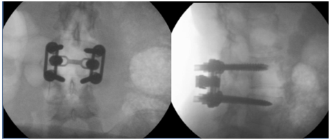
In the group of patients treated by surgical resection, the mean age was 20 years old (16-28), with the osteoid osteoma located at L2 lamina (Figure 1), L2 pediculum (Figure 2), L3 lamina (Figure 3) and L4 superior articular process (Figure 4). In the first case, only curettage was performed while in the others, posterior arthrodesis at two levels was also performed. After 4 years of follow-up, all the patients were asymptomatic, with no signs of relapse of the disease.

Figure 2 Axial CT-scan and bone scintigraphy of an Osteoid osteoma of L2 right pediculum and plain radiograph after L1-L3 posterior spine instrumentation.

Figure 3 Plain radiograph after a L3 lamina osteoid osteoma curettage and L3-L4 posterior spine instrumentation.

Figure 4 CT-scan of an Osteoid osteoma at L4 superior articular process and plain radiograph after L3-L4 posterior spine instrumentation.
In RFA group, the mean age was 19 years old (11-28), and the osteoid osteomas were located at the vertebral body of D8 (Figure 5), L2 pediculum (Figure 6), L4 pediculum (Figure 7) and at the second sacral vertebra (Figure 8). In the last case, the patient presented an irradiated pain to the ipsilateral limb that resolved completely after 48 hours. No other complications were found. After 1 year of follow-up, all patients were asymptomatic.
Complete resection, although historically considered the treatment of choice for OO with very good success rates, can present complications. To minimize surgical damage a precise location of the nidus is crucial. Conventional resection surgery is an effective option associated with low morbidity and low local recurrence rates, however RFA is emerging as an alternative method with obvious advantages.15,16 RAF is efficient, safe, less invasive, performed under local anaesthesia and allows for a faster recovery, lower costs and shorter hospital stays. Postoperative complications are similar in both methods. New protective techniques allow RFA of OO located near neurological structures with increased confidence. To safely approach tumors located near central canal or neural foramen an epidurogram can be obtained by the injection of air or carbon dioxide. The gas will outline the epidural space and increase the distance between the noble structures and the tumor. Since the heat is also a concern, techniques with the infusion of fluid at room temperature through the epidural space with a constant flow presented as a solution. Therefore, currently RFA can be used in all anatomic segments.17
Our results are according to the literature and demonstrate the safety and efficiency of both techniques.
Percutaneous CT-guided radiofrequency ablation of osteoid osteomas is a less invasive, time saving and economic technique with excellent long term results when compared with surgical resection.
None
Authors declare there are no conflicts in publishing the article.

©2018 Serrano, et al. This is an open access article distributed under the terms of the, which permits unrestricted use, distribution, and build upon your work non-commercially.