MOJ
eISSN: 2374-6939


Editorial Volume 18 Issue 1

Department of Pediatric Orthopaedics, “G. Gennimatas” Hospital, Thessaloniki, Greece
Correspondence: NK Sferopoulos, P. Papageorgiou 3, 546 35, Thessaloniki, Greece, Tel 00302310963270, Fax 00302310968265
Received: December 13, 2025 | Published: January 26, 2026
Citation: Sferopoulos KN. The painful, swollen, nontraumatic pediatric knee. MOJ Orthop Rheumatol. 2026;18(1):9-12. DOI: 10.15406/mojor.2026.18.00732
This editorial presents a literature review on the differential diagnosis of the painful, swollen, nontraumatic pediatric knee. It also includes trends in diagnosis and treatment within our pediatric orthopaedic practice, as well as illustrative case studies.
In the immature skeleton, the growth plate serves as a firm barrier between the epiphyseal and metaphyseal vessels after the age of 18 months. In children up to the age of four years and during preadolescence, blood vessels anastomose the two networks via the perichondrial complex, located at the edge of the physis. Transphyseal vessels appear in adolescents, and in early adulthood, their density through the epiphyseal scar is increased.1–3
Acute metaphyseal hematogenous osteomyelitis can be complicated by septic arthritis in children up to four years old due to the aforementioned vascular anastomoses. However, osteomyelitis involving an intra-articular metaphysis, such as the hip, shoulder, or ankle, can cause septic arthritis at any age. The distal femoral and the proximal tibial metaphyses are extraarticular. As a result, acute septic knee arthritis is usually found in children under four years old.4 In older children, extension of metaphyseal infection to the epiphysis after physeal damage5 or a missed diagnosis of acute hematogenous osteomyelitis of the patella should be considered.6
The differential diagnosis of a swollen, painful, nontraumatic knee between septic arthritis and nonseptic knee effusions is a significant diagnostic challenge even for pediatric orthopaedic experts. A thorough history, vital signs, systemic features, a basic systematic check, and the fundamental physical examination techniques, such as inspection, palpation, and mobility assessment, should all be obtained by the clinician. Inspection includes the patella’s contour, skin color, and thigh swelling. It is important to distinguish the intra-articular swelling from prepatellar effusions and cellulitis (Figure 1).7 In the absence of a significant joint effusion, we milk the fluid from the medial to the lateral side and then apply lateral pressure to observe the bulging medial sign. In younger children, we prefer to perform the palpation for local heat and to localize discomfort, and the passive range of motion while they are asleep, taking care not to wake them.
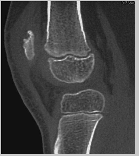
Figure 1 A 6-year-old girl presented with a right prepatellar swelling, night pain, and walked with a limp. Symptoms and signs appeared 6 weeks earlier but deteriorated significantly a few days ago. Computed tomography confirmed the radiographic findings, showing a lytic lesion of the proximal pole of the patella with benign characteristics. The lesion was curetted, and the histopathological diagnosis was subacute osteomyelitis.
The septic knee is typically warm, swollen, and erythematous with a painful restricted range of motion, and the child is unable to bear weight. Compared to the hip, the Kocher criteria are less accurate for ruling out septic arthritis of the knee. A child under five with a painful, swollen knee and a CRP above 2.0 mg/dl has a 95% chance of septic arthritis.8,9 Imaging is useful, but it cannot reliably distinguish between septic and nonseptic arthritis in children under six.10 Aspiration (arthrocentesis) remains the most urgent and crucial test. It helps differentiate between hemorrhagic, nonseptic yellowish, and septic yellow-green synovial fluid.11 However, in our practice, intra-articular pus is usually too thick for needle evacuation.
Pediatric nontraumatic, nonseptic (noninfectious) knee effusions can be either acute or chronic, and either inflammatory or noninflammatory. They typically manifest secondary to distant infections, immune system diseases, hematologic disorders, systemic illnesses, vasculitides, neoplasms, and other causes.12 However, we have not detected pediatric knee effusions due to acute leukemia, unlike the hip joint.
Juvenile idiopathic arthritis (JIA) most frequently affects the knee joint. Early on, synovial hypertrophy and joint effusion are seen due to a vicious cycle of inflammatory cells and cytokines interacting with the synovium. Chronic synovitis leads to cartilage and bone erosions, enhancing the binding of synovial inflammatory cells.13 There is a debate whether monoarticular JIA should be considered as an oligoarticular subtype or as a distinct entity because oligoarticular JIA typically has a monoarticular knee onset.14 The diagnosis is based on the history and physical examination. The laboratory studies typically yield no specific results. Radiography and ultrasound aim to detect soft tissue swelling, synovial thickness, joint fluid, and periostitis. Magnetic resonance imaging (MRI) is the most sensitive modality to diagnose bone marrow edema, synovial inflammation with hypertrophy, enthesopathy, and epiphyseal defects. The clinical onset of JIA is linked to the development of synovial thickening on knee MRI. Synovial thickness greater than 2 mm is deemed abnormal. The retropatellar, suprapatellar, and cruciate ligament regions of the knee are the most frequently impacted (Figures 2 & 3).15,16
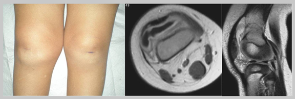
Figure 2 A 21-month-old girl presented with a painful right knee swelling and refusal to walk (left view). MRI showed joint effusion and enhancing synovial hypertrophy in the suprapatellar recess on the T1-weighted axial view after gadolinium contrast administration (middle view). No epiphyseal erosions or subchondral cysts were detected on the T2-weighted sagittal view (right view).
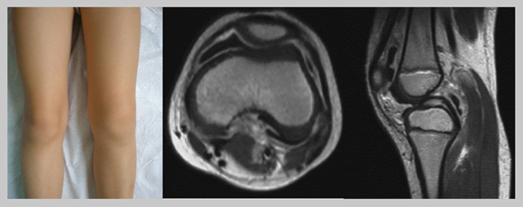
Figure 3 A 5-year-old girl presented with a painful left knee swelling and walked with a limp (left view). She had acute tonsillitis a month ago. Axial (middle view) and sagittal (right view) T1-weighted MRI after gadolinium contrast administration showed joint effusion and enhancing synovial thickening in the anterior and posterior compartments. No bone erosions, subchondral cysts, or bone bruising were evident.
Pigmented villonodular synovitis (PVNS) is a benign proliferative disease. Hemosiderin-laden villonodular synovitis is its hallmark. Usually affecting the knee, it is a non-acute lesion manifesting as a significant single-joint effusion. It may be restricted to Hoffa’s fat pad, the suprapatellar bursa, and the cruciate ligaments (Figure 4). Rheumatologists should consider the diagnosis of PVNS in patients who have not responded to treatment for presumed oligoarticular JIA.17,18
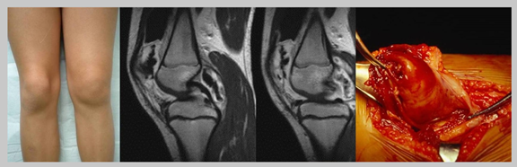
Figure 4 A 10-year-old girl presented with a painful left knee (left view) after an injury 2 days ago. She noticed a swelling in her knee almost 3 months ago. Knee joint aspirations were negative. Sagittal T1-weighted MRI after gadolinium contrast administration showed joint effusion and nodular synovial thickening in the anterior and posterior compartments compatible with PVNS (middle views). She was treated with open partial synovectomy (right view).
Primary synovial chondromatosis, osteochondromatosis, or chondrometaplasia is a tumor-like lesion, usually involving the knee joint as a chronic primary lesion. It is characterized by the formation of multiple round nodules of hyaline cartilage in the synovium and subsynovial connective tissue of the joint. It is followed by secondary calcification, ossification, and detachment that results in the presence of intra-articular loose bodies (Figure 5). An identical process may involve the synovium that extends along tendons and bursae, which is referred to as tenosynovial and bursal chondromatosis, respectively. It does not present with morning stiffness, which is usually found in JIA. Secondary pediatric synovial osteochondromatosis may be due to JIA.19 Children with primary synovial osteochondromatosis of the knee may initially resemble oligoarticular JIA20,21 or coexist with PVNS.22
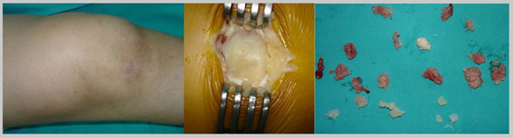
Figure 5 A 14-year-old boy presented with a severely swollen knee (left view). He was diagnosed with synovial chondromatosis. Through a minimal medial arthrotomy, the distended synovium was opened (middle view), and multiple loose bodies were removed (right view).
Pediatric acute septic knee arthritis is a medical emergency requiring urgent hospitalisation, intravenous antibiotics, and joint drainage (washout) within hours, not days. Older pediatric orthopaedic surgeons have been traditionally taught that it was preferable to surgically drain a hip or knee joint that might ultimately turn out to be nonseptic than to postpone a diagnostic and therapeutic arthrotomy of an acute septic arthritis for even an hour. In our practice, we consider the elevated erythrocyte sedimentation rate (ESR>50 mm/hr) as a useful marker of septic arthritis in children with typical findings, clinical and laboratory (complete blood count, C-reactive protein, and procalcitonin). Regarding our treatment of choice for pediatric septic knee, it includes drainage and joint lavage with antibiotics through a minor medial parapatellar arthrotomy under general anaesthesia, intravenous antibiotic therapy, and joint immobilisation. Arthrocentesis for synovial fluid analysis is tried only in effusions with a questionable diagnosis. Synovial fluid is sent for bacterial culture and sensitivity. Synovium is routinely sent for biopsy. We empirically use ceftriaxone postoperatively and modify it, if necessary, following fluid sample cultures. We switch from intravenous to oral antibiotics after the ESR returns to normal levels.
Current internet sources suggest that ‘in abscess treatment, antibiotics alone often are not enough’. This statement is misleading and may imply that antibiotics alone can treat acute or hot abscesses or septic arthritis without rupture or surgical drainage of the pus.23 Antibiotics cannot reach the center of an acute abscess or the intra-articular pus due to a lack of vascularisation.24
Young children with monoarticular JIA may be misdiagnosed with septic arthritis. This can lead to early invasive surgical treatment. Nowadays, pediatricians and rheumatologists are concerned about the devastating consequences of an ‘unreasonable’ arthrotomy. Although this view has not been presented in recent literature, it is scientifically valid. It aims not accelerate the inflammatory process, which is most aggressive in the initial stage and may cause joint deformities.
In conclusion, acute septic arthritis stands at the top of the pediatric orthopaedic emergency list. Acute onset, age, typical systemic and local clinical findings, and blood inflammation markers are the frontline for an accurate early differential diagnosis of septic from nonseptic, either inflammatory or noninflammatory knee effusions. Considering that the outcome depends on the hours elapsed from treatment, open surgery should not be delayed for sophisticated imaging, cultures, and sensitivity tests.
None.
The author certifies that he has no commercial associations (such as consultancies, stock ownership, equity interest, patent/licensing arrangements, etc) that might pose a conflict of interest in connection with the submitted article. The author received no financial support for this study.

©2026 Sferopoulos. This is an open access article distributed under the terms of the, which permits unrestricted use, distribution, and build upon your work non-commercially.
