MOJ
eISSN: 2374-6939


Research Article Volume 14 Issue 5
1Resident Surgeon (Orthopaedic Surgery), Sher E Bangla Medical College Hospital, Bangladesh
2Registrar (Orthopaedics Surgery), Sher E Bangla Medical College Hospital, Bangladesh
3Medical Officer (OSD), DGHS, Mohakhali, Bangladesh
Correspondence: Md Ferdous Rayhan, Resident Surgeon (Orthopaedic Surgery), Sher E Bangla Medical College Hospital, Bangladesh
Received: September 10, 2022 | Published: September 27, 2022
Citation: Rayhan F, Rezwan M, Sarker M. Study on lumbar interbody fusion and posterior instrumentation in spondylolisthesis. MOJ Orthop Rheumatol. 2022;14(5):158-161. DOI: 10.15406/mojor.2022.14.00598
Background: Spondylolisthesis presenting as low back pain is not an uncommon condition. Most of the patients are treated conservatively. Few patients require surgery after failing conservative treatment. Currently obtainable options are Posterior lumbar interbody fusion (PLIF), Anterior lumbar interbody fusion (ALIF) and Transforaminal lumbar interbody fusion (TLIF). Each technique has produced satisfactory outcome with both benefits and disadvantages. Posterior lumbar interbody fusion allows the surgeon to insert bone graft and cage into the disc space and simultaneous stabilization by pedicle screws and rod through single posterior approach. Thus restores the sagittal balance and improve quality of life.
Objective: To determine the outcome of lumbar decompression, interbody fusion and posterior instrumentation in obtaining clinical and radiological favorable outcome of spondylolisthesis.
Materials and methods: This is a prospective interventional study conducted for two years at Sher E Bangla Medical College Hospital, Barishal and private medical institutes of Barishal between July 2017 to June 2019 with total number of 18 patients who underwent operative procedure. The study analyses the influence of lumbar decompression, lumbar interbody fusion by bone graft with cage and stabilization by pedicle screws and rods on patients with lumbar Spondylolisthesis.
Results: Out of 18 patients, 7(39%) were male and 11(61%) were female. The mean age was 90±13.7 years and range between 23-70 years. The commonest involved level L4 over L5 15(83.3%). According to Meyerding grading, grade II were 11(61%) and grade I were 7 (39%). According to types of Spondylolisthesis lytic 10(55.5%), degenerative 7(39%) and dysplastic 1 (5.5%). Excellent outcome was seen in 14(78%) and good result in 4(22.0%) cases. Probably not fused after surgery was observed in 2(11.1%) patients. The preoperative Oswestry Disability Index was 57.78±2.25 and postoperative ODI 16.56±3.53.
Conclusion: This study revealed that a higher fusion rates and a better clinical outcome have been obtained by instrumented fusion. In this study patient number is limited, studied only 18 cases, and more longitudinal study would emphasize same conclusion.
Keywords: outcome, lumbar interbody fusion, posterior instrumentation, spondylolisthesis
Spondylolisthesis refers to displacement of a vertebral body on the one below most commonly the lowest lumber vertebra on the sacrum.1 It was first described in 1782 by Belgian obstetrician Dr. Herbinauz.2 He reported a bony prominence anterior to the sacrum that obstructed the vagina of a small number of patients. The term Spondylolisthesis derived from Greek roots spondyl meaning spine and olisthesis meaning to slip, refers to displacement of a vertebral body on the one below. On the basis of aetiology, the classifications of Spondylolisthesis illustrated 6 different type that is congenital, isthmic, degenerative, traumatic, pathologic and post-surgical.3 The prevalence of Spondylolisthesis in adult population is 5%.4 Among them, in 85%-90% cases conservative therapies may be helpful and 10-15% will fail conservative therapy requiring surgical intervention.5 As an effective treatment modality for proper patients with low back and leg pain suffering from degenerative lumbar spinal disorder, lumbar spinal fusion has been established. Numerous efforts have been undertaken to fulfill the aim of spinal fusion procedure, which is a solid arthrodesis of unstable segment finally relieving patients from pain and restoring their global spinal function. Among various techniques introduced so far, posterior lumbar interbody fusion (PLIF) with instrumentation is considered as one of most solid and biomechanically sound methods for fusion.6There are several procedures that have been described for interbody fusion with or without instrumentation such as posterior lumbar interbody fusion (PLIF), anterior lumbar interbody fusion (ALIF), circumferential 3600 fusion (front and back) and more recently, the transforaminal lumbar interbody fusion (TLIF).7 A posterior lumbar interbody fusion (PLIF) has the advantages of spinal canal decompression, anterior column reconstruction, decompression of foraminal stenosis, and reduction of the sagittal slips from a single posterior approach. The PLIF using double cage has been a standard practice till recently. However, there are many studies now with PLIF using single cages with comparable results and lesser complications.8 During the last decade, posterior lumbar interbody fusion (PLIF) has been widely used in arthrodesis for segmental instability of the lumbar spine. With additional instrumentation and posterolateral fusion, the overall fusion rate has been high, ranging from 96% to100%, and the clinical success has been satisfactory as reported in the literature.9 In practice, several kinds of bone grafts have been used for interbody fusion. Autologous iliac bone graft is a suitable choice with good biological healing ability but may cause considerable donor site morbidity, such as local pain, increased operation time and blood loss, and infection. Local lamina bone and facet joint auto
graft obtained from the decompression procedure are also good sources of bone grafts in PLIF and have the advantage of not increasing morbidity.9 Lumbar interbody fusion by bone graft with cage can be combined with decompression of the spinal canal and instrumented posterior stabilization with pedicle screws and rods through a single posterior incision. It was the aim of the present study to evaluate clinical and functional outcome, fusion rate, structural restoration and complications in treatment of Spondylolisthesis.
Prospective Interventional study was done from July 2017 to June 2019 (24 months) in the Department of Orthopedics Surgery, Sher –E-Bangla Medical College Hospital, Barishal and spine surgery OT of private medical institutes of Barishal. Total 18 patients were selected, among them 11 female and 7 male.
Inclusion criteria:
Exclusion criteria:
Operative techniques
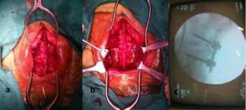
Indication for surgical intervention Symptomatic Spondylolisthesis after adequate conservative treatment. Progressions of a functionally disabling neurologic deficit or caudaequina syndrome, although rarely associated with lumbar Spondylolisthesis, are two indications for urgent operative intervention (Figure 1-4).10

Figure 1 A) Intraoperative photograph showing adequate exposure. B) Shows the four requisite pedicle screws inserted and rods distracted.C) Shows C-arm picture after instrumentation.
A total of 18 patients with lumbar Spondylolisthesis were managed surgically by decompression and Posterior lumbar interbody fusion by cage with autogenous bone graft and stabilization by transpedicular screws and rods. The studied was carried out between July 2017 and June 2019. All patients underwent surgical procedure by posterior midline approach. The level of involved vertebra was identified preoperatively by a skin marking under radiological guidance. All the patients underwent surgery under general anesthesia and in prone position on special designed padded operating table for the purpose of abdomen hang free, intravenous pressure was reduced and per operative blood loss was decreased as a result of collapse of the epidural venous plexus. A longitudinal incision was made in posterior midline of lumbosacral region.
To expose the lamina, the facets and transverse process a standard sub periosteal dissection was carried out using self-retaining retractors to maintain tension on soft tissues during exposure and any bleeding was secured by proper haemostasis using bipolar diathermy. Posterior decompression was done by laminectomy and discectomy.End plate was removed by end plate curette. Then pedicle was identified and guide pins were inserted. The position of guide pins were determined by C-arm.
After correcting direction and level, pedicle screws of adequate length and diameter were inserted. The screws placement were rechecked by C-arm. Local bone grafts previously collected from lamina, spinous process, facet were mashed up and packed in an adequate size of titanium cage. Some bone grafts were placed anterior part of the disc space. A curved cage specially designed for the PLIF technique was filled with bone chips and inserted into the posterior or central part of the disc space. The shape of the cage and the 40° angle of the introducer enable a controlled cage positioning.Finally compression was done after placing two rods. The wound was closed in layers with drain kept in situ.
Among 18 patients mean age was 46.90.0±13.7 years, maximum patients (27.8%) age 51-60 and 41-50 years followed by 22.2% age range 31-40 years. Among 18 patients 61% were female and 39% were male. Among occupational distribution housewife comprised main bulk 66%. Other occupants were sedentary 28.0% and manual worker 11%. In this series most involved L4 over L5 level was found 15 (83.3%). Among 18 patients, in this series grade II slippage was 11 (61%) followed by grade I 7(39%) (Table 1 & 2, Figure 5).
Age |
Number |
Percentage |
21-30 |
2 |
11.10 |
31-40 |
5 |
27.8 |
41-50 |
5 |
27.8 |
51-60 |
2 |
11.10 |
61-70 |
4 |
22.2 |
Table 1 Age distribution of the patients (N=18)
Macnab criteria |
Number of patients |
Percent |
Excellent |
14 |
78 % |
Good |
4 |
22 % |
Table 2 Functional Outcome of the study measured by Macnab criteria (n=18)
Regarding the modified Macnab criteria of the study patients, 14(78.0%) was found excellent in final follow up and only 4(22.0%) found good.
So, among the study population we will find almost 58.8% to 97.1% satisfactory result by this procedure. So, this procedure can say an effective procedure.
In present study, Out of 18 patients, 2 (11.10%) was 21-30 years old, 4(22.20%) was 31-40 years old, 5(27.8%) was 41-50 years old, 5(27.8%) was 51-60 years old and 2(11.1%) was 61-70 years old. The mean age was 46.9±13.7 years and the lowest and highest ages were 23 and 70 years respectively. Male was found in 7(39%) cases and female was found in 11(61%) cases. Alam et al.11 reported in a related study mean age 56.6years and 06 male and 22 female.In this study, the posterior lumbo sacral interbody fusion with cages and bone graft with instrumentation technique in treatment of Spondylolisthesis resulted in significant clinical and functional improvement, structural restoration, fusion, and stability had been associated with low rates of intraoperative neural complications.In this series improvement of pain status measured by Visual Analogue Score (VAS) is, back pain improvement from (6.83±0.49 to 2.27±0.57) and leg pain improvement from (6.75±0.60 to 01.28±0.46), p value of both of which are 0.0001 which is statistically significant. In initial series of the improvement of VAS score of back pain was (07.18±01.09 to 01.84±0.91) and leg pain improvement was (06.88±01.21 to 01.34±0.97) both of which is comparable to this study.9 In this series improvement of disability measured by Oswestry Disability Index (ODI)is (57.78±02.25 to 16.56±3.53) after 6 months of follow-up, here also p valueis0.0001 which is statistically significant. In the study of Hackenberg et al.12 Itwas showed that, in 54 patient series Oswestry Disability Index (ODI %) was 60.00 ±01.21 pre-operatively and 17.09± 0.97 after 6 months of follow-up, which iscomparable to this study.According to Alam et al.11 excellent outcome had been observed around 92.86%cases in posterior lumbar interbody fusion by using Macnab criteria, which was alsocomparable to this study where 14(78%) was found excellent in final follow up andonly 4(22%) found good.Development of Pseudarthrosis is one of the most common (range, 05-45%)complication of interbody fusion. In this study, we have achieved 100% fusion rate byusing Hackenberg,.12 criteria which is comparable to Mehta et al.,13 where qseudarthrosis was present in two (2.60%) patients in their series.In terms of complications, 11.1% of the patients developed minor complications incurrent series such as superficial infection which was managed by regular dressingand oral antibiotics. Dantas et al.14 reported 6.6% of infection in his study onposterior lumbo sacral interbody fusion group and reported wound complications ratewas 0.6% to 5%, which was comparable with our result.The criteria used to analyze the overall outcome was proposed by Modified Macnabcriteria which is based on relief of back and leg pain, return of employment, restriction of physical activities and use of analgesics for lumbar spine fusion. In thisseries 14 patients (78%) got excellent results, 4(22%) belonged to the good results. Periasamy et al.,15 in 2008 got 85.3% excellent and good results with satisfactoryclinical outcome which is comparable with our results.To conclude, Posterior Lumbosacral Interbody fusion method is effective in relievingsymptoms, achieving stability and fusion and lesser complication rates in surgical management of Spondylolisthesis.
This study may be concluding that lumbar interbody fusion, decompression and posterior instrumentation is an effective procedure for the treatment of spondylolisthesis. This method enhances symptoms, reduces pain and efficiently improve functional outcome. In this study patient number is limited, studied only 18 cases, and more longitudinal study would emphasize same conclusion.
None.
The authors declare no conflicts of interest.

©2022 Rayhan, et al. This is an open access article distributed under the terms of the, which permits unrestricted use, distribution, and build upon your work non-commercially.