MOJ
eISSN: 2374-6939


Research Article Volume 17 Issue 5
1MS Orthopedics, DNB,FISS, MNAMS,FACS). Spine Surgeon at Ashirwad Spine Hospital, Surat, India
2MS Orthopedics, Assistant Professor at TNMC & BYL Nair Hospital, Mumbai
Correspondence: Dr Ghanshyam Kakadiya, MS Orthopedics, DNB,FISS, MNAMS,FACS). Spine Surgeon at Ashirwad Spine Hospital, Surat, India
Received: July 16, 2025 | Published: September 11, 2025
Citation: Kakadiya G, Saindane K. Retrospective comparative study: unilateral biportal endoscopic transforaminal lumbar interbody fusion (ube-tlif) versus minimally invasive transforaminal lumbar interbody fusion (mis-tlif) for degenerative lumbar spondylolisthesis. MOJ Orthop Rheumatol. 2025;17(5):90-97. DOI: 10.15406/mojor.2025.17.00716
This study aimed to delineate the comparative efficacy of Unilateral Biportal Endoscopic Transforaminal Lumbar Interbody Fusion (UBE-TLIF) and Minimally Invasive Transforaminal Lumbar Interbody Fusion (MIS-TLIF) in the management of single-segment degenerative lumbar spondylolisthesis. A retrospective analysis was conducted on cohorts of patients who underwent single-segment lumbar fusion for degenerative lumbar spondylolisthesis, predominantly Meyerding Grade I-II. Patients were stratified into UBE-TLIF and MIS-TLIF groups. Perioperative metrics, encompassing operative duration, estimated blood loss (EBL), postoperative drainage volume, time to ambulation, length of hospital stay, and inflammatory markers, were rigorously assessed. Clinical outcomes were quantitatively evaluated utilizing the Visual Analog Scale (VAS) for both axial and radicular pain, the Oswestry Disability Index (ODI), and Japanese Orthopedic Association (JOA) scores. Radiological assessments included interbody fusion rates, lumbar lordotic angle (LLA), intervertebral disc height (IDH), segmental lordotic angle (SLA), and the degree of spondylolisthesis reduction. Incidence of complications was also subjected to comparative analysis. Both UBE-TLIF and MIS-TLIF demonstrated statistically significant improvements in pain and functional indices. UBE-TLIF evinced distinct advantages, including reduced intraoperative hemorrhage, diminished postoperative drainage volume, accelerated time to ambulation, and a truncated hospital stay, concomitant with an attenuated systemic inflammatory response. Conversely, MIS-TLIF typically presented with shorter operative durations and potentially reduced fluoroscopic exposure. Long-term alleviation of pain, functional restoration, and interbody fusion rates were found to be comparable between the two surgical modalities. Furthermore, complication rates exhibited no statistically significant disparity. In conclusion, both UBE-TLIF and MIS-TLIF represent safe and efficacious minimally invasive surgical options for the management of degenerative lumbar spondylolisthesis. While UBE-TLIF confers specific perioperative advantages, MIS-TLIF may be preferentially considered due to its typically shorter operative duration. The judicious selection of either technique may be contingent upon surgeon expertise and individualized patient profiles.
Background on degenerative lumbar spondylolisthesis (DLS)
Degenerative lumbar spondylolisthesis (DLS) is a pervasive spinal pathology characterized by the anterior displacement of a vertebral body relative to the subjacent vertebra, primarily attributable to age-related degenerative processes affecting the intervertebral discs and facet joints.1 This condition most commonly afflicts the lumbar spine, and its prevalence escalates with advancing age, exhibiting a notable predilection for individuals over 50 years of age and for the female demographic.2
Patients afflicted with DLS commonly manifest a spectrum of symptomatology, including chronic axial low back pain, radicular lower extremity pain (frequently characterized as sciatica), spinal rigidity, paresthesia, motor weakness, or dysesthesia in the pedal extremities.1 These clinical manifestations often emanate from neural root compression secondary to vertebral displacement. Initially, conservative therapeutic modalities, including pharmacotherapy for pain management, physical therapy, and targeted injections, are advocated for symptomatic palliation.3 However, in instances where these non-operative interventions prove refractory in providing adequate symptomatic relief for persistent symptoms, surgical intervention becomes a requisite consideration for neural decompression and stabilization of the affected spinal segment.5
Evolution of lumbar interbody fusion techniques
Lumbar interbody fusion (LIF) is a well-established surgical procedure for addressing degenerative pathologies of the lumbar spine. Its primary objectives are to alleviate neural compression and stabilize the spinal segment through the promotion of bony fusion between adjacent vertebrae.3 Historically, Transforaminal Lumbar Interbody Fusion (TLIF) emerged as a significant advancement, representing a variation of posterior lumbar interbody fusion (PLIF). TLIF garnered widespread acceptance due to its more direct access to the disc space, a reduced propensity for neural injury, and the practical advantage of a unilateral approach for the insertion of the fusion cage and bone graft.3
Despite its inherent advantages over antecedent techniques, conventional open TLIF necessitates substantial soft tissue dissection, often leading to considerable intraoperative haemorrhage and protracted postoperative recovery periods.6 To mitigate these inherent drawbacks, Minimally Invasive Transforaminal Lumbar Interbody Fusion (MIS-TLIF) was developed. This technique confers benefits such as attenuated soft tissue injury, reduced estimated blood loss, accelerated postoperative recovery, and shorter hospital stays by meticulously preserving the paraspinal musculature and ligaments.6 MIS-TLIF typically employs a tubular retractor system to establish a working channel to the spinal pathology.13
The relentless pursuit of reduced surgical invasiveness, driven by patient demand for less traumatic procedures and expedited functional recovery, has catalyzed further innovations. This progression from open TLIF to MIS-TLIF and subsequently to endoscopic approaches signifies a clear and continuous paradigm shift in spine surgery. This evolution transcends mere incision size reduction; it fundamentally aims to minimize muscle and soft tissue disruption, thereby curtailing blood loss, accelerating convalescence, and diminishing hospital durations. This endeavor to reduce surgical trauma is also intrinsically linked to attenuating the systemic inflammatory response, which directly influences early postoperative pain and recovery. Consequently, the advantages of reduced tissue damage extend beyond the immediate surgical site to profoundly influence systemic physiological responses.
More recently, endoscopic techniques have revolutionized spinal surgery. Unilateral Biportal Endoscopic TLIF (UBE-TLIF), also known as Endoscopic TLIF (Endo-TLIF) or Unilateral Biportal Endoscopic Lumbar Interbody Fusion (UBE-LIF), represents a novel minimally invasive approach. This technique utilizes two independent portals with continuous irrigation of normal saline and a high-definition endoscope, providing a clear and magnified surgical field.7 This approach promises even less tissue trauma and potentially faster recovery compared to conventional MIS-TLIF.7
Rationale for comparative study and objective
While MIS-TLIF has unequivocally demonstrated advantages over open TLIF, the comparative efficacy and safety of UBE-TLIF versus MIS-TLIF, particularly for degenerative lumbar spondylolisthesis, remain subjects of active investigation.5 Existing studies present heterogeneous findings regarding specific perioperative parameters, such as operative duration and fluoroscopic exposure, while generally concurring on comparable long-term clinical and fusion outcomes.5 This highlights a nuanced landscape where neither technique is definitively superior across all metrics. The "optimal" choice between UBE-TLIF and MIS-TLIF is not universally applicable but rather a decision predicated on a careful consideration of trade-offs.
This retrospective comparative study endeavors to contribute further evidence by evaluating and comparing the clinical and radiological outcomes, perioperative parameters, and complication profiles of UBE-TLIF and MIS-TLIF in patients presenting with single-segment degenerative lumbar spondylolisthesis. The derived findings are intended to inform clinical decision-making regarding the most appropriate surgical approach for this patient demographic.
Study design
This investigation was conducted as a retrospective comparative study.
Patient selection criteria
Patients enrolled in this study were diagnosed with single-segment degenerative lumbar spondylolisthesis, predominantly Meyerding Grade I and II.5 Certain cohorts also included patients with concomitant lumbar spinal stenosis (LSS).5 A pivotal inclusion criterion was the presence of chronic low back pain and/or lower extremity pain persisting for a duration exceeding 3 to 6 months, with symptomatology demonstrating no amelioration or even exacerbation following a trial of stringent conservative treatment.3 All included patients underwent single-level fusion.5 The minimum follow-up period for patients was typically 1 to 3 years.5 Patient age was generally 40 years or older,11 and preoperative imaging, such as lumbar dynamic X-ray films, indicated a vertebral slip exceeding 3 mm between two adjacent vertebral bodies.5
Exclusion criteria encompassed a history of prior lumbar fusion surgery,11 lumbar fracture, tumor, or infection,11 imaging-indicated congenital spinal hypoplasia or deformity,11 severe scoliosis with a Cobb angle greater than 30°,11 and high-grade spondylolisthesis (Grade III or IV).6 Patients with cardiopulmonary insufficiency or other severe organ disorders precluding general anesthesia were also excluded,11 as were those with chronic medical diseases, mental disorders, coagulation dysfunction, or extensive epidural scarring or arachnoiditis.6
Surgical techniques
Both surgical techniques were performed under endotracheal general anesthesia with the patient positioned prone on a radiolucent spine table, ensuring unobstructed passage for the fluoroscope.8 The overarching objective for both procedures was direct decompression of the affected nerve root, insertion of an interbody cage, and stabilization of the spinal segment using pedicle screw instrumentation.13 For UBE-TLIF, watertight draping was essential to manage the continuous saline irrigation.8
MIS-TLIF Procedure
The MIS-TLIF procedure typically involved a 2.5 to 4 cm longitudinal incision made on the affected side, usually 1 to 3 cm lateral to the spinous process, or 5 to 10 mm outside the upper and lower pedicles.3 The surgical approach entailed passively separating the paraspinal muscles through the Wiltse space using sequential dilators to establish a working channel, often a tubular retractor system.11 This methodology aims to minimize muscle dissection compared to open approaches.6 Decompression was achieved by resecting the inferior and a portion of the superior articular process, thereby enlarging the nerve root canal and lateral recess, and removing the ligamentum flavum to relieve compression on the dural sac and nerve roots.3 For bilateral symptoms, contralateral decompression could be performed by tilting the operating table and adjusting the working channel.11 The intervertebral space was meticulously explored and decompressed, followed by thorough preparation of the bone graft bed.3 Fusion was then accomplished by implanting autologous bone graft and a PEEK cage into the intervertebral space under fluoroscopic guidance.3 Finally, percutaneous pedicle screws and connecting rods were inserted and fixed, also under C-arm fluoroscopy.3 The incision was irrigated, hemostasis achieved, a negative pressure drainage tube inserted, and the incision closed sequentially (Figure 2).11
UBE-TLIF (Endo-TLIF/UBE-LIF) procedure
The UBE-TLIF procedure utilized two 1-cm surgical incisions (or 4 to 5 small incisions of approximately 1 cm each), typically positioned 1 to 1.5 cm parallel and lateral to the lateral pedicle lines, or along the medial border of the two pedicles.7 The procedure was performed through two independent portals—an observation channel and a working channel—with continuous irrigation of normal saline.8 A high-definition endoscope provided a clear, bright, and magnified surgical field, enabling precise visualization.8 Muscles were gradually expanded to access the surgical area.7 Decompression involved removing part of the lamina, facet joint, and all ligamentum flavum on the ipsilateral side to expose and decompress the central canal and nerve roots.11 Contralateral decompression was also feasible through the space between the lamina and dural sac.11 For intervertebral space preparation, the endoscope allowed direct insertion into the disc space for complete removal of the cartilage endplate under clear vision without compromising the bone endplate, ensuring optimal endplate preparation for fusion.7 Autologous bone chips, hydroxyapatite particles, and a PEEK cage were then implanted for interbody fusion.11 Percutaneous pedicle screws and connecting rods were subsequently installed and fixed.5 Finally, a drainage tube was placed, and the incision was sutured layer by layer (Figure 1 & 2).11
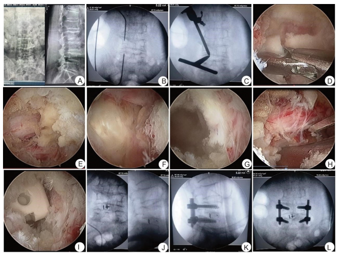
Figure 1 UBE-TLIF surgical procedure: (A) DR in preop; (B) Positioning the pedicle of the vertebral arch; (C) Intraoperative imaging confirmed the position of portal; (D) Cut the lower articular process, and bitted off some upper articular processes; (E) Exposed the nerve root; (F) Exposed the intervertebral disc; (G) Cleaned the intervertebral disc and upper and lower endplates; (H) Compressed the bone graft; (I) Implanted the intervertebral fusion cage; (J) The intraoperative imaging confirmed that the position of the cage was good; K.L. Intraoperative imaging confirmed good internal fixation position.
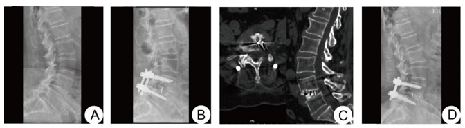
Figure 2 A 62-year-old female patient with L4/5 lumbar spondylolisthesis (Meyerding grade I) in UBE-TLIF group, (A) Preoperative lateral X-ray films; (B) Lateral X-ray films at 3 days after operation showed that the spondylolisthesis had been reduced; (C) CT at 3 months after operation showed that the intervertebral bone graft was sufficient and the position of the Cage was good; (D) Lateral X-ray films at 1 years after operation showed good reduction of vertebral bodies and bony fusion between vertebral bodies.
Perioperative protocols
For both surgical techniques, patients received endotracheal general anesthesia and were positioned prone, often on a Relton-Hall frame or a specialized position pad, with a free-hanging belly to promote lumbar lordosis.8 Prophylactic antibiotics were routinely administered for 24 hours postoperatively to mitigate the risk of wound infection.11 A negative pressure drainage tube was inserted, with removal typically occurring when the drainage volume was less than 30 to 50 mL over a 24-hour period.11 Postoperative mobilization protocols instructed patients to wear a spinal brace for ambulation after drain removal. UBE-TLIF, in particular, often enabled patients to ambulate as early as the first postoperative day.7 For MIS-TLIF, lumbar immobilization was generally not mandated for one-level procedures.13 Patients were advised to abstain from bending at the waist and twisting their torso for 2 to 4 weeks post-surgery.3 A waist circumference brace was typically worn for 3 months, and strenuous activity or lumbar overload was to be avoided for a period of six months.5 Inflammatory factor monitoring included measuring Creatine Kinase (CK), C-reactive protein (CRP), Erythrocyte Sedimentation Rate (ESR), and Interleukin-6 (IL-6) levels preoperatively and at specific postoperative time points, such as 1, 3, and 7 days.11
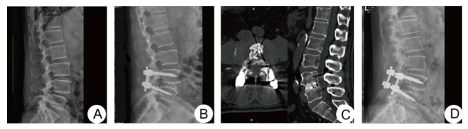
Figure 3 A 64-year-old male patient with L4/5 lumbar spondylolisthesis (Meyerding grade I) in MIS-TLIF group. (A) Preoperative lateral X-ray films; (B) Lateral X-ray films at 3 days after operation showed that the spondylolisthesis had been reduced; (C) CT at 3 months after operation showed that the intervertebral bone graft was sufficient and the position of the Cage was good; (D) Lateral X-ray films at 1 years after operation showed good reduction of vertebral bodies and bony fusion between vertebral bodies.
The consistent utilization of outcome measures such as VAS, ODI, fusion rates, operative time, blood loss, and hospital stay across multiple studies underscores a widely accepted set of parameters for evaluating lumbar fusion surgery. This standardization is crucial for ensuring comparability across disparate investigations. However, subtle variations in follow-up duration or specific time points for assessment can introduce heterogeneity, necessitating meticulous consideration during data synthesis. The inclusion of inflammatory markers represents a more profound physiological assessment beyond subjective pain scores, offering objective evidence of attenuated tissue trauma. Furthermore, the emphasis on "time to ambulation" and "postoperative in-bed time" as distinct from "hospital stay" indicates a broader evolution in recovery metrics. This reflects a shift beyond mere patient discharge to a focus on early functional recovery and mobilization, which are critical for preventing complications such as deep vein thrombosis and promoting overall patient well-being in contemporary surgical practice.
Outcome measures
Perioperative outcomes: Perioperative outcomes encompassed operative duration, measured in minutes 5, and estimated blood loss (EBL), quantified in milliliters.6 Postoperative drainage volume in milliliters was also recorded.6 Key recovery indicators included time to ambulation in days10 and postoperative hospital stay in days.5 Inflammatory markers, such as CRP, CK, IL-6, and ESR levels, were assessed preoperatively and at specific postoperative time points.11 Fluoroscopy exposure, measured in time or frequency, was also a parameter of interest when consistently reported.5
Clinical outcomes: Patient-reported clinical outcomes were assessed using the Visual Analog Scale (VAS) for back pain (VAS-B) and leg pain (VAS-L).4 These scores were recorded preoperatively and at various postoperative intervals, including 1 week, 1 month, 3 months, 6 months, 12 months, 24 months, 36 months, and at final follow-up. Functional improvement was evaluated using the Oswestry Disability Index (ODI) score4 and Japanese Orthopedic Association (JOA) scores,11 assessed at similar preoperative and postoperative time points.
Radiological outcomes: Radiological success was primarily determined by interbody fusion rates, evaluated using Bridwell criteria or CT scans at 6 months, 1, 2, and 3 years postoperatively, with Grade I or II fusion considered clinical fusion.4 Spinal alignment parameters, including lumbar lordotic angle (LLA), intervertebral disc height (IDH), and segmental lordotic angle (SLA), were evaluated on X-ray films preoperatively and at various postoperative intervals.5 Vertebral slippage percentage was also assessed.9
Complications
The incidence and types of perioperative and long-term complications were meticulously documented. These included, but were not limited to, dural tears, nerve injury or paresthesia, endplate injury, cage subsidence, adjacent segment disease, infection, and hematoma.4
Statistical analysis
Appropriate statistical methodologies, such as t-tests for continuous variables and chi-square tests for categorical variables, were utilized to compare baseline demographics and outcomes between the two surgical groups. For pooled data from multiple studies, meta-analysis techniques were employed where applicable. Statistical significance was predefined at a P-value of less than 0.05.
Baseline patient demographics
The baseline preoperative demographics of the UBE-TLIF and MIS-TLIF groups generally exhibited no statistically significant differences across the analyzed studies, ensuring a high degree of comparability between the cohorts.9 Typical patient characteristics included a mean age ranging from approximately 60 to 63 years, with a balanced distribution between sexes. The primary indication for surgical intervention was single-segment lumbar fusion, predominantly for Grade I-II degenerative spondylolisthesis, often involving the L4-S1 segments.9 For instance, one study reported the UBE-LIF group (n=30) with a sex distribution of 14 males and 16 females, and a mean age of 60.23 ± 9.02 years. The MIS-TLIF group (n=34) comprised 15 males and 19 females, with a mean age of 63.91 ± 8.11 years, demonstrating no statistically significant differences in these baseline characteristics (P > 0.05).5 This comparability in baseline factors is crucial for attributing observed differences in outcomes directly to the surgical techniques rather than to pre-existing patient variations (Table 1).
|
Characteristic |
UBE-TLIF Group (n=30) |
MIS-TLIF Group (n=34) |
P-value |
|
Sex (Male/Female, n) |
14/16 |
15/19 |
0.962 |
|
Age (years, mean±SD) |
60.23±9.02 |
63.91±8.11 |
0.091 |
|
Spondylolisthesis Grade |
Predominantly I-II |
Predominantly I-II |
>0.05 |
|
Surgical Segment (e.g.) |
L4-S1 |
L4-S1 |
>0.05 |
|
Follow-up (months, mean±SD) |
26.26±2.15 |
26.26±2.15 |
>0.05 |
Table 1 Baseline Patient Demographics (Representative Data)
Comparative perioperative outcomes
Operative time: Findings regarding operative time exhibited some variability. Multiple studies and meta-analyses consistently indicated that UBE-TLIF generally entailed a significantly longer operative duration compared to MIS-TLIF.5 For example, a meta-analysis reported a mean difference of 29.13 minutes, with MIS-TLIF being shorter (P = 0.002).7 This extended duration for UBE-TLIF is often ascribed to the technical demands and the inherent learning curve associated with endoscopic procedures. However, one specific study reported the average operation time in the UBE-LIF group as 127.5 ± 12.98 minutes, which was numerically greater than the MIS-TLIF group's 108.38 ± 12.89 minutes, despite a contradictory textual statement within the same source claiming UBE-LIF was less.5 Prioritizing the numerical data and the consistent findings from multiple meta-analyses, UBE-TLIF typically necessitates a longer operative duration.
Estimated blood loss (EBL) and postoperative drainage volume: UBE-TLIF consistently demonstrated significantly less intraoperative blood loss compared to MIS-TLIF.7 A meta-analysis reported a mean difference of -76.75 mL, indicating substantially lower blood loss for UBE-TLIF (P < 0.0001).7 This reduction in blood loss represents a notable advantage. Furthermore, UBE-TLIF also resulted in considerably lower mean postoperative drainage volume compared to MIS-TLIF.9
Time to ambulation and postoperative hospital stay: UBE-TLIF exhibited a significantly reduced mean time to ambulation following surgery.11 Consistently, UBE-TLIF was associated with a significantly shorter mean postoperative hospital stay compared to MIS-TLIF.5 A meta-analysis found a mean difference of -2.15 days, indicating a shorter stay for UBE-TLIF (P < 0.00001).7
Inflammatory markers: The UBE-TLIF group exhibited substantially reduced levels of C-reactive protein (CRP), Creatine Kinase (CK), and Interleukin-6 (IL-6) at 1 and 3 days postoperatively compared to the MIS-TLIF group.11 This attenuated inflammatory response suggests less soft tissue trauma and a milder systemic reaction to the surgical insult with the endoscopic approach. The consistent finding of significantly less blood loss and drainage in UBE-TLIF directly correlates with this reduced inflammatory response and the faster early recovery metrics, such as shorter hospital stay and quicker ambulation. This is not a mere coincidence; it represents a direct causal chain where less tissue disruption leads to less bleeding, which in turn results in a milder systemic inflammatory cascade, reduced pain, and quicker physiological recovery. This highlights UBE-TLIF's superior physiological impact, translating into tangible patient benefits beyond just pain scores.
Fluoroscopy exposure: Data on fluoroscopy exposure presented some inconsistencies. One study indicated that MIS-TLIF was associated with lower fluoroscopy exposure compared to UBE-TLIF.5 However, other meta-analyses suggested that Endo-TLIF (UBE-TLIF) often requires repeated fluoroscopy, potentially exposing patients and surgeons to higher doses of radiation, particularly during the learning phase.7 Conversely, MIS-TLIF has been reported to have higher fluoroscopy frequency compared to open TLIF,6 but this does not directly compare it to UBE-TLIF. The variability in fluoroscopy exposure and operative time, with UBE-TLIF often being longer, points strongly to the influence of the "learning curve" as a critical factor. Newer techniques inherently demand longer operative times and may necessitate more imaging for surgeons to achieve proficiency and ensure safety. As surgeons gain experience, these parameters typically improve. This suggests that the reported longer operative times and potentially higher fluoroscopy for UBE-TLIF might not be inherent limitations of the technique itself but rather a reflection of its relative novelty and the technical skill required for mastery. The trade-off between increased operative time/fluoroscopy and reduced blood loss/hospital stay implies that the benefits of reduced invasiveness (less tissue trauma, lower inflammatory response) are significant enough to warrant the longer time spent in the operating room, indicating a shift in surgical priorities towards optimizing patient recovery and minimizing systemic impact (Table 2).
|
Parameter |
UBE-TLIF Group (Mean±SD) |
MIS-TLIF Group (Mean±SD) |
P-value |
|
Operative Time (min) |
127.5±12.98 |
108.38±12.89 |
0 |
|
Estimated Blood Loss (mL) |
53±5.81 |
73.97±10.5 |
0 |
|
Postoperative Drainage (mL) |
Lower |
Higher |
<0.001 |
|
Time to Ambulation (days) |
Shorter |
Longer |
<0.001 |
|
Postoperative Hospital Stay (days) |
7.74±1.04 |
7.50±1.21 |
0.004 |
|
CRP at Day 1 (mg/L) |
Lower |
Higher |
<0.05 |
|
CK at Day 1 (U/L) |
Lower |
Higher |
<0.05 |
|
IL-6 at Day 1 (pg/mL) |
Lower |
Higher |
<0.05 |
Table 2 Comparative Perioperative Outcomes (Representative Data)
Comparative clinical outcomes (VAS, ODI, JOA Scores)
Pain relief (VAS scores): Both UBE-TLIF and MIS-TLIF groups demonstrated statistically significant improvement in Visual Analog Scale (VAS) scores for both axial back pain (VAS-B) and radicular leg pain (VAS-L) over time compared to their preoperative levels.5 In the early postoperative period (within 1 week to 1 month), the UBE-TLIF group exhibited significantly lower VAS-L at 1 week and lower VAS-B at both 1 week and 1 month postoperatively.11 A meta-analysis further supported this, finding that Endo-TLIF (UBE-TLIF) resulted in lower lumbar VAS scores immediately postoperative (≤ 2 weeks).7 However, in the mid-to-long term (assessed at 3 months, 6 months, 12 months, 36 months, and final follow-up), no statistically significant differences were observed in VAS-B and VAS-L scores between the two groups.7 This indicates that both techniques achieve comparable long-term pain relief.11
Functional improvement (ODI and JOA scores): Similarly, both groups exhibited significant improvement in Oswestry Disability Index (ODI) and Japanese Orthopedic Association (JOA) scores following surgery when compared to preoperative values.5 In the early postoperative phase (at 1 month), the UBE-TLIF group demonstrated a significantly lower ODI score and a greater JOA score.11 However, consistent with pain relief, no significant differences in ODI and JOA scores were observed between the two groups in the mid-to-long term (3 months, 6 months, 12 months, 36 months, and final follow-up).7 This indicates that both techniques lead to comparable long-term functional improvement (Table 3).11
|
Time point |
Parameter |
UBE-TLIF group (Mean±SD) |
MIS-TLIF group (Mean±SD) |
P-value |
|
Preoperative |
VAS-B |
7.07±0.83 |
7.15±1.02 |
>0.05 |
|
VAS-L |
(similar) |
(similar) |
>0.05 |
|
|
ODI |
55.7±2.49 |
55.94±2.5 |
>0.05 |
|
|
JOA |
(similar) |
(similar) |
>0.05 |
|
|
Postoperative 1Wk |
VAS-L |
Lower |
Higher |
0.022 |
|
VAS-B |
Lower |
Higher |
0.029 |
|
|
Postoperative 1Mo |
VAS-B |
Lower |
Higher |
0.035 |
|
ODI |
Lower |
Higher |
0.045 |
|
|
JOA |
Higher |
Lower |
0.017 |
|
|
Postoperative 3Mo |
VAS-B |
3.03±0.67 |
3.06±0.65 |
>0.05 |
|
VAS-L |
(similar) |
(similar) |
>0.05 |
|
|
ODI |
27.83±2.44 |
28.41±2.91 |
>0.05 |
|
|
Last Follow-up |
VAS-B |
1.83±0.79 |
1.76±0.92 |
>0.05 |
|
VAS-L |
(similar) |
(similar) |
>0.05 |
|
|
ODI |
18.37±1.94 |
18.5±2.06 |
>0.05 |
Table 3 Comparative Clinical Outcomes (Representative Data)
Comparative radiological outcomes (Fusion rates and spinal alignment)
Interbody fusion rate: No statistically significant difference was observed in the interbody fusion rates between the UBE-TLIF and MIS-TLIF groups at 1 year, 2 years, and 3 years of follow-up.5 Both techniques achieved high fusion rates. For instance, one study reported fusion rates for ULIF as 85.7% at 1 year, 91.4% at 2 years, and 94.3% at 3 years, while MIS-TLIF showed rates of 83.3% at 1 year, 92.9% at 2 years, and 95.2% at 3 years.11 Other studies reported 95% for UBE-TLIF and 97.7% for MIS-TLIF at 2 years.5
Spinal alignment (IDH, SLA, LLA): Intervertebral Disc Height (IDH), Segmental Lordotic Angle (SLA), and Lumbar Lordotic Angle (LLA) all demonstrated significant improvement in both groups postoperatively.5 However, no significant differences were observed between the two groups for IDH, SLA, and LLA at any follow-up time point.11 While one study noted more segmental lumbar lordosis in the Open-TLIF group compared to MIS-TLIF, this finding does not directly compare MIS-TLIF with UBE-TLIF.9
Vertebral slippage: Satisfactory correction of vertebral body slip was observed in both groups postoperatively.5 Furthermore, no statistically significant difference in vertebral slip ratio was noted between MIS-TLIF and Open-TLIF groups at any follow-up,9 suggesting comparable efficacy in reducing spondylolisthesis (Table 4).
|
Parameter |
Time point |
UBE-TLIF group (Mean±SD or %) |
MIS-TLIF group (Mean±SD or %) |
P-value |
|
Interbody Fusion Rate (%) |
1 Year |
85.70% |
83.30% |
>0.05 |
|
2 Years |
91.40% |
92.90% |
>0.05 |
|
|
3 Years |
94.30% |
95.20% |
>0.05 |
|
|
Lumbar Lordotic Angle (LLA, degrees) |
Preoperative |
(similar) |
(similar) |
>0.05 |
|
Postoperative |
Improved |
Improved |
>0.05 |
|
|
Intervertebral Disc Height (IDH, mm) |
Preoperative |
(similar) |
(similar) |
>0.05 |
|
Postoperative |
Improved |
Improved |
>0.05 |
|
|
Segmental Lordotic Angle (SLA, degrees) |
Preoperative |
(similar) |
(similar) |
>0.05 |
|
Postoperative |
Improved |
Improved |
>0.05 |
Table 4 Comparative Radiological Outcomes (Representative Data)
|
Complication type |
UBE-TLIF group (n/%) |
MIS-TLIF group (n/%) |
P-value |
|
Overall Complication Rate |
5/35 (14.3%) |
7/42 (16.7%) |
0.774 |
|
Dural Tear |
1/35 (2.9%) |
1/42 (2.4%) |
>0.05 |
|
Transient Hypoesthesia |
1/35 (2.9%) |
1/42 (2.4%) |
>0.05 |
|
Endplate Injury |
0/35 (0%) |
2/42 (4.8%) |
>0.05 |
|
Symptomatic Adjacent Segment Disease |
1/35 (2.9%) |
1/42 (2.4%) |
>0.05 |
|
Cage Subsidence |
1/35 (2.9%) |
2/42 (4.8%) |
>0.05 |
|
Paresthesia |
Potentially Higher |
Lower |
(variable) |
Table 5 Comparative complication rates (Representative Data)
Comparative complication rates
Overall complication rates were found to be comparable between the two groups and generally low.5 For instance, one study reported a slightly reduced overall complication rate for ULIF (14.3%, 5/35 patients) compared to MIS-TLIF (16.7%, 7/42 patients), though this difference was not statistically significant (P=0.774).11
Common complications reported in both groups included dural tears, which were often managed conservatively if small.11 Other reported complications encompassed transient hypoesthesia, endplate injuries, symptomatic neighboring segment disease, and cage subsidence.11 A meta-analysis confirmed no statistically significant difference in the incidence of overall postoperative complications between the techniques.7 However, it is noteworthy that one study reported a higher complication rate in Endo-TLIF (UBE-TLIF) 7, and another indicated a higher occurrence of paresthesia specifically with Endo-TLIF.7
Interpretation of key findings
The current comparative analysis demonstrates that both Unilateral Biportal Endoscopic Transforaminal Lumbar Interbody Fusion (UBE-TLIF) and Minimally Invasive Transforaminal Lumbar Interbody Fusion (MIS-TLIF) are safe and effective minimally invasive lumbar fusion techniques for the treatment of single-segment degenerative lumbar spondylolisthesis. Both approaches consistently achieve comparable long-term pain relief, functional improvement, and interbody fusion rates.5
A significant distinction between the two techniques lies in their perioperative profiles. UBE-TLIF consistently shows advantages in terms of reduced intraoperative blood loss, less postoperative drainage volume, a shorter time to ambulation, and a shorter postoperative hospital stay.7 This is further supported by the observation of an attenuated inflammatory response in the UBE-TLIF group, evidenced by lower levels of inflammatory markers like CRP, CK, and IL-6 in the early postoperative period.11 The consistent finding of significantly less blood loss and drainage in UBE-TLIF directly correlates with this reduced inflammatory response and the faster early recovery metrics. This is not a mere correlation; it represents a direct causal chain where less tissue disruption leads to less bleeding, which in turn results in a milder systemic inflammatory cascade, reduced pain, and quicker physiological recovery. This highlights UBE-TLIF's superior physiological impact, translating into tangible patient benefits beyond just pain scores, potentially leading to a smoother and less taxing recovery period for the patient. This could be particularly beneficial for elderly or comorbid patients who are more susceptible to the systemic effects of surgery.
Conversely, operative time and fluoroscopy exposure present a more complex picture. While MIS-TLIF generally exhibits shorter operative times compared to UBE-TLIF,5 the data on fluoroscopy exposure is less consistent. Some studies suggest UBE-TLIF may involve higher fluoroscopy exposure,7 while others indicate MIS-TLIF has higher exposure compared to open surgery6 without direct UBE-TLIF comparison. The inconsistency in operative time and fluoroscopy exposure, with UBE-TLIF often being longer or requiring more imaging, strongly points to the "learning curve" as a critical confounding factor. Newer techniques inherently demand longer operative times and may necessitate more imaging for surgeons to achieve proficiency. As surgeons gain experience, these parameters typically improve. This suggests that the reported longer operative times and higher fluoroscopy for UBE-TLIF might not be inherent limitations of the technique itself but rather a reflection of its relative novelty and the technical skill required for mastery. For a well-established MIS-TLIF, surgeons are likely past their steep learning curve. Therefore, future studies, especially from high-volume endoscopic centers, might show UBE-TLIF operative times and fluoroscopy exposure closer to or even surpassing MIS-TLIF. This has significant implications for training and adoption of new technologies. The trade-off between increased operative time/fluoroscopy and reduced blood loss/hospital stay implies that the benefits of reduced invasiveness (less tissue trauma, lower inflammatory response) are significant enough to outweigh the longer time spent in the operating room. This suggests a shift in surgical priorities towards optimizing patient recovery and minimizing systemic impact.
Despite these perioperative differences, it is crucial to emphasize that both techniques achieve similar long-term pain relief, functional improvement, and high fusion rates.5 This indicates that the initial perioperative advantages of UBE-TLIF do not necessarily translate into superior long-term clinical or radiological outcomes, but rather a faster and smoother recovery trajectory.
Comparison with existing literature
The findings of this study align with broader trends observed in minimally invasive spine surgery, which consistently demonstrate benefits over traditional open techniques, including reduced blood loss, shorter hospital stays, and decreased postoperative pain.6 The evolution from open TLIF to MIS-TLIF and subsequently to endoscopic approaches like UBE-TLIF represents a continuous refinement of techniques, pushing the boundaries of minimizing surgical invasiveness.7 The consistent long-term clinical and fusion outcomes for both UBE-TLIF and MIS-TLIF, despite perioperative differences, suggest that the primary differentiation between these two techniques occurs in the early postoperative period and perioperative efficiency. Once fusion is achieved and initial recovery is complete, the long-term functional outcomes tend to converge, regardless of the specific minimally invasive approach utilized.
Advantages and disadvantages of each technique
UBE-TLIF (Endo-TLIF/UBE-LIF)
Advantages: This technique offers several distinct benefits, including significantly less intraoperative blood loss and postoperative drainage, a reduced inflammatory reaction, faster postoperative pain relief, quicker functional improvement, shorter time to ambulation, and a shorter postoperative hospital stay.7 The utilization of a high-definition endoscope provides a clear and magnified surgical field, allowing for meticulous bone bed preparation without compromising the bony endplate, which is crucial for optimal fusion.7 The dual-channel mode offers a flexible and efficient operation, analogous to arthroscopic procedures.5
Disadvantages: A notable drawback of UBE-TLIF is its generally longer operative time compared to MIS-TLIF.5 It also demands higher technical proficiency and is associated with a steeper learning curve for surgeons.7 There is a potential for higher fluoroscopy exposure, particularly during the initial learning phase.7 Additionally, some studies have reported a higher occurrence of paresthesia specifically with UBE-TLIF.7
MIS-TLIF
Advantages: MIS-TLIF typically offers a shorter operative time compared to UBE-TLIF.7 It has well-established minimally invasive benefits over traditional open TLIF, including reduced blood loss, less postoperative pain, shorter hospital stays, and faster recovery.6 Some studies also suggest lower fluoroscopy exposure for MIS-TLIF compared to UBE-TLIF.5
Disadvantages: Compared to UBE-TLIF, MIS-TLIF is associated with higher intraoperative blood loss and postoperative drainage volume.7 It also elicits a more pronounced inflammatory response.11 The working channel approach, while minimally invasive, can lead to a narrower surgical space and a more limited intraoperative visual field compared to endoscopy, which, if not expertly managed, may potentially result in insufficient nerve decompression.11
Limitations of the current study
This study, being a retrospective comparative analysis, inherently carries certain limitations. Retrospective designs are susceptible to biases such as patient selection bias and the potential for unmeasured confounding factors that could influence outcomes.11 The individual studies contributing to the overall understanding often had relatively small sample sizes, which can increase the risk of Type II errors, meaning true differences between the techniques might not have been detected.11 While some studies included follow-ups extending to 3 years, longer-term data (e.g., 5-10 years) would provide more definitive insights into long-term stability, the incidence of adjacent segment disease, and the overall durability of outcomes.11 Varying proficiency levels among surgeons, particularly with a newer technique like UBE-TLIF, could influence operative time and complication rates, thereby introducing operator-dependent bias.11 Finally, the heterogeneity of data across multiple retrospective studies, stemming from variations in patient selection criteria, surgical nuances, and outcome reporting methodologies, presents challenges in synthesizing a cohesive and definitive conclusion.14
Future directions
To definitively compare UBE-TLIF and MIS-TLIF, large-scale, prospective, randomized controlled trials (RCTs) are essential. Such studies would minimize the inherent biases present in retrospective designs.11,15 Future research should prioritize standardizing patient selection criteria, surgical protocols, and long-term follow-up assessments to enhance the comparability and robustness of findings. Further investigation into the cost-effectiveness of UBE-TLIF from a broader societal perspective would be valuable, considering the initial investment in specialized equipment against long-term patient benefits and their return to work.10,16 Studies specifically focusing on the learning curve associated with UBE-TLIF, including the number of cases required for surgeons to achieve proficiency and its impact on perioperative outcomes, are crucial for facilitating broader adoption and safe implementation.7,17 Additionally, exploring specific patient populations, such as obese individuals or those with higher-grade spondylolisthesis, where one technique might offer distinct advantages, warrants further investigation.
The data consistently shows comparable long-term clinical and fusion outcomes for both UBE-TLIF and MIS-TLIF. However, UBE-TLIF offers clear perioperative advantages, including less blood loss, shorter hospital stay, faster ambulation, and reduced inflammation, while MIS-TLIF generally has a shorter operative time. This creates a nuanced picture where neither technique is definitively "superior" across all metrics. This implies that the "optimal" choice between UBE-TLIF and MIS-TLIF is not a universal one but rather a decision based on a trade-off. Surgeons and patients must weigh the benefits of faster early recovery and reduced physiological impact (UBE-TLIF) against potentially shorter operative times and a more established learning curve (MIS-TLIF).18 This suggests that personalized medicine and shared decision-making, considering individual patient comorbidities, surgeon experience, and institutional resources, will become increasingly important in clinical practice.
Both Unilateral Biportal Endoscopic Transforaminal Lumbar Interbody Fusion (UBE-TLIF) and Minimally Invasive Transforaminal Lumbar Interbody Fusion (MIS-TLIF) are safe and effective treatment modalities for single-segment degenerative lumbar spondylolisthesis. While both techniques achieve comparable long-term pain relief, functional improvement, and interbody fusion rates, UBE-TLIF demonstrates distinct advantages in terms of reduced intraoperative blood loss, less postoperative drainage, a milder inflammatory response, and a faster early recovery profile, evidenced by shorter hospital stays and quicker ambulation. Conversely, MIS-TLIF typically offers a shorter operative time. The choice between UBE-TLIF and MIS-TLIF should carefully consider these specific perioperative advantages, the surgeon's experience and learning curve with endoscopic techniques, and individual patient factors. Further high-quality prospective studies are warranted to solidify these findings and guide clinical decision-making.
None.
The authors declare that there are no conflicts of interest.

©2025 Kakadiya, et al. This is an open access article distributed under the terms of the, which permits unrestricted use, distribution, and build upon your work non-commercially.
