Research Article Volume 13 Issue 4
Questions of the current state of treatment of post-traumatic deformity of the distal radial metaepiphysis
AS Botasheva,1 NG Shikhaleva,1 KI Novikov,1,2 MM Bari,3 
Regret for the inconvenience: we are taking measures to prevent fraudulent form submissions by extractors and page crawlers. Please type the correct Captcha word to see email ID.

S Bari3
1National Medical Research Center of Traumatology and Orthopedics named after Academician G.A. Ilizarov “of the Ministry of Health of the Russian Federation, Kurgan, Russia
2Federal State Budgetary Educational Institution of Higher Education “Tyumen State Medical University” of the Ministry of Health of the Russian Federation (FSBEI HE Tyumen State Medical University of the Ministry of Health of Russia)
3Bari-Ilizarov Orthopaedic Centre, Dhaka, Bangladesh
Correspondence: Bari MM, Bari-Ilizarov Orthopaedic Centre, 1/1, Suvastu Shirazi Square, Lalmatia Block E, Dhaka-1207, Bangladesh, Tel +8801819211595
Received: July 30, 2021 | Published: August 13, 2021
Citation: Botasheva AS, Shikhaleva NG, Novikov KI, et al. Questions of the current state of treatment of post-traumatic deformity of the distal radial metaepiphysis. MOJ Orthop Rheumatol. 2021;13(4):85-88. DOI: 10.15406/mojor.2021.13.00554
Download PDF
Abstract
Based on the analysis of modern scientific Russian and foreign literature, this article includes information about the current state of the treatment of post-traumatic deformity of the distal metaepiphysis of the radius. The statistical information on the number of registered cases with this pathology, gender characteristics of injury, as well as some of the diagnostic radiological aspects is briefly considered.
Keywords: deformities of the distal metaepiphysis of the radius, post-traumatic deformity, traumatization
Introduction
Anatomically, the distal part of the radius is presented as an irregularly shaped figure, an important role of which is the formation of the wrist and radioulnar joints. An incorrectly chosen algorithm for conservative treatment of fractures of the distal metaepiphysis (DM) of the radius, errors in the choice of tactics for surgical treatment of fresh injuries lead to the formation of post-traumatic deformities.1
The fracture of the distal radius was first described in 1814, but for a long time it remained a poorly understood and complex phenomenon in the orthopedic community.2 Modern scientific withStatistical data show that the unfavorable outcome of the treatment of forearm injuries varies from 13 to 66% and is expressed by the occurrence of pathological formations such as pseudoarthrosis, bone deformities, etc,3–7 a wide range of complications can be associated with the lack of a single algorithm for the provision of medical care. The age indicators of people who have received this type of injury can be divided into two large groups, according to the characteristics of frequency, namely: from 40 to 60 years old - 51%, over 60 years old - 43%.8 It should be noted that both age categories are included in the definition of able-bodied citizens, therefore, a fracture of the distal metaepiphysis of the radius carries complex medical and social consequences and requires careful research and development. Getting injured at a young age is most often due to extreme sports or high-energy loads, while at an older age, injuries occur as a result of falling, for example, from a height.9–11 There is a difference in the gender characteristics of trauma, for example: women have an increased risk of a fracture of the distal metaepiphysis of the radius after 50 years, and by the age of 80, the peak of this type of trauma is reached.12 This was noted on the materials of the study of Norwegian scientists, which are also confirmed by other works.13–16 Thus, the relevance of the topic under consideration is undeniable and important in the context of the listed factors.
Material and methods
The literature review includes publications from open Internet resources, namely: Cochrane Library, PubMed, eLIBRARY, release dates are 1997 and 2019. The selection of articles was carried out by the method of content analysis. Also, general scientific methods were used, such as analysis, comparison, generalization, classification, and the principles of objectivity, consistency, integrity.
Research results
Incorrectly healed DMLK fractures significantly impair the patient's quality of life, which leads to the need for surgical treatment. Let us dwell in more detail on the clinical picture and the functional state of the patient.
- Shortening of the radius and impaction of the ulna in the wrist lead to wrist and mid-carpal instability, which, in turn, causes an uneven distribution of the load on the ligamentous apparatus and articular cartilage of the wrist and radioulnar joints.17 (Figure 1).
- Incorrect treatment tactics, which contributed to the appearance of significant deformities of the distal metaepiphysis of the radial bone, leads to a discrepancy between the articular surfaces of the wrist and distal radioulnar joints, pronounced disorders in the biomechanics of movements of the wrist bones, which leads to the formation of secondary adaptive collapse of the wrist. In this case, conservative treatment is unpromising.1
- With an increase in the time of immobilization, during the treatment of fractures of the distal metaepiphysis of the radius, contractures develop in the wrist joint and finger joints.18,19
- As a result of a violation of the physiological position, due to a fracture of the DMLK and subsequent long-term, forced fixation, changes occur in the anatomy and functional state of the carpal canal and median nerve.20 Conditions for resection of a bone or bone in order to eliminate a defect in the nerve trunk occur in rare, isolated cases: extensive damage to the nerves, combined with comminuted bone fractures or their consequences; limb replantation.21 After combined injury of tendons and nerves in the distal third of the forearm, motor function disorders develop due to damage to the flexors of the fingers and paralysis of the own muscles of the hand. After delayed and repeated operations on the tendons and nerves, it is possible to restore the function of the long flexors of the fingers, the sensitivity of vegetative-trophic functions in the autonomous zone of innervation.22
- Clinical manifestations are expressed by the following aspects: limitation of movements in the wrist and distal radioulnar joints in different planes, a decrease in the grip force of the hand, the development of complex regional pain syndrome type 1 and post-traumatic arthrosis of the wrist joint, which leads to a pronounced dysfunction of the upper limb.1 Decreased grip strength in the wrist joint; deformity of the lower third of the forearm - more often radial deviation of the hand; - post-traumatic neuropathy with the development of tunnel syndrome; Zudeck's syndrome.23

Figure 1 Radiographs of the wrist joint of patient D., 42 years old, a - before the operation; B - one year after the operation.17
Discussion
Treatment of post-traumatic deformity of DMLK is of two types: conservative and operative. Mulders M., considering the results of his practice, is convinced that the effectiveness of both surgical and conservative treatment is on an equal level.24 Recently introduced internal fixation devices have provided hand surgeons with additional options for treating intra-articular fractures of the distal radius. However, modern technologies of X-ray examinations and surgical support, as well as the availability of new means of bone osteosynthesis, are the impetus for surgical operations that improve the quality of life. This process is common to orthopedic traumatologists around the world.25 However, visualization of the articular surface of the distal radius after the plate has been applied can be difficult on standard straight and lateral radiographs due to the location of the plate directly below the articular surface. Although several studies of cadaveric material have shown that angular radiographs can better demonstrate the articular surface. No study comparing the efficacy of standard straight and lateral distal radial images with angular radiographs for evaluating fixation devices dorsally placed on the distal radius has not been performed.26 Thus, improperly fused DMLK fractures lead to a number of negative consequences for the functioning of the hand, wrist, and radioulnar joints. The lack of surgical treatment will lead to irreversible consequences, however, an important role is played by rehabilitation measures, which allow preventing the development of contractures, edema, and neurodystrophic syndromes. Alsothe presence of an intra-articular fracture of the distal joint can lead to the development of premature arthritis of the wrist, elbow and distal wrist joints. Numerous studies have shown that residual articular deformity of 1-2 mm during fracture healing, attributed to signs of arthrosis according to X-ray studies, leads to negative results.27 On the contrary, in additional studies,28,29 concluded that symptomatic arthritis is rare after intra-articular fractures of the distal radius, despite radiological signs of arthrosis. Since each of these studies has shown that persistent residual joint misalignment at the time of fracture connection leads to radiological signs of premature arthrosis (but not necessarily arthritis), it seems reasonable that the treating surgeon should achieve as close anatomical fusion as possible in order to minimize the development of early degenerative changes. Accurate assessment of the distal radial articular surface (pre-reduction and post-reduction) requires careful radiographic evaluation.
Conclusion
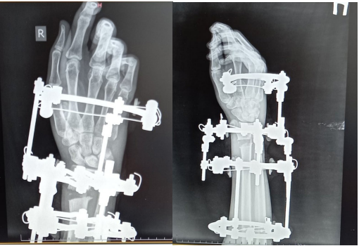
The existing progress in medical science allows us to revise the established views on the issue of DMLK treatment, suggesting the use of technically improved equipment, other methodological aspects at the preoperative stage of working with a patient, and much more. However, it should be noted that, despite all of the above, the use of external osteosynthesis, despite its popularity, does not solve the issues of minor post-traumatic deformity (1-2 mm), which causes osteoarthritis and arthritis of the affected segment, and also complicates the X-ray observation of DMLK and postpones the start of rehabilitation measures. Thus, the use of transosseous osteosynthesis according to Ilizarov has a number of immediate advantages (Figure 2 & 3).

Figure 2 Functional state of the wrist joint of patient D., 19 years old, before surgery.

Figure 3 Radiographs of the wrist joints of the patient D. 19 years old at the stage of treatment with the device G.А. Ilizarov.
Acknowledgments
Conflicts of interest
The authors declare no conflicts of interest.
References
- Malets VL. Method of bone autotransplantation in the surgical treatment of post-traumatic deformity of the distal metaepiphysis of the radial bone. 2017;3.
- Colles A. On the fracture of the carpal extremity of the radius. Edinb Med Surg J;1814;10:181. Clin Orthop Relat Re. 2006;445:5–7.
- Borzunov D Yu. Replacement of long bone defects by polylocal lengthening of fragments. Traumatology and Orthopedics of Russia. 2006;4(42):S24–S29.
- Afaunov AI, Afaunov AA, Afaunov AI, et al. Nat. Congress. SPb., 2003.S. 36.
- Kolchanov KV, Sokolova MN, Borzunov DYu. The functional state of the muscles of the forearm and hand of patients with acquired defects and pseudarthrosis of the forearm bones at the stages of rehabilitation using the method of transosseous osteosynthesis. The genius of Orthopedics. 2010;4:90.
- VI Shevtsov, VD Makushin, LM Kuftyrev, et al. Pseudoarthrosis, defects of the long bones of the upper limb and contractures of the elbow joint. Kurgan, 2001.406 p.
- Slobodsky AB, Popov AB. Osteosynthesis of the forearm bones with external fixation devices.Man and his health: orthopedics-traumatology-prosthetics-rehabilitation: collection of articles. thesis. 9 Ros. nat. Congress. SPb., 2004. S. 99.
- Bartl C. The treatment of displaced intra-articular distal radius fractures in elderly patients. Dtsch Arztebl Int. 2014;111(46):779–787.
- Azad A. Epidemiological and treatment trends of distal radius fractures across multiple age groups. J Wrist Surg. 2019;8(4):305–311.
- Channareddy H. Epidemiological profile of articular fractures of distal radius. Nat J Clin Orthop. 2018;2(3):17–20.
- Vosbikian MM. Optimal Positioning for Volar Plate Fixation of a Distal Radius Fracture: Determining the Distal Dor-sal Cortical Distance. Orthop Clin North Am. 2016;47(1):235–244.
- Solvang HW. Epidemiology of distal radius fracture in Akershus, Norway, in 2010–2011. J Orthop Surg Res. 2018;13(13):199.
- Abe Y. Management of intra-articular distal radius frac-tures: volar or dorsal locking plate - which has fewer complications. 2017;12(6):561–567.
- Jo YH. Incidence and Seasonal Variation of Distal Radius Fractures in Korea: a Population-based Study. J Korean Med Sci. 2018;33(7):e48.
- Stirling ERB. Epidemiology of distal radius fractures in a geographically defined adult population. J Hand Surg Eur. 2018;4(9):974–982.
- DH Toon, RAX Prem-chand, J Sim, et al. Outcomes and financial implications of intra-articular distal radius fractures: a comparative study of open re-duction and internal fixation (ORIF) with volar locking plates versus non operative management. J Orthop Traumataol. 2017;18:229–234.
- Semenkin OM, Izmalkov SN. A method for eliminating post-traumatic deformity of the distal metaepiphysis of the radial bone. Traumatology and Orthopedics of Russia.
- Rushai AK, Lisunov SV. Prevention and early treatment of post-traumatic neurodystrophic syndrome in fractures of the distal metaepiphysis of the radius in patients with diabetes mellitus. Trauma. 2016;17(6):40.
- Kirillov VI. Experience of surgical treatment of fractures of the distal metaepiphysis of the radius in elderly patients. All-Russian Congress of the Society of Hand Surgeons (June 2-3, 2016, Nizhny Novgorod). Materials of the Congress: FSBI "PFMITs" of the Ministry of Health of Russia, 2016. 63 p.
- Golubev IO, Maksimov AA, Merkulov MV, et al. Bone grafting in the treatment of patients with incorrectly fused fractures of the distal metaepiphysis of the radial bone. M. CITO. 2014. p. 24.
- Sushkov AN. Surgical treatment of chronic damage to the peripheral nerve trunks at the level of the forearm, hand and fingers. Author dis. Cand honey sciences. 2010.S. 5.
- Volkova AM. Hand surgery. Vol 1. Yekaterinburg. P. 209.
- Ugleev OI, Fedorov VN, Ivanov VL, et al. Surgical treatment of post-traumatic deformity of the distal metaepiphysis of the radial bone in the conditions of the Department of Traumatology and Orthopedics of the Republican Clinical Hospital of the Ministry of Health of Chuvashia. Health of Chuvashia. 2018:26.
- Mulders MAM. Classification and treatment of distal radius fractures: a survey among orthopedic trauma surgeons and residents. Eur J Trauma Emerg Surg. 2017;43(2):239–248.
- Armstrong KA. Stable rates of operative treatment of distal radius fractures in Ontario, Canada: a population-based retrospective cohort study (2004–2013). Can J Surg. 2019;62(6):386–392.
- Martin I Boyer, Kenneth J Korcek, Richard H Gelberman, et al. Anatomic Tilt X-Rays of the Distal Radius: An Ex Vivo Analysis of Surgical Fixation. J Hand Surg Am. 2004;29(1):116–122.
- Louis W Catalano, O Alton Barron, Steven Z Glickel. Assessment of Articular Displacement of Distal Radius Fractures. Clin Orthop Relat Res. 2004;423:79–84.
- Catalano LW, Cole RJ, Gelberman RH, et al. Displaced intra-articular fractures of the distal aspect of the radius. J Bone Joint Surg. 1997;79(9):1290–1302.
- McKay SD, MacDermid JC, Roth JH, et al. Assessment of complications of distal radius fractures and development of a com-plication checklist. J Hand Surg. 2001;26(5):916–922.

©2021 Botasheva, et al. This is an open access article distributed under the terms of the,
which
permits unrestricted use, distribution, and build upon your work non-commercially.


