MOJ
eISSN: 2374-6939


Case Report Volume 16 Issue 1
1Pediatric Orthopedic Surgeon at Instituto de Ortopedia e Traumatologia do Hospital das Clínicas of School of Mediciine, Universidade de Sao Paulo, Brazil
2Pediatric Orthopedic Surgeon at Hospital Israelita Albert Einstein, Sao Paulo, Brazil
Correspondence: Nei Botter Montenegro, Pediatric Orthopedic Surgeon at Instituto de Ortopedia e Traumatologia do Hospital das Clínicas of School of Mediciine, Universidade de Sao Paulo, Brazil
Received: December 30, 2023 | Published: January 3, 2024
Citation: Montenegro NB, Grangeiro PM, Junior AR. New posterior distal femoral hemiepiphysiodesis for treatment of recurvatum knee in a patient with cerebral palsy: a case report. MOJ Orthop Rheumatol. 2024;16(1):1-4. DOI: 10.15406/mojor.2024.16.00653
Hyperextension of the knee (Genu recurvatum) is a rare deformity in the sagittal plane which can impact on walking by pelvis’, ankle’s and foot’s biomechanics changes. It can be caused by cerebral palsy, arthrogryposis poliomyelitis, traumatic tibial tuberosity arrest and generalized ligamentous hyperlaxity. Specially in cerebral palsy, factories like hamstrings lengthening and equinus deformity of the foot can produce this knee condition. The treatment methods to correct this deformity are more aggressive to date, such as quadricepsplasty, hamstring tenomyoplasty and femur or tibia osteotomies.
We describe here the treatment of a patient with mild hemiparetic hypertonic cerebral palsy GMSCF I, with unilateral genu recurvatum, using a safe and minimally invasive technique with posterior hemiepiphysiodesis of the distal femur, performed with two transphyseal cannulated screws for correction. This technique has great potential for correcting the recurvatum knee in the immature skeleton during growth and can be a good alternative to the more aggressive methods currently used for the treatment of this deformity.
Keywords: bone retrovertion (c05.116.214.750) / orthopedic procedures (e02.718) / minimally invasive surgical procedures (e04.502) / knee joint (a02.835.585.475)/ growth and development (q000254) / growth plate (a02.835.232.251.352)/ cerebral palsy
Hyperextension of the knee (Genu recurvatum) e is a rare deformity of this joint in the sagittal plane which can impact on patient walking changing the proper knee, pelvis’, ankle’s and foot’s biomechanics. It must be treated when results on limping and can leads to complications, such as knee pain, pelvis tilting and precocious gonarthrosis. At the extreme of the deformity, there may even be anterior dislocation of the knee.1–3
Recurvatum knee is a less usual deformity at immature skeleton. Cerebral palsy,4–7 arthrogryposis,8 poliomyelitis, bone infection sequelae, some syndromes with ligament hyperlaxity generalized joint hypermobility,9 anterior femoral distal physis arrest and tibial tuberosity fracture sequelae10–12 and iatrogenic complication following eight-Plate epiphysiodesis13 care causes of this deformity.
It is important to emphasize that the treatment of the knee in hyperextension is challenger. When the deformity is significant and there is indication of correction surgery, procedures like soft tissue surgeries, such as quadricepsplasty,14 hamstring tenomyoplasty15 were done unsuccessing. Osteotomies of the distal femur and proximal tibia with internal 16 or external fixation17 for correction of bone alignment are not indicated for immature skeleton. These kinds of surgeries are offensive and require a long recovery time, dare may be neurovascular injury, infection and compartment syndrome.
Searching for less aggressive correction surgical treatment to sagittal knee hyperextension, it was used a posterior hemiepiphysiodesis of the distal femur to the recurvatum correction by guided growth. Here we intend to describe the surgical technique performed with two guided cannulated steel screws inserted in the posterior part of the distal femoral growth plate and show the clinical and radiographic outcomes of a mild hemiparetic cerebral palsy patient (GMSCF I) presenting an unilateral left recurvatum knee, treated with this method.
The operative treatment was done with the patient placed in horizontal dorsal decubitus. On the anterior aspect of the distal thigh of the affected limb, two 1 cm longitudinal incisions were made. The quadriceps muscle was crossed through blunt dissection to reach the anterior aspect of the distal femur, proximal to the growth plate.
Using images in the coronal and sagittal planes with the fluoroscopy aid, two guide wires were inserted percutaneously (one for each incision), from proximal to distal and anterior to posterior, crossing the femoral distal physis with a 45 degrees angle at its posterior third, close to the subcortical limit of the medial and lateral femoral condyles. Two steel cannulated screws 4.5 mm thick were then inserted using two guide wires (Followmed®), with the screws tips fully located in the distal medial and lateral femoral condyles (Figures 1A & 1B). Then, suture of the subcutaneous tissue and the skin was performed.
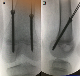
Figure 1A,B Intraoperative control of posterior hemiepiphysiodesis of the distal femur, with two cannulated steel screws guided by metallic wires (1A profile view; 1B AP view) for treatment of deformity in recurvatum knee in a 9-year-old patient due to hemiparetic cerebral palsy.
After surgery, the patient was released for walking with no knee immobilization immediately. The deformity angle was evaluated clinically and radiographically every four month until its hyperextension knee complete correction, when the screws were then removed.
Casuistic
One male 9 years old patient with unilateral genu recurvatum at the left knee, caused by mild hemiparetic hypertonic cerebral palsy GMSCF I, (Figure 2A-2E) was treated to correct surgically the described deformity. He was also operated before recurvatum knee treatment due to an ipsilateral foot deformity.
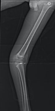
Figure 2A Panoramic radiograph of the left lower limb in profile, male patient, 9 years old, demonstrating recurvatum deformity knee of 32 degrees, due to cerebral palsy.
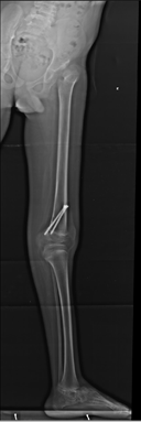
Figure 2B Panoramic profile X-ray of the left lower limb in profile, 25 months after surgical treatment for distal posterior epiphysiodesis of the femur with 2 cannulated 4.5 mm screws, showing correction of recurvatum knee deformity.
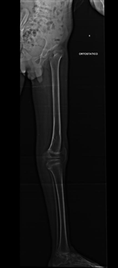
Figure 2C Panoramic profile radiography two years after the two screws removal from distal femoral physis, showing correction maintenance of the left knee recurvatum deformity.
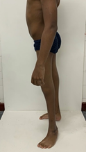
Figure 2E
Figure 2D & 2E Pre- and post-operative clinical photographs of the left lower limbs in profile, showing correction of the recurvatum deformity by posterior epiphysiodesis of the distal femur in a patient with cerebral palsy.
All legal guardians of the patients signed a consent form (TCLE) before surgical treatment and the procedures followed the rules of the Ethical Committee on Human Experiences of the institutions, with protocol having been approved by the Research Ethics Committee of the Hospital das Clínicas of the Faculty of Medicine at the University of São Paulo, under number 4,334,540.
After the surgical treatment the patient was followed clinically and radiographically each 4 months, until the knee deformity was corrected. The time to active the hyperextension knee deformity on both evaluations was 25 months. There were no peri or postoperative complications and no recurrence of the deformity. The correction of the femorotibial angle in the sagittal plane was 36 degrees (figures 2, 3 and 4). The follow-up time is about 2 years.
The hyperextension knee deformity, or recurvatum, can be present on many pathologies like cerebral palsy,4 bone deformities of the distal femur or proximal tibia caused by traumatic anterior hemiepiphysiodesis, joint or bone infection, iatrogenesis after epiphysiodesis to treat leg length discrepancy,11 congenital malformations due to arthrogryposis,8,18 and ligamentous hypermobility.
The clinic appearance and gait are characteristic, with excessive posterior knee angulation, that can be uni or bilateral according to the pathogic cause. When the deformity impacts on the patient walking, we can see a characteristic limp, with a posterior bump in the support phase of the affected limb. The deformity can be confirmed and analyzed by panoramic orthostatic profile radiograph with knees in maximum extension, which can define the place of the deformity (bone, joint or mixed) and measure the angle deformity by goniometry. The indication for surgical correction depends on this clinical and radiographic analysis.
The non-surgical treatment modalities to recurvate knee like physiotherapy, serial casting and bracing2,6,19 have failed. Surgical treatment is rarely indicated, just for situations on that the deformity is the principal reason or part the overall cause of clinical limping, included on situations of many inferior limb joint deformities (together with foot7 and hip4 anomalies). In the past, sort tissues surgical options like quadriceps tenotomies, quadricepsplasty,14 tight tenomyoplasty21 have been done, but failed to treat this clinical conduction.
Until nowadays, the best choice of surgical treatment to recurvatum knee were the osteotomies of the distal femur, with a posterior wedge ressection20 or proximal tibia osteotomy opening wedge, with addition of a bone graft.16,17 When bone deformities are present on adults, the supracondylar femoral osteotomies with removal of a posterior wedge, aiming keeping the angle between the femoral diaphysis and the intercondylar groove regular, is a described surgical option.20
A more frequent surgical treatment option to mature skeleton patients used is the tibial anterior opening wedge osteotomy, with insertion of bone graft, behind the tibial tuberosity.16,17 It is important to say that to avoid the distal displacement of the patella, the tibial anterior tuberosity must be replaced proximally17 mainly indicated in cases in which the recurvatum deformity was caused by premature closure of the anterior proximal tibial growth plate.11
One of the pathologies that leads to hyperextension knee deformity is cerebral palsy, a neurologic condition caused in different diseases, leading to multiple development lower limb joint deformities, due to tonus and athetoid movements present on cerebral palsy. Clinical and surgical treatment for hyperextension of the knee in cerebral palsy used to be made with soft-tissue operation or femoral or tibial osteotomies 18. These kinds of procedures may have ha high risk of complications, for which we can demand searches for less invasive surgical methods, safer and effective.
The surgical technique described in this study to correct the recurvatum knee by guided growth using two screws, leading to posterior hemiepiphysiodesis of the distal femur, in an alternative treatment used to a patient with mild hypertonic cerebral palsy. This is not indicated for situations in which there was premature closure of the anterior femoral or tibial growth plate.10,11 Eventually, in these conditions, the posterior distal femur hemiepiphysiodesis could be indicated with to avoid the progression of the recurvatum deformity during the residual growth.
Based on Metaizeau22 techniques to guided growth, who described the correction of varus and valgus knee using one screw crossing the growth plate on the convex aspect of the deformity, we have used two parallels transphyseal cannulated steel screws inserted in the sagittal plane to increase distal femoral anterior growth. The patient was stimulated to immediate walking, loading the operated limb and performing free knee movements. Though scheduled clinical and radiographic evaluations every four months (three times a year), the knee deformity was evaluated until achieved full correction. At this time, the screws were removed to release the linear residual growth of the distal treated femur.
We have considered the posterior femoral guided growth technique described in this study certainly a less harmful surgical technique, minimally invasive, reversible method presenting a low rate of complications, in addition to not require the knee immobilization, allowing the patient to walk immediately after the surgical treatment and returning to the pretreatment regular activities.
Hemiepiphysiodesis is used as a treatment technique for valgus and varus knee deformities by Metaizeau,22 Jorneau23 and Klatt24 and Stevens,25 who also described the use of guided growth in the sagittal plane to correct of the antecurvatum knee with guided growth of the distal femur using two Eight-Plates placed on the anterior aspect of growth plate, a possible treatment in mild flexion deformity in cerebral palsy.
In 2021, Stevens et al.25 published the guided growth technique to proximal tibial recurvatum after treatment to leg length discrepancy, by posterior epiphysiodesis of the proximal tibia using two Eight-Plates initially positioned too anteriorly, with good outcomes moving the two plates posteriorly. Kievit et al.26 reported also a case of recurvatum knee as complication of treatment of lower limb length discrepancy at the distal femur, with temporary epiphysiodesis using two Eight-Plates. The hypothesis was similar to Stevens25 report, so the recurvatum deformity was caused by a very anterior positioning of the plates, and the correction obtained with the surgical reapproach and posterior replacement of them.
In addition to cerebral palsy, other pathologies that cause the recurvatum knee deformity like arthrogryposis, a congenital condition that is described in different diseases, with the presence of stiffness and multiple joint deformities. The clinical presentation is diverse, like the functional prognosis, which leads to therapeutic options from case to case. Indeed the knee involvement is very common in arthrogryposis with amyoplasia, ranging from soft tissue contractures (flexion or hyperextension) to subluxation or femorotibial dislocation. Flexion contractures are more common and disabling, with difficult treatment and a high recurrence rate.18 The prognosis for ambulation is better with recurvatum knee deformities. According to the literature, nonoperative treatment of hyperextended knee in arthrogryposis with passive mobilization and orthoses fails in about one third of cases. Other patients present recurvatum knee due to hyperlaxity of the ligaments, with no physical therapy or surgical treatment options to soft tissue to correct it, being the osteotomies reserved for patients with bad gait. The recurvatum knee present in this pathologies can be treated by the method described in this article, an undoubtedly less aggressive surgical alternative, presenting progressive and perennial correction after removing the screws, with lower risk to complications during the treatment. It is important to emphasize the need for follow-up at short intervals, to define the exact moment when the screws should be removed, avoiding overcorrection with inversion of the deformity.
No studies have been found on treatment of genu recurvatum using posterior hemiepiphysiodesis of the distal femur with transphyseal screws, as described in this study.
The guided growth by hemiepiphysiodesis of the posterior distal femur with two steel screws crossing the growth plate proved to be a safe and very useful alternative of treatment to recurvatum knee deformities caused by cerebral palsy in patients with immature skeleton. This technique has great potential for correction of recurvatum knee also for the treatment of this deformity caused by many other pathologies.
The author contributed individually to the development of this article.
NBM: intellectual concept, performing surgeries, writing and revising the article; ARJ: intellectual concept, performing surgeries, writing and revising the article; TOG: writing and revision of the article; BLCS: wording and article review.
None.
None.

©2024 Montenegro, et al. This is an open access article distributed under the terms of the, which permits unrestricted use, distribution, and build upon your work non-commercially.