MOJ
eISSN: 2374-6939


Research Article Volume 3 Issue 3
1Chief Consultant, Bari-Ilizarov Orthopaedic Centre, Visiting and Honored Prof., Russian Ilizarov Scientific Centre, Russia
2Bari-Ilizarov Orthopaedic Centre, Bangladesh
3Jessore Medical College & Hospital, Bangladesh
Correspondence: Mofakhkharul Bari, Chief Consultant, Bari-Ilizarov Orthopaedic Centre, Visiting and Honored Professor, Russian Ilizarov Scientific Centre, Kurgan, Tel +88 01819 211595
Received: September 03, 2014 | Published: September 21, 2015
Citation: Bari MM, Islam S, AHMA R, Rahman M (2015) Management of Segmental Fracture Tibia by Ilizarov Technique. MOJ Orthop Rheumatol 3(3): 00097. DOI: 10.15406/mojor.2015.03.00097
Cubitus valgus, Ilizarov, Osteotomy
Segmental fracture is defined as two level fractures with an intact circumferential cortex of the intermediate segment. It is usually follows high energy trauma as is often associated with a significant soft tissue injury. Sometimes it may combine with comminuted fracture of the fragments.1 The use of the Ilizarov method for treating segmental tibial fractures is an attractive concept as it offers many advantages over existing techniques of fracture stabilization. It minimizes surgical trauma at the fracture site, with a percutaneous approach that does not further compromise the biological condition of the fracture site, therefore utilizing the full capacity of the bone and soft tissue to achieve the bone healing.
The tibia has many unique fractures that make it vulnerable to many complications. It is a subcutaneous bone with poor soft tissue coverage on the medial border with a high incidence of open fractures. Its blood supply is also limited particularly, at the distal end. These factors results in a higher incidence of significant soft tissue damage and susceptibility to infection, wound breakdown and non union.2
The Ilizarov apparatus is a universal, stable and yet dynamic constructs that permits functional axial loading of the injured limb. This in turn stimulates bone angiogenesis and promotes osteogenesis, leading to quicker remodeling.3 Its versatility allows correction of any residual deformity. These characteristics allow the Ilizarov method to be used to treat segmental tibial fractures with minimal interference at the fracture site, minimizing the deep infection and non union.
36 patients with segmental tibial fractures (25 male, 11 female) were treated using Ilizarov fixator, 26 were open with a mean age of 35 years (range 20-65 years). 6 patient were initially treated by interlocking nail, 10 patients were treated conservatively in plaster and 2 with uniaxial fixator.4-7 The mean length of intermediate segment was 9.5 cm (range 5.5 to 16 cm) soft tissue coverage was required in 6 cases; intra-articular involvement occurred in 8 cases (6 plateau and 2 pilon). 6 cases of compartment syndrome were treated by fasciotomies, 2 cases with vascular injury required vascular repair. 8 cases of infection 4 of them after nailing which required nail removal and excision of non viable
segment and bone transport, and other 4 cases were superficial infection treated by debridement and antibiotics2 (Table 1).
Correction of residual displacement can be done in OPD, without the need of anaesthesia. Angulation, translation, rotation and shortening can be corrected by the traditional Ilizarov adjustment techniques.
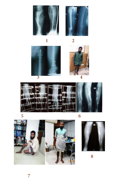
The mean time of union of the proximal segment was 36.5 weeks and 38.2 weeks for the distal segment. Non-union in 3 cases required reapplication of Ilizarov, this happened due to the lack of proper follow up of the patient. Knee ROM less than 90° was observed in 2 cases (Figure 1 & 2).

Figure 1 Case-I:

Figure 2 Case-2:
In our series (total 36 patients), the treatment of segmental fractures was challenging for us due to associated high energy trauma and interrupted blood supply to the intermediate segment. Segmental fracture is almost always associated with high incidence of non-union and infection. But we treated all the patients meticulously by Ilizarov technique with Ilizarov external fixator, better results are being reported even in severe injuries. Today this Ilizarov fixator is versatile, modular and allows free wire placement. It allows secondary correction of segmental fracture during the course of treatment. Open fractures of G-II and III with compartment syndrome is definitely a treatment of choice with the Ilizarov fixator. This fixation does not further damage the vascularity at the segmental fracture sites. By providing it allows in growth of the new capillary buds, increasing the vascularity. The frame configuration allows for the proper care of the injured soft tissues and also for the subsequent cross leg flaps, and other vascular procedures.8 The early wound management in open segmental fractures allowed by Ilizarov fixator makes it possible to save a limb from being amputated. It helps in aligning the comminuted fracture and allows soft tissue to heal. Plate fixation is not recommended in majority of cases. Bone grafting should be carried out early in cases, around 3 to 6 weeks time, where union does not progress satisfactorily, more so with Ilizarov fixation.9-11
|
Initial Treatment |
No. of Cases |
|
Interlocking nail |
6 |
|
Conservatively in plaster |
10 |
|
Uniaxial fixator |
2 |
|
Intra-articular involvement occurred |
8 |
|
Vascular injury required |
2 |
|
Infection |
4 |
|
Superficial infection |
4 |
|
Total |
36 |
Table 1 Initial treatment
The Ilizarov method and frame is very versatile and empowers the skilled surgeon to treat all types of tibia fractures.12 The method can be used to treat the most complex tibia fractures including open fractures, fractures with bone loss, segmental fractures, tibia with open growth plates and with small intramedullary canals. Advanced techniques of bone and soft tissue transport or temporary intentional deformation to enable wound closure can be implemented early to optimize the clinical results. The use of Ilizarov device and technique is an excellent, effective method simultaneously to overcome all above mentioned problems where alternate methods are expected to fail.
None.
None.

©2015 Bari, et al. This is an open access article distributed under the terms of the, which permits unrestricted use, distribution, and build upon your work non-commercially.