MOJ
eISSN: 2374-6939


Research Article Volume 15 Issue 2
Department of Orthopaedic Surgery, J. N. Medical College, Faculty of Medicine, India
Correspondence: Yasir Salam Siddiqui Assistant Professor, Dept. of Orthopaedic Surgery, J. N. Medical College, Faculty of Medicine, A.M.U, Aligarh, Uttar Pradesh, India, Tel +919837343400
Received: February 10, 2023 | Published: March 16, 2023
Citation: Yadav A, Abbas MB, Siddiqui YS, et al. Management of complex idiopathic clubfoot using modified Ponseti method. MOJ Orthop Rheumatol. 2023;15(2):37-42. DOI: 10.15406/mojor.2023.15.00615
Clubfeet with rigid equinus, severe plantarflexion of all the metatarsals, a transverse crease in the sole of the foot, short and hyperextended great toe, forefoot adduction and supination and a deep posterior crease are categorized under the heading of complex clubfeet, which do not respond to the conventional Ponseti technique. Rather treatment of such feet with conventional Ponseti method results in the development of secondary deformities and thus failure of treatment. Hence modification of the technique is warranted for optimal correction of these feet as described by Ponseti. The aim of our study was to evaluate effectiveness of Modified Ponseti technique in the management of complex clubfoot. Thirty two complex clubfeet in 19 patients were managed by Modified Ponseti technique through the study period. Four patients with 7 complex clubfeet were lost to follow-up. At the end of study 15 patients with 25 complex clubfeet were available for final follow up assessment and evaluation. Pirani and Dimeglio score was allotted to each foot at every visit. There after each foot was manipulated and casted as per the Modified Ponseti technique at an interval of one week. Average number of casts required for deformity correction was 7.68. Tendo-achilles tenotomy was obligatory in 23 (93.33 %) feet. The mean follow-up duration was 12.35 months (Range 8-21 months). The mean pre-treatment Pirani score (initial PS) was 5.60±0.54 and mean post-treatment Pirani score (PS at SFAB) was 0.70±0.38. The change in mean score post intervention was found to be statistically significant. The mean pre-treatment Dimeglio score (initial DS) was 15.80±2.02 and mean post-treatment Dimeglio score (DS at SFAB) was 3.68±1.35. The change in mean score post intervention was found to be statistically significant. Relapse rate was 4% (n=1), which responded to re-casting with modified Ponseti technique and re-tenotomy of the tendo-achilles. Based on our study results and existing literature we recommend modified Ponseti technique as the first line initial treatment for these complex feet. However such feet require higher number of plaster casts with higher rate of tendo-achilles tenotomy, with high relapse rate than their classical idiopathic counterparts, nonetheless the eventual outcome is reasonable correction of deformity, negating the necessity of multifaceted operating procedures.
Keywords: Complex clubfeet, Modified Ponseti method, Conventional Ponseti technique, Pirani score, Dimeglio score, SFAB (Steenbeek Foot Abduction Brace)
Congenital idiopathic clubfoot is one of the commonest foot deformities with an occurrence of one to two per thousand live births; males are affected two-fold as often as females and the condition is bilateral in 2/3rd of the patients.1,2 There is consensus on the initial treatment of the deformity by non-operative means using Ponseti technique.3–5 Majority of the idiopathic clubfeet respond to conventional Ponseti technique of manipulation and serial casting treatment protocol. However, some clubfeet do not respond to the conventional Ponseti technique owing to the multi-faceted nature of the deformity. Such feet have been termed as complex clubfeet.6
Clubfeet with rigid equinus, severe plantarflexion of all the metatarsals, a transverse crease in the sole of the foot, short and hyperextended great toe, forefoot adduction and supination and a deep posterior crease are categorized under the heading of complex clubfeet.6 As these feet do not respond to the conventional Ponseti technique, hence complex clubfeet must be documented from the more typical idiopathic clubfeet as an alteration in the technique of manipulation and casting is obligatory for successful outcome. Treatment of complex feet with conventional Ponseti method of manipulation and serial casting results in the development of a secondary deformity of foot with hyperflexion and disproportionate abduction of the metatarsals at the tarso-metatarsal joint (Lisfranc articulation) rather than abduction of calcaneum.6 Hence modification of the technique is warranted for preventing development of secondary deformity and thus failure of treatment. The overall occurrence of complex clubfoot is not clear on account of the limited literature and research work. However, in the last few years, the occurrence of complex clubfoot has significantly augmented owing mainly to the faulty manipulative techniques6 and also due to recognition of the entity among the orthopaedicians. Ponseti (2006) revolutionized the management of complex clubfoot by pioneering the concept of “Modified Ponseti technique”, which started a new chapter in the management of these short, stubby and rigid feet. To the best of our knowledge there are only few published results of this method, particularly in the Indian population, therefore we conducted a prospective research to assess effectiveness of Modified Ponseti technique in the management of complex clubfeet.
Study design
This was a prospective study conducted in the department of orthopaedic surgery, Jawaharlal Nehru Medical College, A.M.U., Aligarh from November 2018 to October 2020 after approval by the Institutional Ethical committee (D. No-242/FM/IEC). All the study participants were briefed about the study and written informed consents were obtained.
Case definition of complex clubfoot
Clubfeet with rigid equinus, severe plantarflexion of all the metatarsals, a transverse crease in the sole of the foot, short and hyperextended great toe, forefoot adduction and supination and a deep posterior crease are categorized under the heading of complex clubfeet.6
Inclusion and exclusion criteria
Patients satisfying the criteria of case definition of complex clubfoot were included in the study. Patients with non-idiopathic clubfoot (secondary to neuromuscular disorders & those associated with syndromes) and classical Idiopathic clubfoot were excluded. Thirty two complex clubfeet in 19 patients were managed by Modified Ponseti technique throughout the study period. Out of 19 patients, four patients with 7 complex clubfeet were lost to follow-up. At the end of study 15 patients with 25 complex clubfeet were available for final follow up assessment and evaluation.
Pre-treatment evaluation
All study participants were thoroughly examined as per the predetermined study protocol. After inclusion in study, Pirani7 and Dimeglio8 score was allotted to each foot at every visit. There after each foot was manipulated and casted as per the Modified Ponseti technique at an interval of one week. The modification as described by Ponseti was application of counter pressure on lateral aspect of head of talus and posterior aspect of lateral malleolus (figure 1). The hyper-plantarflexed metatarsals and rigid equinus were corrected concurrently; wherein the deformed foot was grasped by the ankle with index finger of both the hands while thumbs under the metatarsals helped the foot into dorsiflexion. The knee was immobilized in 110° to 120° flexion in contrast to 90 degree in conventional method. Clinical improvement in deformity (Pirani and Dimeglio score) and complications were monitored and recorded at each visit.
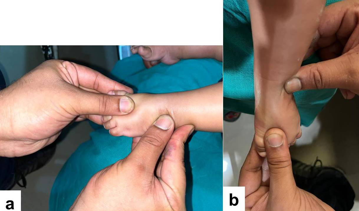
Figure 1 A & B) Clinical photograph showing the precise hand location for holding and manipulating the complex deformed foot as described by Ponseti. For correction of the mid-foot inversion and heel varus, the thumb is placed over the lateral aspect of the talar head, and the index finger of the ipsilateral hand is placed over the posterior aspect of the lateral malleolus as shown in photograph. The forefoot is abducted with the other hand while counter-pressure on the lateral aspect of the talar head and posterior aspect of the lateral malleolus is upheld.
Manipulation and serial casting with Modified Ponseti technique
Casting in complex clubfoot was done as per the Modified Ponseti method.6 The modification from the classical method was firstly to correct the mid-foot inversion and heel varus, followed by correction of hyperflexion of metatarsals and the rigid equinus. For correction of the mid-foot inversion and heel varus, the thumb is placed over the lateral aspect of the talar head, and the index finger of the ipsilateral hand is placed over the posterior aspect of the lateral malleolus. The forefoot is abducted with the other hand while counter-pressure on the lateral aspect of the talar head and posterior aspect of the lateral malleolus is upheld (Figure 1). It should be kept in mind never to abduct the forefoot beyond 30° to 40°. If one tries to gain more abduction, it will lead to further flexion of metatarsals and toes as well as excessive abduction of the metatarsals at the tarso-metatarsal joint (Lisfranc articulation). This is owing to short and taut deep plantar muscles. The subsequent plaster casts were applied till abduction of 30° was achieved and hind-foot varus was corrected. Second modification was to correct the hyperflexion of metatarsals and the rigid equinus concurrently. For achieving this, both the index fingers are positioned on either side of the head of the talus, and both the thumbs are positioned on the sole of the foot on the heads of first and fifth metatarsals. The hyperflexed metatarsals are pushed in dorsiflexion concurrently pressure being applied on both the metatarsal heads to produce the extension of all the metatarsals. While doing this maneuver the knee is steadied in flexion. The outcome of the technique will position the forefoot in mild abduction and heel in mild valgus. Care should be taken not to produce a rocker bottom foot deformity. After correction of the hyperflexed metatarsals and the equinus, the well molded above knee plaster cast is applied in about 110⁰ to 120⁰ flexion at the knee. Once 30⁰ to 40⁰ of abduction have been accomplished, and the hyperflexion of metatarsals has been corrected, but the equinus is still persisting, tenotomy can be performed in complex clubfeet. The tenotomy should be done 1.5 cm above the heel crease to avoid injury to the calcaneal tuberosity. If desired correction was achieved then a plaster cast was given in maximum correction for 3 weeks else the post-tenotomy cast is changed every 4 to 5 days so as to achieve at least 5⁰ of dorsiflexion and 40⁰ of abduction. Thereafter, SFAB (Steenbeek Foot Abduction Brace) with 40° External rotation and 15° dorsiflexion was given to all the patients 3 weeks after tenotomy to maintain correction. It was applied 23 hours a day for first 3 months and then at night/nap time for another 3 years. The brace used in modified technique should only have an abduction of about 40°, when compared to usual 70° in idiopathic variety of clubfoot. However, the timings and duration of bracing protocol is essentially the same. Figure 2 & 3 depicting the pre-treatment and post-treatment clinical photograph of the two patients.
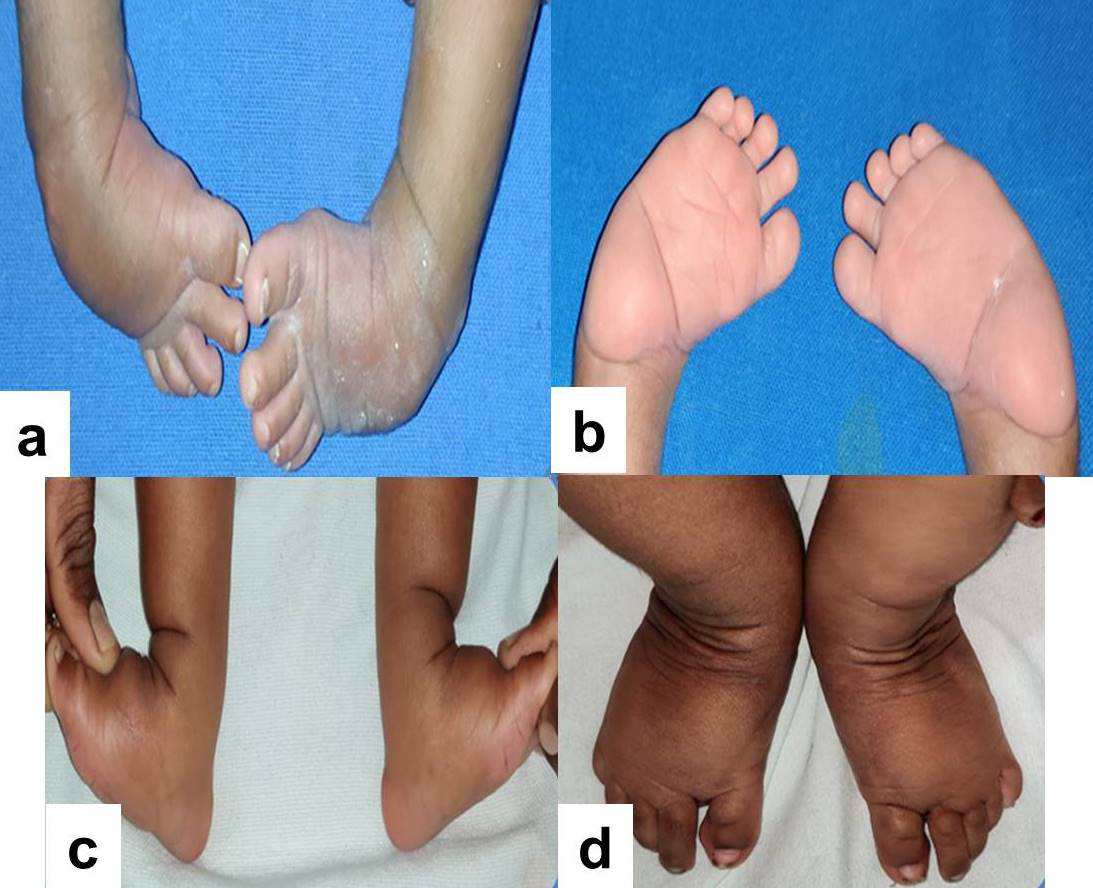
Figure 2 A & B) Pre-treatment clinical image of the patient’s feet showing bilateral complex clubfeet. C & D) Post-treatment clinical image showing plantigrade feet.
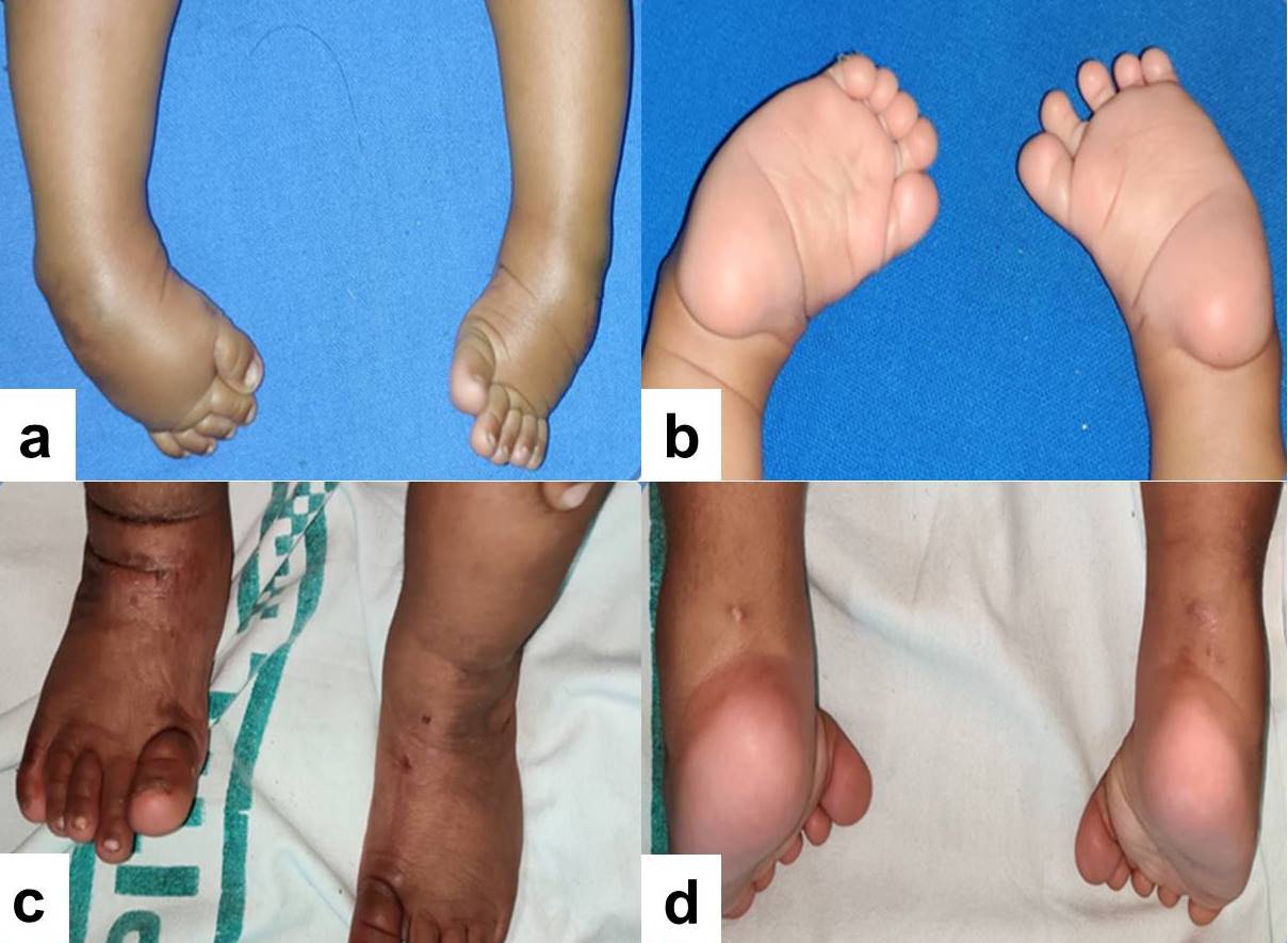
Figure 3 A & B) Pre-treatment clinical image of the patient’s feet showing bilateral complex clubfeet. C & D) Post-treatment clinical image showing plantigrade feet.
Statistical analysis
Descriptive statistics such as mean, median, mode, standard deviation, frequency and percentage were used to describe the data. Inferential statistics such as Wilcoxon signed rank test was used to analyze the difference in means before and after intervention. Also Spearman Correlation test (rho) was used to find the relationship between two quantitative variables. Data was analyzed by using Statistical package for Social Sciences (SPSS) version 20.0. P value less than 0.05 was considered significant.
Study population and demographic characteristics
Thirty two complex clubfeet in 19 patients were managed by modified Ponseti technique during the study period of 2 years (November 2018 to October 2020). Four patients with 7 complex clubfeet were lost to follow-up. Finally, fifteen patients with 25 complex clubfeet were included in the study and were available for final follow up assessment and evaluation. Male to female ratio was 2.7:1. Mean age at the time of presentation was 1.12 months (Range 0.5 to 5 months). Of the total 15 patients, majority were bilateral (n=10) and rest 5 were unilateral. Out of the 5 unilateral cases, 3 were left sided and 2 were right sided. Mean follow-up duration was 12.35 months (Range 8 to 21 months). The distribution of age, sex and laterality is presented in Table 1– 3 respectively.
|
Age (months) |
No. of patients |
% |
|
0-1 |
6 |
40 |
|
1-2 |
4 |
26.6 |
|
2-3 |
1 |
6.6 |
|
3-4 |
2 |
13.3 |
|
4-5 |
2 |
13.3 |
Table 1 Age Incidence
|
Sex |
No. of patients |
% |
|
Male |
11 |
73 |
|
Female |
4 |
27 |
Table 2 Sex distribution
|
Laterality |
No. of patients |
% |
|
Bilateral |
10 |
66.6 |
|
Right sided |
2 |
13.3 |
|
Left sided |
3 |
20 |
|
Total |
15 |
100 |
Table 3 Laterality
Post-treatment outcome in study population
The mean pre and post-correction Pirani score (PS) in our study was 5.6 (Range 4-6) and 0.7 (Range 0-1) respectively. The mean pre and post-correction Dimeglio score (DS) in our study was 15.8 (Range 12-19) and 3.6 (Range 1-6) respectively. Majority of the feet (n=18; 72 %) were Grade IV according to the Dimeglio criteria. The mean pre-treatment Pirani score (initial PS) was 5.60±0.54 and mean post-treatment Pirani score (PS at SFAB) was 0.70±0.38. The change in mean score post intervention was found to be statistically significant (Table 4). The mean pre-treatment Dimeglio score (initial DS) was 15.80±2.02 and mean post-treatment Dimeglio score (DS at SFAB) was 3.68±1.35. The change in mean score post intervention was found to be statistically significant (Table 5). There was a significant positive correlation between pre-treatment Pirani and initial Dimeglio scores with Spearman rho – 0.445, P – 0.026 (Figure 4). There was also a strong positive correlation between post-treatment Pirani and Dimeglio scores. With increase in post-treatment PS, the value of DS also increased, with Spearman rho – 0.814, P – 0.0001 (Figure 5).
|
|
Mean |
SD |
Wilcoxon signed Rank test |
P-value |
|
Pre-treatment PS |
5.60 |
0.54 |
-4.406 |
0.0001 |
|
Post-treatment PS |
0.70 |
0.38 |
|
|
Table 4 Inferential statistical change in Pirani score (PS) before and after intervention
|
|
Mean |
SD |
Wilcoxon signed Rank test |
P-value |
|
Pre-treatment DS |
15.80 |
2.02 |
-4.386 |
0.0001 |
|
Post-treatment DS |
3.68 |
1.35 |
|
|
Table 5 Inferential statistical change in Dimeglio score (DS) before and after intervention
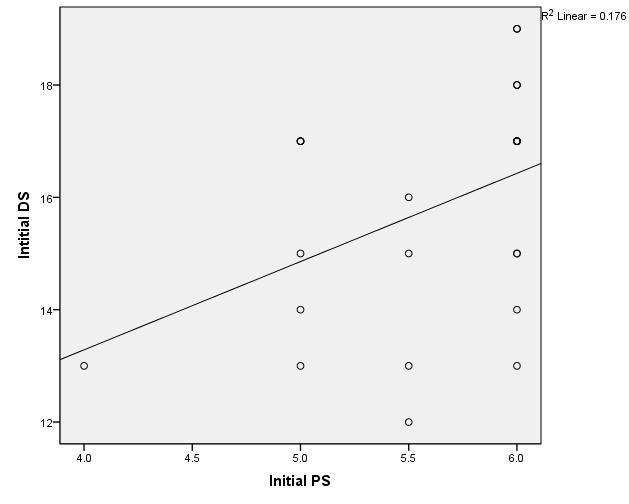
Figure 4 Shows significant positive correlation between initial Pirani Score (pre-treatment PS) and initial Dimeglio Score (pre-treatment DS).
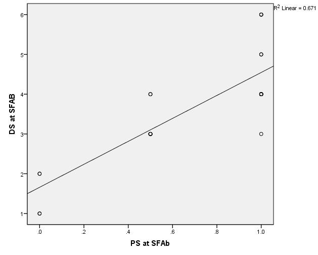
Figure 5 Shows significant positive correlation between Pirani Score at SFAB (post-treatment PS) and Dimeglio Score at SFAB (post-treatment DS).
Average number of cast required for achieving correction was 7.68 casts (Range 5 to 11). Tendo-achilles tenotomy was required in 23 (93.33 %) feet. An average of 0.9 casts (Range 0 to 2) was required after tenotomy (Table 6).
|
No. of post-tenotomy casts |
No. of feet |
|
0 |
10 |
|
1 |
8 |
|
2 |
7 |
Table 6 Number of additional casts required after tenotomy (0 cast indicates no additional cast was required after the 1st post-tenotomy cast)
Relapses and other complications
Out of fifteen patients, one patient (4%) had a relapse. There was relapse of forefoot adduction and equinus, noticed after 3 months of initial correction. This relapse was managed with re-casting with Modified Ponseti technique and re-tenotomy of the tendo-achilles. Major complications including tenotomy related complications (excessive blood loss or wound complications) were not observed. Plaster cast slippage was noted in 4 patients (16%). Minor dermatological complications observed in our study are tabulated in table 7. In 2 feet, we noticed the development of plaster sore at the dorso-medial aspect of the base of great toe, possibly due to hyperextension of great toe in these feet (figure 6). All these dermatological complications were managed conservatively with Neosporin powder, Emollients, topical steroids and extra padding over the lesion.
|
Complication |
No. of feet |
|
Plaster cast slippage |
4 |
|
Plaster sore over dorsolateral aspect of foot |
1 |
|
Eczematization with scaling |
1 |
|
Skin discoloration |
1 |
|
Plaster sore over dorsomedial aspect of great toe |
2 |
|
Pressure sore due to tight abduction brace with Paronychia |
1 |
|
Superficial skin laceration due to plaster cutter |
1 |
|
Desquamation over heel |
1 |
Table 7 Complications/Pitfalls
The purpose of this study was to assess the role of modified Ponseti technique in management of complex idiopathic clubfoot. Complex clubfoot is short, stubby and rigid feet that do not respond to the conventional Ponseti method of manipulation and serial casting. Therefore such feet must be differentiated and documented from the more usual idiopathic variety, as modification of the casting technique is warranted for successful correction of deformities.
Incidence
The overall incidence of complex clubfoot is not clear on account of limited literature and research work. Its aetio-pathology and management is still debatable. However, in the last few years, the occurrence of complex clubfoot has greatly increased owing mainly to the faulty manipulative techniques.6 The proportion of patients of complex clubfeet presenting to our institute was 8.67% (15/173; number of patients with complex clubfoot/Total number of patients with clubfoot). Incidence of complex clubfoot as reported by Ignacio V Ponseti, M Dragoni, S Yoshioka was 6.5%, 17% and 13% respectively. Our incidence of complex clubfoot was comparable to benchmark multi-centre study done by Ponseti. The incidence of complex clubfeet seems to be under reported owing to lack of knowledge and difficult aspect of differentiation from its classical idiopathic variety among the medical fraternity. Consequently the conventional Ponseti technique if employed will result in development of secondary deformities and treatment failure, eventually leading to frustration among the parents and the phenomenon of Doctor-shopping. Hence prompt diagnosis of the complex clubfoot is essential to ensure prompt and adequate treatment of the disease by simple modification of the technique.
Aetiology
While studying the clubfeet, Turco9 noted the intractable nature of some clubfeet to the usual conservative and operative treatment. He designated such feet as atypical. The deformities in the complex clubfeet may be attributable to shortening and tightness of the deep plantar intrinsic muscles, of the analogous nature and extent as was observed in the calf muscles of classical idiopathic clubfoot. Severe fibrosis in the quadratus plantae inserted into the long toe flexors explains the persistent hyperabduction of the metatarsals after faulty manipulations. In complex clubfeet, the gastro-soleus and the plantar intrinsic muscles and ligaments are more severely involved.1,9,10 Another proposed mechanism is via action of an anomalous muscle; Flexor digitorum accessories longus (FDAL), which leads to more flexion of the lateral 4 toes as compared to the great toe.1,6,9,10
Effectiveness of modified Ponseti technique
A functional, plantigrade foot is considered as an acceptable outcome in these children. Prior to the description of the modified Ponseti technique, operative as well as non-operative treatment strategies were employed for treatment of such feet with variable results. Turco in 1994 reported that the results with early soft tissue surgery were unpredictable and early surgical intervention usually results in a grotesquely deformed foot.9 Ponseti in their benchmark multicenter study in 2006 defined a modified protocol for the correction of these complex clubfeet and named it as Modified Ponseti technique.6 Since then, this technique has been the standard protocol in the management of Complex clubfeet.
Ponseti studied the treatment of complex idiopathic clubfeet in fifty patients (75 clubfeet) with modified Ponseti protocol for correcting these complex clubfeet. By using this modified technique, he achieved correction in all the patients with average of 5 corrective casts. Two patients (4%) underwent posterior soft tissue release procedure with tendo-achilles lengthening. There were 7 relapses that responded to re-casting with the same technique. Three patients underwent a second tendo-achilles tenotomy due to recurrent equinus deformity. The authors concluded that once a complex clubfoot is corrected using Modified technique, the rigidity of the soft tissues diminishes, the skin creases and puffiness disappear, and the foot matures normally.6
H. E. Matar in 201611 studied the behavior of complex idiopathic clubfoot by means of the Modified Ponseti method in 11 children (M:F = 9:2) with 17 Complex feet. Correction was attained in all the children with an average number of 7 casts (range 5-10). Tendo-achilles tenotomy was required in all the feet. Rate of relapse observed was 53% (n=9 feet). A satisfactory and desirable outcome was achieved in 13 of 17 clubfeet (76.5%) at the final follow-up. The authors concluded that Modified Ponseti technique is an effective first line treatment modality for complex idiopathic clubfeet. P. Mandlecha12 studied the evaluation of Modified Ponseti technique in the management of complex clubfeet in 16 children, out of which 11 had bilateral deformity and 5 had unilateral deformity. Average number of corrective casts required for the thorough correction of the deformity with modified Ponseti method was 7.44 (range 6-10). All 27 clubfeet required tenotomy. In their study, percutaneous tenotomy was required in 19 feet while 8 feet required mini-open tenotomy. Relapse was seen in 3 clubfeet (11.11%) which was further managed by re-application of corrective casts by modified Ponseti technique and re-tenotomy of the tendo-achilles. The authors concluded that the modified Ponseti method of casting is a successful and effective method for treatment of complex clubfeet. Furthermore the technique also reduces the need of additional surgical procedures in such patients.
In the present study, the average number of casts required for deformity correction was 7.68 which were more as compared to the studies done by other authors, possibly due to the learning curve we had to go through in the application of casts by this relatively newer technique (Modified Ponseti technique) in the initial phase of our study. Secondly, as most of these casts were applied by trainee junior residents with rotating posting in different clinics including paediatric orthopaedic clinic, which in due course lead to increase in the number of casts required for correction. The mean follow-up period was 12.35 months (ranging from 8-21 months). When compared to other studies, the mean follow-up duration of our study may not be long enough to assess the relapse pattern and functional outcome and the shape of the foot when the child becomes an adult. One patient (4 %) with left-sided complex CTEV had a relapse after initial successful correction. There was relapse of forefoot adduction and equinus, noticed after 3 months of initial correction. This patient was non-compliant to abduction bracing protocol. The parents of the child reported problems like difficulty in keeping the shoes, ill-fitting shoes and slippage of shoes at night while the child was asleep. This relapse was managed with re-casting with modified Ponseti technique and re-tenotomy of the tendo-achilles. We do realize that the relapse rate in our study was lower as compared to the other authors may be due to a relatively short term follow-up of the patients. Table 8 compares the observations and results of our study with the available literature.
|
Author |
Incidence |
Age (months) |
Laterality |
M:F |
Pre-treatmentPS |
Post-treatment PS |
No. ofCasts |
Need of tenotomy |
Post-tenotomy casts |
Follow-up duration (months) |
Complications |
Relapse |
|
Ponseti6 |
6.5 % |
3 |
|
1.63:1 |
5.5 |
|
5 (1-10) |
100% |
1-4 |
23 m (6-46) |
22 % |
14 % |
|
Matar11 |
|
1.2 |
B/L 54.5% R 18.18% L 27.27% |
4.5:1 |
|
|
7 (5-10) |
100% |
|
7 yrs (3-11) |
|
53 % |
|
Mandlecha12 |
|
4.77 |
B/L 68.75% R 12.5% L 18.75% |
4.3:1 |
5.57 |
0.18 |
7.44 (6-10) |
100% |
1.3 (0-4) |
14.76 m (6-22) |
29.63 % |
11.11% |
|
Our Study |
8.67 % |
1.12 |
B/L 66.67% R 13.33% L 20% |
2.75:1 |
5.6 |
0.70 |
7.68 (5-11) |
93.33% |
0.9 (0-2) |
12.35 m (8-21 m) |
32 % |
4 % |
Table 8 Comparison of our study with available literature on Modified Ponseti Technique
Strengths, limitations and future recommendations
Our study evaluates the role of modified Ponseti technique in management of complex idiopathic clubfoot. The strengths of the study are prospective nature of study, inclusion of complex feet as per the case definition and definite treatment protocol. Our study has several limitations. Firstly, the patients with recurrent history of slippage of casts; we do not have the precise data for the number of casts that had slipped and for how long were those casts kept in slipped position, trapping the foot in a forced plantarflexed position with the digits disappearing inside the plaster. Secondly, there was no radiographic follow-up, which limits the comparison of clinical (cosmetic and functional) correction with that of bony correction delineated with routine radiographs of foot. Thirdly, average duration of follow-up was short (range 8-21 months), which may not be long enough to assess relapse pattern and functional outcome and the cosmetic look of the foot when the child attains skeletal maturity. Hence future research with large sample and comprehensive follow-up are indispensable for appraising the role of modified Ponseti technique in management of complex clubfeet.
Based on our study results and existing literature we recommend modified Ponseti technique as the first line initial treatment for these complex feet. However such feet require higher number of plaster casts with higher rate of tendo-achilles tenotomy, with high relapse rate than their classical idiopathic counterparts, nevertheless the eventual outcome is reasonable correction of deformity, negating the necessity of multifaceted operating procedures.
None.
The authors declare no conflicts of interest.

©2023 Yadav, et al. This is an open access article distributed under the terms of the, which permits unrestricted use, distribution, and build upon your work non-commercially.