MOJ
eISSN: 2374-6939


Research Article Volume 15 Issue 5
1Department of Surgery, School of Medicine, Federal University of Health Sciences of Porto Alegre (UFCSPA), Brazil
2Head of Orthopaedics, Irmandade Santa Casa de Misericórdia de Porto Alegre (ISCMPA), Brazil
3Department of Orthopaedics, Irmandade Santa Casa de Misericórdia de Porto Alegre (ISCMPA), Brazil
4Graduate Program in Rehabilitation Sciences, Federal University of Health Sciences of Porto Alegre (UFCSPA), Brazil
Correspondence: Carlos Roberto Schwartsmann, Department of Surgery, School of Medicine, Federal University of Health Sciences of Porto Alegre (UFCSPA), Porto Alegre, Rio Grande do Sul, Brazil
Received: August 20, 2023 | Published: September 8, 2023
Citation: Schwartsmann CR, Zambra Wink LH, Mendes Eggers EK, et al. Macroscopic analysis of the subchondral bone of the femoral head during total hip replacement. MOJ Orthop Rheumatol. 2023;15(5):182-185. DOI: 10.15406/mojor.2023.15.00642
Objective: The present research aims to determine areas of necrosis in the subchondral bone of femoral heads removed from patients undergoing total hip replacement (THR) for primary coxarthrosis.
Methods: The femoral heads of 154 patients were analysed during the THR surgical procedure following femoral neck osteotomy. The heads were divided into four quadrants: anterosuperior (AS), posterosuperior (PS), anteroinferior (AI), and posteroinferior (PI). The heads were examined for foci of subchondral bone necrosis.
Results: In 30 femoral heads, the arthrosis was restricted to cartilaginous lesions and the subchondral bone was considered fully vascularis In 124 heads, there were eburnated areas in the subchondral bone. Of these, 95 cases were eburnated in the AS region, 86 in the PS quadrant, 57 in the AI, and only 37 in the PI. Hips with more severe coxarthrosis had a 5.2 times higher chance of presenting femoral head necrosis.
Conclusion: The analysis of 154 femoral heads during total hip replacement for osteoarthritis revealed foci of subchondral bone necrosis in 124 cases. The anterosuperior and posterosuperior quadrants showed a significantly larger devitalised area of subchondral bone, of 76% and 70% respectively. Coxarthrosis is a disease that not only affects the articular cartilage, but also substantially affects the subchondral bone, as observed in this work. There was a positive correlation between the higher severity of the disease by the Tönnis classification and the presence of subchondral necrosis.
Keywords: hip/surgery, femur head, osteoarthritis
Hip osteoarthritis (OA) is a pathology with a high prevalence, estimated to affect up to 25% of the population in its symptomatic form by the age of 85, and leading 7.1% to 11.6% of the population by the age of 50 to undergo total hip replacement (THR) surgery.1–3 In large prevalence studies of OA with X-ray and symptomatology, the percentage of the affected population varies from 1% to 10%.3-8 OA mainly occurs in weight-bearing joints, most commonly affecting the elderly population. The hip joint is one of the most affected in the body by this pathology, second only to knee OA.3,5,9 There are also secondary etiologies such as inflammatory, post-traumatic, infectious, or metabolic diseases.9
We still do not understand the precise pathophysiology responsible for the development of coxarthrosis. It is currently understood that OA is the result of a mechanical and biological imbalance that destabilises the normal homeostasis of cartilage and subchondral bone, which, combined with the body's innate inability to repair damaged tissue, leads to OA. The disease is characterised by the degeneration of cartilage, sclerosis, and necrosis of the subchondral bone, and osteophyte formation.9–14 The vascularisation of the femoral head appears to play an important role in the development of OA.10 When discussing vascularisation of the femoral head, the consolidated literature considers that the femoral head is primarily perfused by the medial circumflex femoral artery (MCFA). The lateral circumflex femoral artery (LCFA) is responsible for irrigating the tissues adjacent to the hip joint. There is also the participation of two other vessels: the piriform branch of the inferior gluteal artery and the obturator artery through the artery of the round ligament.15–20
Imaging studies using magnetic resonance imaging (MRI) have gained ground and significance in the study of OA. It is possible to verify bone oedema associated with OA. In histological analysis, it is possible to confirm that this edema is characterised by the presence of necrosis, fibrosis, and microfractures.21,22 Given the significant functional limitation imposed by joint wear, combined with the patient's loss of quality of life, a large part of physicians recommend THR as the treatment of choice in most cases. It is estimated that one million THRs are performed worldwide each year.9 The objective of this study was to perform a macroscopic analysis of the femoral head during hip replacement surgery for OA, seeking to assess the vascularisation and areas of necrosis of the femoral head.
A transversal study was performed considering the analysis of 154 femoral heads from patients who underwent total hip replacement for osteoarthritis during the year 2021 at our Medical Residency Program/ University Hospital. All cases were of primary coxarthrosis, excluding patients with secondary changes that justified the degenerative process. None of the operated patients were taking anticoagulants or platelet antiaggregants. The research was approved by the Ethical Committee Board of our institution. Patients were initially analysed by radiograph and classified by the degree of OA according to the Tönnis classification for hip OA.23 The grading of the classification was made after consensus among the four orthopedic surgeons involved in the procedure.
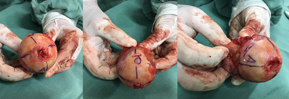
The surgical approach used in all cases was a posterior approach with lumbar level block by spinal anaesthesia. Patients were positioned in contralateral lateral decubitus. After opening the fascia lata, we repaired the tendons of the external hip rotators and exposed the posterior capsule. The hip dislocation was then performed in internal rotation and adduction, followed by femoral neck osteotomy with an electric saw. The femoral head was then removed and split with an electric saw into four quadrants: anterosuperior (AS), anteroinferior (AI), posterosuperior (PS), and posteroinferior (PI) (Figure 1).

Figure 1 Division of a femoral head subjected to total hip arthroplasty for primary osteoarthritis into four quadrants: anterosuperior (AS), anteroinferior (AI), posterosuperior (PS), posteroinferior (PI).
After the femoral head was divided into 4 parts, the presence or absence of vascularisation was analysed through the colour presented. It was considered vascularised when the predominant colour was reddish, as in Figure 2A. On the other hand, we considered areas of ischemia when the predominant color was eburnated, compatible with necrotic and ischemic areas, as in Figure 2B. The decision to be considered vascularised or not was made by the surgeons team involved in the procedure (one head surgeon and 3 assistants). There was no quantitative precision of necrotic or non-necrotic areas present in each fragment since the analysis was of predominance and performed macroscopically.

Figure 2 (A) Femoral head of a 55-year-old female patient with primary coxarthrosis, who underwent total hip replacement. (B) Femoral head divided into 4 parts from a 64-year-old male patient. Despite the appearance of small red areas in the PS (posterosuperior) and PI (posteroinferior) quadrants, all four quadrants were considered non-vascularized.
A total of 154 femoral heads from patients undergoing total hip replacement for the treatment of primary coxarthrosis were analysed. Of these, 86 were female and 68 were male. Ages ranged from 36 to 84 years with an average of 67.9 years and BMI ranged from 21.6 to 38.4 with an average of 28.6 kg/m². In terms of laterality, 80 of the femoral heads analysed were from the right side and 74 from the left. In the radiographic evaluation, analysed according to the Tönnis classification for osteoarthritis, grades I to III were divided into mild, moderate, and severe. There were no grade I patients. Of the 154 femoral heads analysed, 35 were classified as Tönnis II and 119 as Tönnis III.23
Of the 35 cases classified as Tönnis II, 20 showed necrosis and the other 15 showed no macroscopic vascular change. In the 119 cases classified as Tönnis III, there was a significant increase in the presence of ischemic areas, with 104 femoral heads showing eburnated areas and only 15 being fully vascularised (Table 1). The average age at which necrosis was found when compared with fully vascularized femoral heads was slightly higher, with an age of 68.7 years versus 65.1 years respectively.
|
Tönnis Classification |
Presence of Necrosis |
Without Necrosis |
Total |
|
Tönnis 2 |
20 (57,1%) |
15 (42,8 %) |
35 (22,7%) |
|
Tönnis 3 |
104 (87,3%) |
15 (12,6%) |
119 (77,3%) |
|
Total |
124 (80,5%) |
30 (19,5%) |
154 (100%) |
Table 1 Cross-referencing of femoral heads with the presence or absence of necrosis according to the Tönnis classification.
We used the chi-square test calculated in SPSS 26 software, which showed a statistically significant difference (p < 0.001) in favour of the appearance of necrotic areas when the cases were classified as Tönnis III. When analysing the odds ratio, we concluded that there is a 5.2 times greater chance (95% CI) of finding eburnated subchondral areas in patients classified as Tönnis III compared to those classified as Tönnis II. We also analysed the presence of eburnated areas in the 4 fragments of the 124 femoral heads. Eburnated areas were observed in 94 of them in the anterosuperior quadrant (76%) and, 86 in the posterosuperior quadrant (70%). When examining the inferior quadrants, we noticed a lower presence of ischemic areas. Of the 124 femoral heads, 57 had them in the anteroinferior quadrant (46%) and 37 in the posteroinferior quadrant (30%) (Figure 3).
In this study, we assessed the presence or absence of eburnated areas in the femoral head in patients diagnosed with coxarthrosis. We also analysed the quadrant in which osteonecrotic areas presented themselves. Coxarthrosis is highly prevalent, particularly when considering only diseases involving the hip, causing limitations and decreased quality of life.1–8 It is characterised by the degeneration of the articular cartilage, the formation of osteophytes, and subsequent subchondral bone degeneration, as we can confirm in our study.9–14 When reviewing the vascularisation of the femoral head, we found that it occurs through three distinct routes: intraosseous, foveolar, and retinacular. Most authors consider the medial circumflex artery, a branch of the deep femoral artery, as the main vascular support of the femoral head. This artery originates from 2 to 4 superior retinacular branches and occasionally inferior retinacular branches, with the superior branches being the most important, as they are capable of nourishing the femoral head alone. The lateral femoral circumflex artery is not significant in the vascularisation of adults, as described by Ganz et al.17 It was only shown to have anastomosis with the MFCA in individuals under one year of age. Of lesser importance, there is also the piriform branch of the inferior gluteal artery and the obturator artery through the round ligament artery.15–20,24–26
The articular cartilage, in turn, is avascular, being nourished by imbibition from the capillaries beneath it, provided by intermittent weight pressure. With the reduction of the joint space associated with the degeneration of the articular cartilage and increased pressure on certain points of the femoral head, it leads to retraction of the capillaries, forming areas with ivory-colored eburnated bone and common cysts of osteoarthritis.10 That said, there is no evidence in the literature indicating a change in the pattern of circulation in the hip joint when it presents with arthritis. In our study, we found that the severity of arthritis, assessed by the Tönnis classification in radiographs, is related to the prevalence of necrotic areas of subchondral bone.23
We found that the anterosuperior and posterosuperior quadrants, which bear the most load pressure, present a greater presence of affected subchondral bone. This finding corroborates the study by Trueta et al.10 which discusses the association between areas of greater pressure and the formation of eburnated subchondral bone. Circulatory deficit in the anterior and superior quadrants was similarly demonstrated in the study by Schwartsmann et al.25 in which femoral head perforations were performed during hip arthroplasty. This study also enabled us to demonstrate that osteoarthritis is not merely a disease of the cartilage but also affects the subchondral bone, aligning with findings from magnetic resonance imaging studies.21,22,25
The authors of this study agree that there are limitations regarding the observations. There is no evidence in the literature of a change in the femoral circulation pattern, and further studies can be conducted in this direction. The fact that the analysis was done macroscopically does not provide high precision for the vascularised and non-vascularised areas, but could be an indication of severity of the disease.
The analysis of 154 femoral heads during total hip arthroplasty due to osteoarthritis revealed foci of subchondral bone necrosis in 124 cases. The anterosuperior and posterosuperior quadrants displayed significantly larger areas of devitalised subchondral bone, accounting for 76% and 70%, respectively. Coxarthrosis is a disease that affects not only the articular cartilage but also significantly impacts the subchondral bone, and the study demonstrated that there was a positive correlation between the greater severity of the disease by Tönnis classification and the presence of subchondral necrosis.
None.
The authors declare no conflicts of interest.

©2023 Schwartsmann, et al. This is an open access article distributed under the terms of the, which permits unrestricted use, distribution, and build upon your work non-commercially.