MOJ
eISSN: 2374-6939


Research Article Volume 8 Issue 2
1Chief Consultant, Bari-Ilizarov Orthpaedic Centre, Visiting and Honoured Professor Russian Scintific Centre, Russia
2Bari-Ilizarov Centre, Dhaka, Bangladesh
Correspondence: Bari MM, Chief Consultant, Bari-Ilizarov Orthopaedic Centre, Visiting and Honored Professor Russian Ilizarov Scientific Centre, Kurgan, Russia, Tel 8800000000000
Received: May 09, 2017 | Published: May 12, 2017
Citation: Bari MM, Islam S, Shetu NH, Rahman M (2017) Infected Gap Non-Union of Lower Tibia, Fidelity by Ilizarov Technique. MOJ Orthop Rheumatol 8(2): 00309. DOI: 10.15406/mojor.2017.08.00309
Introduction: We retrospectively studied infected non-union of distal tibia which have deformity, bone gaps and eradication of infection using Ilizarov technique.
Materials and Methods: 41 distal tibial non-union were treated from 2000 to 2015; fifteen presented without active discharge and was treated with Ilizarov ring fixator, 26 presented with draining infection and were treated with debridement, 12 had a bone transport to fill gap of 2.5 to 15.5 cm. Bifocal treatment were in 8 cases. None had bone grafting to achieve union.
Results: All healed with the application of Ilizarov fixator, 5 needed reapplication of Ilizarov to achieve 100% union. 25 were excellent, 10 good and 6 were fair by ASAMI criteria. Mean Ilizarov duration was 366 days (130-250). Mean 8.2 cm length was achieved in the regenerate.
Conclusion: Infected lower tibial non unions have a deformity, bone gaps causing difficulties in management. Debridement and Ilizarov fixator reliably gives good union and eradicates infection and corrects deformity.
Keywords: Bone transport, Distal tibial non unions, Ilizarov fixator, Infected non union
Infected non-union of distal tibia are difficult to treatment because of infection, bone loss, shortening, poor sift tissue over and deformity.1 Step by step management and definitive treatment by Ilizarov fixator was achieved in our cases without bone graft. We are treated 41 patients with INU distal tibia from 2000 to 2015.
41 patients with a distal tibial infected non-union treated by Ilizarov technique from 2000 to 2015. Patients with aseptic non unions of distal tibia were excluded. Mean age of patient was 25.5 years (10-70). 35 were male and 6 were female. Mean size of distal fragment was 5 cm (2-9). Active infection, drainage or a large open wound was seen in 29 patients, 13 presented with implant removal. All patients had at least 3 olive wires that fixed the distal tibia and fibula and we never used the half pins. The fundamental principles of Ilizarov methodology is load and motion. With biocompatible Ilizarov wires the patient can bear the full load day after the surgery. We did not perform any acute compression at the non-union site as has been described by several authors.2,3 We strictly followed the Ilizarov principles that mean everything must be done gradually.
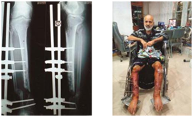
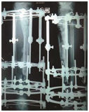
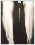
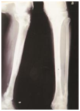
In all 41 patients union were achieved. Union was assessed as per the ASAMI criteria.4 Excellent result were seen in 21 patients (51.21%), good in 12 (29.26%) and fair in 8 (19.51%). Mean Ilizarov fixation for all patients was 366 days (130-250). Mean duration for monofocal compression was 251.2 days (130-320). Mean duration for patients with bifocal simultaneous compression distraction was 340 days (135-360). Mean duration for patients with bone transport was 353 days (321-365). Mean regenerate length of 7 cm (2.5-15.5 cm) was achieved in 15 patients. 16 had shortening ranging from 1 to 2.5 cm; 2 patients had some loss of ankle range of motion (ROM): The mean preoperative valgus deformity of 10.5˚ (9-13.1˚) seen in 8 patients. Mean preoperative varus deformity of 15˚ (10.5-22˚) seen in 9 patients; the mean procurvatum deformity of 15.1˚ (9-20˚) seen in 4 patients. The mean recurvatum deformity of 18˚ (14-25˚) seen in 2 patients (Figure 1 & 2).


a b c d

e f g h
Figure 1 Case 1-Illustration.

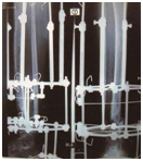
a b c
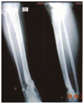
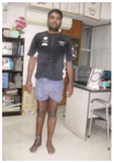

d e f
Figure 2: Case 2
Infected non-union is a very big problem in distal tibia. This happens due to poor vascularity and bad soft tissue cover. High energy trauma, poor muscle envelope and multiple surgeries increases this risk.1 Debridement, remaining, and antibiotic-coated cement rods and beads are also accepted available methods of eradication of infection.5
Infected non-union of distal third is associated with infection, bone loss, shortening and deformity. All these problems create difficulties of achieving union. The Ilizarov fixator is the best tool to perform all these problems. By this wonderful instrument we can go for definitive treatment. Union was achieved and infection eradicated in all our patients without bone grafting, which is against the Ilizarov principles. Poor soft tissue cover over the non-union site cannot accommodate the bone grafts. Ilizarov’s gradual controlled coordinated compression transformed the fibrus tissue into bony tissue.6 Schottel PC et al.7 Schoenleber SJ & Hutson JJ,8 Katsenis D et al.9 Lonner JH et al.10 all treated the infected non-union of distal 1/3 tibia with Ilizarov technique.11 Ilizarov fixation lowered the need for higher antibiotics. Elastic stable fixation of small distal fragments is difficult to perform by either locking plates or rods; Problems with osteoporosis reduces the stability of internal fixation.
Fixation of small fragments is possible with 3 or 4 Ilizarov bio compitable wires attached to a single ring for stability. Stability of fixation is maintained by a foot frame or half ring which we used in 15 of the 41 cases. Ankle stiffness we have seen in 6 patients with distal tibial non unions.
Infected non-union in lower tibia is a challenge to the orthopaedic and reconstructive surgeon. Here we faced problems of infection, deformity, bony gaps and small distal fragment. Step by step treatment by Ilizarov technique allowed us to control infection and to achieve 100% union.
None.
None.

©2017 Bari, et al. This is an open access article distributed under the terms of the, which permits unrestricted use, distribution, and build upon your work non-commercially.