MOJ
eISSN: 2374-6939


Short Communication Volume 17 Issue 5
Department of Orthopaedic and Traumatology, Hospital Carlos Bocalandro, Argentina
Correspondence: Ernesto Bertoni, Department of Orthopaedic and Traumatology, Hospital Carlos Bocalandro, Argentina
Received: August 26, 2025 | Published: October 14, 2025
Citation: Bertoni E, Vecchio MD, Moreno EA, et al. Impact of using the C-arm without traction table in the reduction of lateral hip fractures with locked cephalomedullary nail fixation. MOJ Orthop Rheumatol. 2025;17(5):133-136. DOI: 10.15406/mojor.2025.17.00721
This study investigates the impact of the use of the C-arm without a traction table, proposing an alternative patient positioning for the reduction of hip fractures that require reduction and fixation with a cephalomedullary nail. The motivation arises because in our institution the tool available has limitations regarding patient height, which consequently generates the need for a new technique to achieve optimal results, as it creates an exclusion criterion. The proposed technique seeks to improve anatomical alignment and fracture stability, with the goal of improving clinical outcomes and reducing postoperative complications. Effectiveness was examined through the evaluation of traditional and emerging methods for measuring reduction quality.
Keywords: Lateral hip fractures, traction table, C-arm, cephalomedullary nail
IV
Hip fracture is a frequent injury, especially in elderly patients with poor bone quality. Its surgical treatment is crucial for recovery and restoration of quality of life.1,2 The use of the C-arm with traction table, when available, are practical and efficient tools that are significant in this procedure. However, the choice of the optimal position of the fluoroscope without the use of the table during surgery may significantly influence clinical outcomes.
This analytical, longitudinal, retrospective research focuses on determining the feasibility of performing an
adequate anatomical reduction and fixation of a lateral hip fracture without the use of the traction table, using
an alternative positioning of the C-arm and the patient, with the aim of showing the results, intraoperative and
postoperative complications, and patient recovery. Understanding how the positioning of the image intensifier
without table affects fracture reduction, implant stability, and hip biomechanics is essential to improve surgical
practices and optimize clinical outcomes.
By addressing this issue, the aim is not only to improve surgical care and patient recovery, but also to contribute
to the advancement of knowledge in the field of orthopedic surgery and provide evidence for clinical decision-making based on best practices and available resources within an institution. Optimizing surgical technique to improve clinical outcomes is essential. Lateral hip fracture surgery is common and carries risks. Investigating and determining fluoroscope and patient positions, without the use of traction table, to facilitate the intervention and minimize risks.3 Likewise, to provide a solution in those institutions where a traction table is not available, is broken or presents limitations, as in ours when trying to achieve an optimal patient positioning.
An analytical, retrospective, longitudinal, and observational study was conducted evaluating
patients operated between November 2022 and March 2025, who underwent surgical reduction and fixation with a cephalomedullary nail for lateral hip fracture using the C-arm technique without traction table with an alternative patient and fluoroscope positioning.
|
Inclusion criteria |
Exclusion criteria |
|
Age > 50 years |
Age < 50 years |
|
Height < 1.65 m |
Height > 1.65 m |
|
Lateral fractures |
Medial fractures |
|
Closed fractures |
Open fractures |
|
Tronzo III fractures |
Tronzo I, II, IV, and V fractures |
|
Seinsheimer fractures |
Multiple fractures |
|
Single lesion |
First consultation after 72h |
|
First consultation within 72h of trauma |
Operated after 21 days of diagnosis |
|
Operated within 21 days of diagnosis |
|
|
Indoor walker or greater |
|
|
Postoperative follow-up: 10 days after discharge, monthly until 3 months, at 6 months and annually |
|
The patient was positioned supine under spinal anesthesia, preferably, with a support placed under the gluteal region ipsilateral to the fracture in order to achieve a 15° pelvic elevation to align the femoral neck. According to consulted bibliography, this generates a pathway that correlates with the anatomical curvature of the femur, which must be followed by the guidewire. As counter-traction, a sheet loop was placed around the contralateral groin, secured to the table. The C-arm was positioned at 90°, perpendicular to the axial axis of the body, obtaining a strict anteroposterior hip view. Once correct positioning was achieved, it was fixed with specific
floor markings and then rotated approximately 60° relative to the patient to obtain an optimal lateral profile for
surgery. During the procedure, it was essential to maintain axial traction of the lower limbs during guidewire insertion. In our case, this traction was performed by the assistant, together with abduction and internal rotation to achieve reduction, placement of the cephalic guidewire, and central positioning of the cephalic screw. An incision from the greater trochanter distally (average 5–8 cm) was performed. Access to bone was achieved by retracting the vastus lateralis anteriorly. Under fluoroscopic control, the guidewire was placed, the canal was reamed, and subsequently the cephalic screw inserted. For distal locking, a 15 cm radiolucent box was used, and the fluoroscope was rotated to 180° to visualize the distal hole of the nail. The site was marked with a guide, drilled, and the locking screw inserted accordingly. Clinical and radiological data were collected before and after treatment. Conventional radiographs and CT were used to assess reduction quality (Figures1-8).
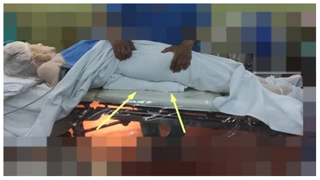
Figure 1 Patient supine with support under ipsilateral gluteal region.
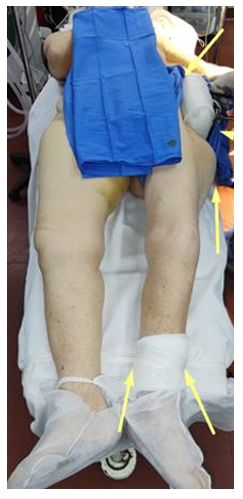
Figure 2 Counter-traction with sheet loop around contralateral groin.
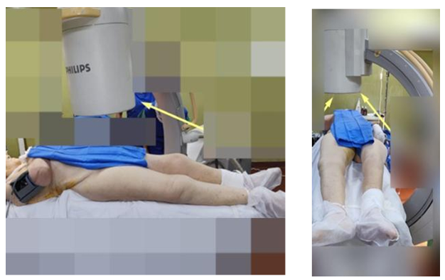
Figures 3 & 4 C-arm at 90° perpendicular to axial body axis, obtaining strict AP view of hip.
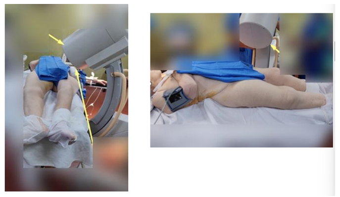
Figures 5 & 6 C-arm rotated approximately 60° relative to patient to obtain optimal lateral profile.
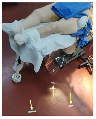
Figures 7 Specific floor markings.
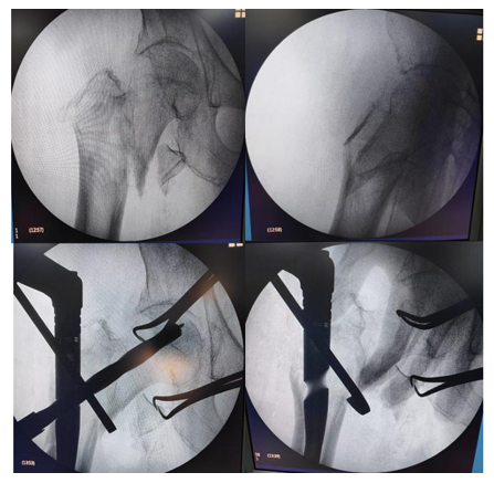
Figures 8 Intraoperative .
Out of 85 patients, 32 were included, 12 male and 20 female (37.5% and 62.5%), with a mean age of 80.1 years (range 64–92 years). The mean cervicodiaphyseal angle was 126.1° in operated hips versus 128.1° in contralateral hips. Mean Tip-Apex Distance was 4.8 mm (AP) and 5.2 mm (lateral). Regarding cephalic screw
position, 50% were located between central and superior quadrants (Cleveland & Bosworth). All patients achieved independent ambulation at 6 months. One surgical site infection and two implant-related complications (“cut-out”, 6%) were recorded. Nevertheless, rehabilitation and reintegration were achieved.
Patient data table
|
Age |
Sex |
Fracture date |
Surgery date |
Cephalic screw position |
Operated hip CDA |
Contralateral hip CDA |
Tip-apex AP |
Tip-apex Lat |
|
81 |
F |
12/11/2022 |
28/11/2022 |
1 |
123 |
122 |
8 |
13 |
|
78 |
F |
27/11/2022 |
5/12/2022 |
5 |
124 |
127 |
4 |
4 |
|
81 |
M |
26/11/2022 |
6/12/2022 |
3 |
127 |
123 |
7 |
9 |
|
75 |
F |
27/12/2022 |
6/1/2023 |
2 |
125 |
127 |
5 |
3 |
|
82 |
M |
30/12/2022 |
5/1/2023 |
2 |
121 |
122 |
5 |
6 |
|
91 |
M |
4/1/2023 |
9/1/2023 |
2 |
114 |
120 |
7 |
3 |
|
64 |
F |
22/12/2022 |
12/1/2023 |
2 |
130 |
133 |
7 |
6 |
|
82 |
F |
24/1/2023 |
31/1/2024 |
1 |
123 |
134 |
2 |
3 |
|
84 |
F |
1/2/2023 |
6/2/2023 |
3 |
123 |
125 |
4 |
5 |
|
66 |
F |
12/4/2023 |
20/4/2023 |
2 |
120 |
127 |
5 |
5 |
|
88 |
M |
26/5/2023 |
1/6/2023 |
1 |
134 |
122 |
5 |
8 |
|
81 |
M |
2/7/2023 |
6/7/2023 |
2 |
123 |
130 |
7 |
6 |
|
67 |
M |
4/8/2023 |
16/8/2023 |
2 |
122 |
124 |
9 |
8 |
|
80 |
M |
4/8/2023 |
17/8/2023 |
1 |
121 |
124 |
6 |
5 |
|
92 |
F |
27/8/2023 |
30/8/2023 |
1 |
123 |
130 |
6 |
9 |
|
86 |
F |
27/8/2023 |
10/9/2023 |
1 |
123 |
120 |
3 |
4 |
|
82 |
F |
25/9/2023 |
1/10/2023 |
2 |
127 |
130 |
3 |
3 |
|
70 |
F |
18/11/2023 |
23/11/2023 |
3 |
124 |
140 |
2 |
2 |
|
82 |
F |
31/10/2024 |
7/11/2025 |
2 |
127 |
130 |
3 |
4 |
|
72 |
M |
9/11/2024 |
14/11/2024 |
3 |
132 |
130 |
4 |
2 |
|
76 |
F |
23/12/2024 |
2/1/2025 |
2 |
118 |
127 |
7.5 |
8.8 |
|
92 |
F |
13/12/2024 |
1/12/2024 |
2 |
133 |
129 |
4 |
7 |
|
67 |
M |
1/2/2025 |
11/2/2025 |
5 |
128 |
130 |
2 |
2 |
|
77 |
M |
12/2/2025 |
28/2/2025 |
1 |
127 |
132 |
3 |
2 |
|
63 |
M |
16/2/2025 |
14/3/2025 |
1 |
130 |
129 |
5 |
6 |
|
78 |
F |
3/3/2025 |
18/3/2025 |
2 |
132 |
130 |
7 |
9 |
|
89 |
F |
7/3/2025 |
19/3/2025 |
3 |
135 |
132 |
5 |
2 |
|
92 |
F |
5/3/2025 |
21/3/2025 |
2 |
128 |
130 |
3 |
4 |
|
91 |
F |
22/3/2025 |
3/4/2025 |
3 |
130 |
132 |
4 |
5 |
|
88 |
F |
2/4/2025 |
25/4/2025 |
3 |
132 |
130 |
4 |
6 |
|
86 |
F |
19/4/2025 |
30/4/2025 |
3 |
128 |
130 |
4 |
6 |
In this analytical, retrospective, longitudinal, and observational study, we described the results of patients who received a cephalomedullary nail without traction table (supine or manual repositioning). Our findings reflect those observed in the literature. We observed congruence with TAD <25mm and equivalent radiological and clinical outcomes.4,5
Reduction quality and radiological parameters such as Tip-Apex Distance (TAD) and cervicodiaphyseal angle (CDA) did not differ significantly between techniques, suggesting outcomes equivalent to the literature.6 This aligns with other studies showing biomechanical and postoperative results similar to those with traction table.
Patients were followed for 29 months, achieving consolidation and full weight-bearing at 6 months, resuming
low-demand daily activities. Unlike some reports of traction table–related complications (perineal compression,
neuropraxia),7–11 we did not observe such events. The 6% cut-out complication rate is comparable to Jiamton et al.7 who reported 15.8%. This method showed positive outcomes with functional recovery and low complication rate.
The presented technique without traction table can be a valid alternative for femoral neck fractures, especially in resource-limited settings. It proved efficient and safe compared with conventional techniques, showing advantages in OR setup time and radiation exposure without compromising reduction or functional outcome.
Although our sample size was small, further data collection will continue.
None.
The authors declare that there are no conflicts of interest.

©2025 Bertoni. This is an open access article distributed under the terms of the, which permits unrestricted use, distribution, and build upon your work non-commercially.
