MOJ
eISSN: 2374-6939


Case Report Volume 17 Issue 4
1Specialist in Orthopedics and Traumatology, attending physician at Hospital III de Emergencias Grau, Peru
2Specialist in Orthopedics and Traumatology, sub-specialist in Hip and Knee Surgery, assistant physician at Hospital III de Emergencias Grau, Peru
Correspondence: Ortiz J, Specialist in Orthopedics and Traumatology, sub-specialist in Hip and Knee Surgery, assistant physician at Hospital III de Emergencias Grau, Peru
Received: June 06, 2025 | Published: July 25, 2025
Citation: Fernandez D, Espinoza C, Laura C, et al. Iliopsoas impingement following total hip replacement. MOJ Orthop Rheumatol. 2025;17(4):86-88. DOI: 10.15406/mojor.2025.17.00707
Iliopsoas impingement is a little-known cause of periarticular pain after total hip replacement surgery, which is why its diagnosis is essential. The following observational, non-analytical, case series study is presented. Between the months of February and April 2024, four post-total hip replacement patients diagnosed with iliopsoas impingement were included, three of these were treated conservatively and one surgically with arthroscopic psoas tenotomy.
In our case, the treatment of this condition was satisfactory; however, further studies on arthroscopic treatment of this pathology are required to demonstrate consistent usefulness when conservative management fails.
Keywords: impingement, iliopsoas, arthroscopy
Iliopsoas impingement is an underrecognized cause of joint restriction and pain after total hip arthroplasty. The incidence of this pathology described in the literature is 4.4% and was first described by Postel et al. in 1991. It consists of inflammation of this myotendinous junction (MTJ), usually as a result of mechanical irritation at the anteroinferior rim of the acetabulum due to the prominence of the acetabular component of the prosthesis. Other recognized causes are excess residual cement and screws of inadequate length passing through the iliac bone.1
The diagnosis is based on the symptoms described, physical examination and complementary imaging. The cardinal symptom is increasing pain in the hip and/or gluteus, beginning from the first month after total hip replacement surgery until several years later. Physical examination often reveals pain on hip flexion, especially when raising the ipsilateral leg in extension against resistance. Additionally, the use of anesthetics and corticosteroids on the fascia of the iliopsoas muscle tendon may help confirm the diagnosis.2
The physical examination findings are complemented by X-ray and CT images. The anteroposterior and lateral views on radiography allow the anteroinferior prominence of the acetabular component to be determined, as well as the axial CT sections. Chalmers et al. reported in their study a significant improvement in symptoms in patients who underwent open tenotomy of the iliopsoas muscle or revision of the acetabular component with release of the same tendon when the anteroinferior prominence was greater than 8 mm.3
Treatment of this condition can be conservative with physical therapy, anti-inflammatory drugs, and/or corticosteroid injections. If satisfactory results are not achieved, surgical treatment is recommended through debridement or tenotomy of the iliopsoas muscle tendon, which can be performed openly or arthroscopically. Another alternative, based on imaging findings and protrusion of the acetabular cup, would be revision of the acetabular cup component.4,5
Between February and April 2024, a case series study was conducted based on the medical records of four patients diagnosed in 2023 with iliopsoas impingement after total hip replacement. Two of these patients were treated at the Hospital III de Emergencias Grau located in Lima. The other two patients are in private practice.
Case 1
A 78-year-old woman with diabetes and hypertension underwent total left hip replacement with uncemented prosthesis due to femoral neck fracture (Figure 1). She presented with clinical symptoms that had progressed for about six months, beginning in the second postoperative month. She was characterized by severe pain in the ipsilateral gluteal region, not associated with pain in the lower back, which limited her walking and daily activities, forcing her to use a wheelchair.
On physical examination the surgical scar was in good condition, with no signs of inflammation. The patient presented severe pain on palpation in the anteroinferior and posteromedial regions of the left hip, which increased with elevation of the limb. The Stinchfield and Log Roll maneuvers were negative. Acute-phase reactants were negative.
Postoperative radiographic images show the prosthetic implant without signs of prosthetic loosening or cup protrusion (Figure 1 A and B). The CT scan shows images suggestive of inflammatory fluid around the iliopsoas muscle tendon at the level of its insertion in the lesser trochanter (Figure 2).
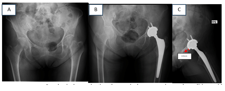
Figure 1 Preoperative radiograph of the patient on admission to the emergency department (A). Postoperative radiographs the day after surgery: AP (B) and lateral with lateral protrusion of the acetabular cup <3mm (C).
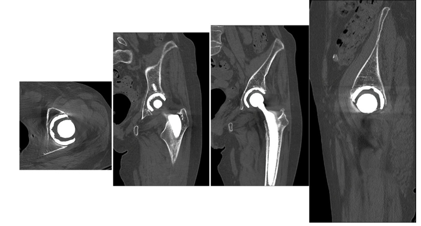
Figure 2 CT scan of the left hip. Images suggestive of fluid can be seen around the iliopsoas muscle tendon at its insertion into the lesser trochanter.
The results with conservative treatment were not encouraging. After three months of inter-daily physical therapy, the patient continued to experience pain, so surgical management with arthroscopic tenotomy of the iliopsoas muscle was chosen (Figures 3 & 4).
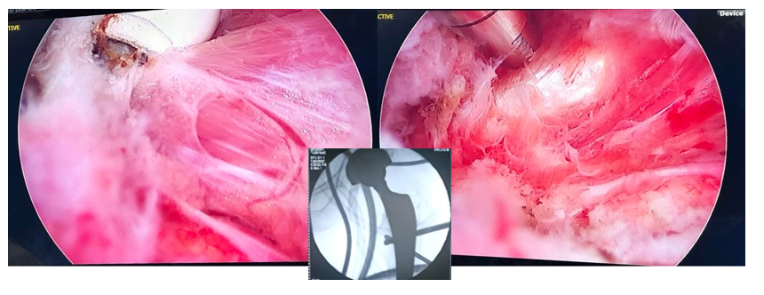
Figure 2 Arthroscopy of the left hip. The iliopsoas muscle tendon is observed with inflammatory characteristics.
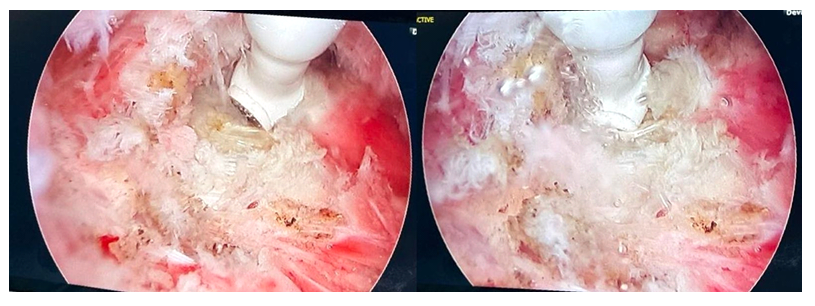
Figure 4 Arthroscopic tenotomy of the left iliopsoas muscle tendon with bipolar radiofrequency probe, under fluoroscopic view.
Two weeks after surgery, the patient presented favorable progress, reporting remission of pain, recovery of range of motion and return to walking with a cane.
Case 2
An 86-year-old male patient with diabetes and hypertension presented to the emergency department for trauma to the left hip. Figure 5 shows the pre and postoperative radiographs of the patient.
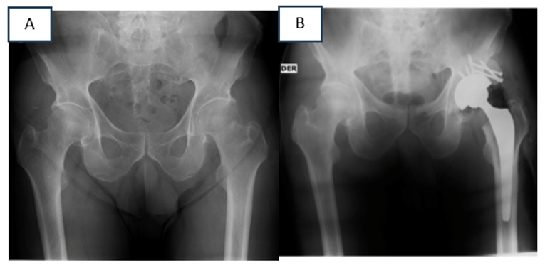
Figure 5 AP radiograph of the patient on admission to the emergency department (A). Postoperative radiograph. Due to the superolateral acetabular defect during reaming, it was decided to reinforce the acetabular cup coverage with a femoral head autograft, which was secured with cancellous bone screws (B).
After surgery, the patient progressed favorably with good pain control. He resumed walking with the support of a walker and started physical therapy. After one month, the patient returned for consultation, walking with a cane and complained of pain in the operated hip. On physical examination the scar was in good condition, and he presented pain in the gluteal and posteromedial area on the operated side. Lasegue and Gowers Bragard signs were negative, and the Stinchfield test came back negative. Postoperative images showed no signs of prosthetic loosening. A repeat CT scan was requested.
Figure 6 shows the repeat CT scan with evidence of anterior protrusion of the acetabular cup of approximately 1 cm, and images of the iliopsoas muscle impingement with the anterior acetabular screw.
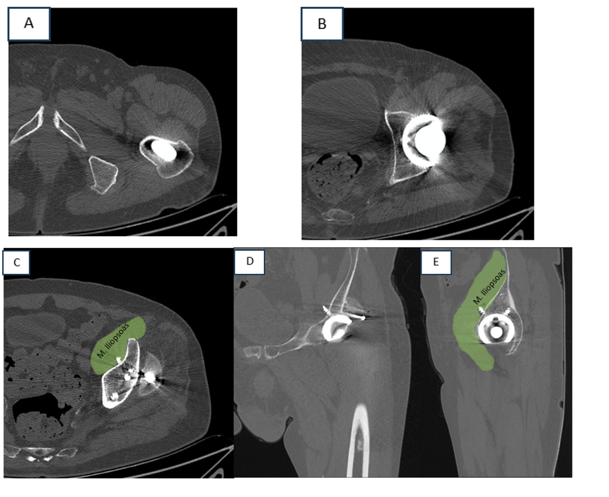
Figure 6 Postoperative control CT scan. The Axial view shows protrusion of the anterior acetabular region (A). Positioning of the femoral component in the medullary canal (B). Protrusion of the anterior acetabular screw is seen in axial (C) coronal (D) and sagittal (E) views.
Case 3
A 57-year-old male patient underwent uncemented primary total left hip prosthesis surgery in September 2023. Figure 7 shows the preoperative X-ray images. Two months postoperatively, he developed gluteal pain and signs of iliopsoas tendon impingement ipsilateral to the surgery, which completely resolved with physical therapy four months postoperatively. The patient is currently pain free. The postoperative radiograph is also shown in Figure 7.
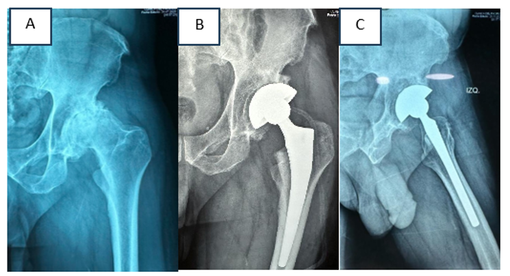
Figure 7 Preoperative radiograph prior to cementless left total hip replacement (A). Postoperative AP (B) and lateral images show evidence of acetabular cup protrusion (C).
Case 4
A 57-year-old male patient with a history of unknown rheumatologic disease underwent surgery in April 2022 for left total hip arthroplasty. The preoperative radiograph is shown in Figure 8. He is scheduled for primary right hip replacement surgery. At the second postoperative month, the patient presented pain in the right gluteus that limited walking. Physical therapy was prescribed for one month. At the third postoperative month the pain resolved. Currently the patient is asymptomatic.

Figure 8 Preoperative radiographs of the right total hip replacement (A). Postoperative radiographs show protrusion of the superolateral acetabular region (B) and (C).
Persistent hip pain following joint replacement can have multiple etiologies: infection, loosening, wear, lumbar spine conditions, etc. Within this range of possibilities is iliopsoas tendon/muscle impingement which, due to its low frequency, represents a challenge to diagnose and treat.6 The physical examination and symptoms of the patient are the initial factors that allow us to suspect this pathology. Traditionally, the literature reports the most frequent symptoms as groin pain associated with climbing stairs and getting in and out of cars. When examining the patient, groin pain is frequently described when flexing the hip against resistance and when elongating the iliopsoas muscle.7
In our case series, the most frequent symptom was pain in the gluteal region on the operated side, associated with hip flexion.
Radiological and tomographic imaging studies allow us to observe the size and positioning of the implants used in surgery, which are related to this pathology. The most common findings are excessive anteversion of the acetabular component with protrusion greater than 8 millimeters, inadequate reaming of the anterior wall, long screws or cementation in equivocal locations.6 In the group of patients presented here, inadequate anteversion, protrusion of the acetabular cup and inadequate length of the screws are consistent with the findings described in the world literature.
This pathology can be treated conservatively with analgesia, injections and physical therapy. However, when these fail - in 50% of the cases, surgical treatment is chosen. Our case series showed a 75% success rate for patients who started physical therapy. Regarding surgical options, a prominence of the acetabular component greater than 8 mm suggests revision of the latter. The success rate for this procedure is approximately 94%. If the prominence of the acetabular component is less than this number, iliopsoas tenotomy can be performed open or arthroscopically, the latter carrying less complications and faster patient recovery.8 In this case series one patient underwent iliopsoas tendon release via arthroscopy with painless gait recovery within a few days of surgery.
Iliopsoas impingement in post-operative total hip replacement patients is an existing and rare condition whose treatment should be staged and should be initiated with anti- inflammatory drugs, physical therapy and, occasionally, corticosteroid injections.
The treatment of this pathology with arthroscopic tenotomy was successful in our experience without postoperative complications and with good functional results. We agree with the scientific evidence in the current literature. It is an effective procedure in the treatment of this pathology when properly indicated. Due to its low frequency, it is essential to first rule out its most frequent causes.
None.
The authors declare that there are no conflicts of interest.

©2025 Fernandez, et al. This is an open access article distributed under the terms of the, which permits unrestricted use, distribution, and build upon your work non-commercially.
