MOJ
eISSN: 2374-6939


Research Article Volume 10 Issue 2
Laboratory of Rheumosurgery and Rehabilitation, V.A. Nasonova Research Institute of Rheumatology, Russia
Correspondence: Byalik Valeiy Evgenievich, Orthopaedist, Russian Federation, Moscow, Kashirskoe schosse, 34a, Russia, Tel 79645109862
Received: February 09, 2018 | Published: March 28, 2018
Citation: Evgenievich BV, Anatolievich MS, Ivanovna AL, et al. High tibial osteotomy in patients with stages 2 and 3 of knee osteoarthritis. Short-term result and factors affecting the outcome. MOJ Orthop Rheumatol. 2018;10(2):120–128. DOI: 10.15406/mojor.2018.10.00399
Introduction: The most common form of osteoarthritis (OA) is osteoarthritis of the knee. Conservative treatment of OA is effective only in stages I and II of the disease. Meanwhile, increasing incidence of knee osteoarthritis and lowering ages of the disease onset makes high tibial osteotomy (HTO) more and more vital, allowing to extend the function of the own knee joint and to postpone or completely avoid total knee replacement (TKR).
The aim of the study was to assess the effectiveness of НTO at 2-3 stages of knee osteoarthritis and to investigate the influence of age, body mass index (BMI) and correction angle on the nearest result of the operation.
Materials and methods: during the period from 2003 to 2016, 35 HTOs were performed in 32 patients. The ratio of men to women was 2:1. The mean age was 59.0±13.1 years, a BMI of 29.04±3.57kg/m² and a correction angle of 12.5±2.78°. A visual analogue scale (VAS) was used to assess the pain severity. The Knee Society Score (KSS) was applied to assess the functional and objective state of the knee joint. The stage of the degenerative process was evaluated according to the X-ray classification of Kellgren-Lawrence.
Results: The HTO was effective in patients with both 2nd and 3rd stages of knee osteoarthritis. One year after the operation, a significant reduction in VAS scores (from 72.27±11.79 mm to 7.72±6.62mm) and an improvement in functional and objective KSS scores (from 43.66±11.5mm and 54, 39±11.77mm to 86.51±10.86mm and 81.93±6.65mm) were observed. We obtained the following results of the HTO: excellent (36.4%), good (57.6%) and satisfactory (6%). The X-ray signs of progression of the disease were not revealed one year after the operation. The connection of BMI with the nearest result of the operation was revealed. (Spearman coefficient=-0.34 at p <0.05).
Conclusion: HTO is more effective at the 2-nd stage of osteoarthritis of the knee compared with the third stage. Age and angle of correction do not affect the nearest result, while increased values of body mass index are associated with worse result and complications.
Keywords: knee joint, osteoarthritis of the knee, high tibial osteotomy, age, BMI, correction angle
BMI, body mass index; DAS, 28 – disease activity score for 28 joints; DMARDs, disease-modifying antirheumatic drugs; GIBP, genetically engineered biological preparations; HTO, high tibial osteotomy; KSS, knee society score; MT, methotrexate; OA, osteoarthritis; RA, rheumatoid arthritis; TKR, total knee replacement; UKA, unicompartmental knee arthroplasty; VAS, visual analogue scale
Osteoarthritis (OA) is a heterogeneous group of diseases of various etiologies with similar biological, morphological, clinical manifestations and outcomes, in which cartilage, subchondral bone, ligaments, synovial membrane and periarticular muscles are involved in the pathological process.1 OA is the leading cause of chronic pain and takes the fourth place as the cause of disability worldwide.2 The most common form of OA is knee osteoarthritis. The frequency of symptomatic OA of the knee joint in general population is 25%.3 Generally, the medial compartment of the knee joint is involved in the pathological process (75%), the patellofemoral joint is on the second place (48%), and the lateral compartment is rarely involved (26%).4
Untreated knee osteoarthritis in stages II and III was diagnosed in 85% of women and 74% of men consulted for pain in the knee joint for the first time.5 Moreover, conservative treatment of OA is more effective at stages I and II of the disease.6 Among the methods of surgical treatment of knee osteoarthritis with predominant lesion of the medial compartment a special place is occupied by high tibial osteotomy (HTO), which is divided into 3 main types: the closing wedge, dome or barrel-vault osteotomy, and the opening wedge.7 Regardless of the method of the operation, any correctly performed HTO results in restoring the mechanical axis of the lower limb, transferring the load from the affected medial compartment of the knee joint to the intact lateral one, slowing the progression of the degenerative process, prolonging the function of own knee joint and procrastinating the total knee replacement [TKR].8–10 Nevertheless, to this day, controversies continue over the factors that affect the outcome of the HTO (age, body mass index (BMI), correction angle), and its survival rates.11–13 This is important due to increased frequency of OA and lowering ages of the disease onset.14,15 The objectives of our study were: to clarify the indications for the operation, to study the influence of age, BMI, and correction angle on the nearest result, and to evaluate the effectiveness of the HTO in primary and secondary knee osteoarthritis in stages 2 and 3.
In the laboratory of rheumosurgery and rehabilitation of the V.A. Nasonova Research Institute of Rheumatology for the period from 2003 to 2016 years 35 HTO were performed in 32 patients. The ratio of men to women was 2 to 1. Mean age of patients was 59.0±13.1 years, mean BMI score - 29.04±3.57kg/m², and mean correction angle - 12.5±2.78°.
The operations were performed in primary and secondary knee osteoarthritis. 25 patients had a diagnosis of osteoarthritis (in 3 cases, both knee joints were operated with an interval of 6-12 months). Three patients with posttraumatic knee osteoarthritis, two with rheumatoid arthritis (RA), one with psoriatic arthritis and one with Koenig disease were also operated using the HTO method (Table 1).
Diagnosis |
Number of patients |
% |
Osteoarthritis |
25 |
78,18 % |
posttraumatic knee osteoarthritis |
3 |
9,38% |
rheumatoid arthritis |
2 |
6,25% |
psoriatic arthritis |
1 |
3,12% |
Koenig disease |
1 |
3,12% |
Total |
32 |
100% |
Table 1 Diagnoses of patients
We used the following indications to the HTO: isolated OA of the medial compartment of the knee joint at any stage without bone defects, no changes or 1-2 stages of OA in the patellofemoral joint, intact lateral compartment, amplitude of movements >100°, BMI <40kg/m², high degree of initial functional activity of the patient, varus deformation less than 17.5°. Contraindications to this operation were: severe concomitant somatic diseases, previous infection, patellofemoral OA of 3-4 stages, OA of lateral compartment of any stage, BMI >40 kg/m², limitation of flexion >25°, absence of lateral meniscus.
In the preoperative period, pain severity was assessed using a visual analog scale (VAS), as well as the clinical and functional state of the knee joint using the Knee Society Score (KSS) scale. These parameters were assessed again after 3 months and a year after the operation. The nearest results were evaluated by changing the indices of the above scales. We estimated the pain values for VAS from 0 to 20 mm as an excellent result, 21-40mm - as good, 41-60mm - as satisfactory, more than 60mm - as an unsatisfactory result. Similarly, the changes in the functional and objective state of the COP on the KSS scale were evaluated. The values of the functional and objective (scales?) less than 50 points corresponded to the unsatisfactory result, 51-70 - to the satisfactory result, 71-85 - to the good score, and 86-100points - to the excellent result. Since the total score of the KSS is the sum of the above parameters, with a value of <100 the result was considered unsatisfactory, 101-140 - satisfactory, 141-170 – good, and >171 - excellent.
A teleradiography of the lower limb was used for preoperative planning. The correction angle was calculated by the Miniaci method. The stage of the degenerative process was evaluated by the X-ray classification of Kellgren-Lawrence. 25 cases (71.5%) were diagnosed with stage 3, in the other 10 (28.5%) cases second stage of the knee osteoarthritis was diagnosed.
12 HTO (34.3%) of 35 were supplemented by arthroscopy (10 patients with stage 3 knee OA and 2 with second stage). Intraoperatively, we assessed the defects of the cartilaginous tissue in accordance with the arthroscopic classification of Outer bridge articular cartilage damage. Damages of the articular cartilage on the medial condyle of the thigh were visualized, corresponding to 2nd stage in Outer bridge in 3 patients, 3rd stage in 7 patients, 4th stage in 2 patients. At the same time, in 6 patient adjacent defects of the medial condyles of the tibia and femur were identified. The methods used for concomitant arthroscopy are presented in Table 2.
Arthroscopy |
Number of operations |
% |
Microfracturing |
6 |
50,0% |
Abrasive chondroplasty + microfracturing |
2 |
16,67% |
Partial resection of the medial meniscus |
2 |
16,67% |
Partial resection of the medial meniscus + microfracturing |
2 |
16,66% |
Total: |
12 |
100% |
Table 2 Methods of concomitant arthroscopy
We preferred the opening wedge HTO technique the most often (28 operations, 80%). The closing wedge and dome-osteotomy were used in 3 (8.57%) and 4 (11.43%) cases, respectively. This is due to the fact that we consider the opening wedge HTO to be less traumatic than the closing wedge and the dome methodology of HTO implementation. Thus, with opening wedge HTO, there is no need for massive dissection of soft tissues, fibular osteotomy and violation of the integrity of the proximal tibiofibular junction, as in closing wedge HTO. Dome-osteotomy is possible to restore the mechanical axis of the lower limb, even with a varus deformation exceeding 20°, by completely separating the proximal epiphysis from the metaphysis of the tibia and subsequent manual repositioning under the X-ray control to the desired correction angle. However, at smaller correction angles, the opening wedge HTO technique, in our opinion, is preferable, since it allows keeping the posterior cortical layer of the tibia intact, which is less traumatic in comparison with the dome-osteotomy.
The postoperative rehabilitation program included, in addition to analgesic, antibacterial and anticoagulant therapy, immobilization in the orthosis with full extension in the knee joint for 8 weeks from the day of surgery. Nevertheless, the patients performed exercises aimed to passively increase the range of motions in the knee joint (on the apparatus of passive robotic mechanotherapy of the joints of Artromot), mobilization of the patella, as well as exercises to strengthen the muscles of the thigh. For walking, patients used crutches for 2 months, then a cane for 2 weeks. Finally, they were allowed to walk with full support on the operated leg.
Statistical processing of the obtained data was carried out on a personal computer using the Microsoft Excel application and statistical data package Statistica 10 for Windows (Stat Soft Inc., USA). Mean and median values, standard deviation, and interquartile range were calculated. To determine the mutual influence of the indicators a correlation analysis was performed using the Spearman and Pearson coefficient. Differences were considered statistically significant at p <0.05.
The nearest result was evaluated after 3 months and a year after the operation (Figure 1). Three months after the HTO, the pain decreased from 72.27±11.79 to 17.87±11.04 (from 5 to 50)mm, and after a year the indicator had values of 7.72±6.62 (from 0 to 20)mm. A similar picture was demonstrated by the objective and functional scores of the KSS: before the operation, the objective score was 43.66±11.50 points, and the functional score was 54.39±11.77 points. 3 months after the operation together with the decrease in pain intensity we noted an improvement in the objective score to 77.72±9.48 points, and a functional score to 80.0±12.86 points. A year after the operation these indicators were respectively 81.93±6.65 points and 86.51±10.86 points. Pearson's correlation coefficient demonstrates a strong association of pain reduction in the VAS with an improvement in the functional and objective state of knee joint (-0.62 at p <0.05).
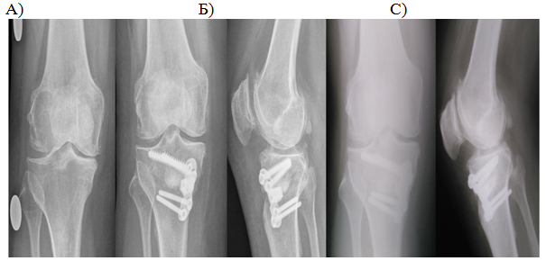
When studying the radiographs in the direct and lateral projections, disclosure of the joint gap was noted 3 months after the operation. Progression of medial compartment OA after a year did not occur, degenerative changes in another compartments were also absent (Figure 2) (Figure 3).

Figure 2 Patient T., 57 years old, 3-rd stage OA of the right knee. Before surgery (A), after 3 months (B), and 1 year (C) after surgery.

Figure 3 Patient M., 54 years old, rheumatoid arthritis. 2-nd stage secondary OA of the right knee. Before surgery (A), after 3 months (B), and a year (C) after surgery.
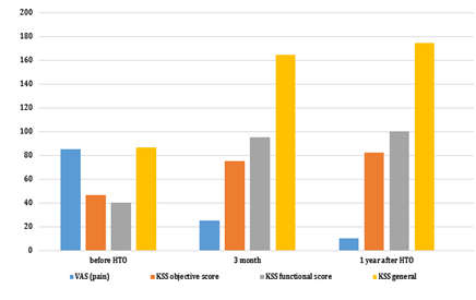
Since RA patients are of particular interest, the dynamics of their pain indices and KSS are presented separately. Assessment of RA activity and efficacy of antirheumatic therapy was carried out using the DAS 28. Both patients were 54 years old, with the the BMI scores of 32.5kg/m² and 31.3kg/m². Both patients had a remission according to DAS 28. Both patients received methotrexate (MT) at 15 mg/week at the time of surgery. The first patient received an additional methylprednisolone 4mg/day. On days without MT patients received folic acid at mg/day. Both patients were made opening wedge HTO without arthroscopy. The correction angle was 9° in both cases.
The fixation was carried out with a plate for the HTO by Osteomed. In the postoperative period, the rehabilitation program described above was carried out. In both cases, the wound healed by primary tension and the sutures were removed on the 14th day from the day of surgery. Later, one of the patients had pain in the projection of the implanted plate. This patient, if necessary, took meloxicam. In general, this complication did not affect the short-term result of the operation. Consolidation in both cases occurred within 3 months from the date of surgery. The plate was removed within 12 to 18 months after the operation. The short-term result of HTO in these patients was assessed as good and excellent and are presented in Figure 4.

Figure 4 Change in pain, functional, objective, and total score of KSS in patients with RA (mean values).
In general, we received 12 excellent (36.4%), 19 good (57.6%) and 2 satisfactory (6%) results. Excellent results were more often noted in the group of patients with stage 2 arthrosis. At the same time, patients with stage 3 OA of the knee had 2 satisfactory results, while in the group of patients with stage 2 OA no satisfactory results were observed (Table 3). Statistical analysis using the Pearson correlation coefficient demonstrates a fairly strong relationship between the nearest result and the stage of the degenerative process at the time of the operation (the Pearson’s r=0.64, p <0.05).
Stage of OA |
Short-term results |
||||
|---|---|---|---|---|---|
Unsatisfactory |
Satisfactory |
Good |
Excellent |
Total |
|
2nd stage |
0 (0,0%) |
0 (0,0%) |
4 (44,4%) |
5 (55,6%) |
9 (100%) |
3rd stage |
0 (0,0%) |
2 (8,3%) |
15 (62,5%) |
7 (29,2%) |
24 (100%) |
Total |
0 (0,0%) |
2 (6,0%) |
19 (57,6%) |
12 (36,4%) |
33 (100%) |
Table 3 Relationship between the results of the HTO and the stage of the degenerative process before operation
Evaluation of the relationships between the nearest result of the operation and sex, age, BMI, concomitant arthroscopy and the size of correction angle of the osteotomy using the nonparametric Spearman coefficient demonstrated reliable association between the nearest result and the BMI scores. In this case, the higher the BMI, the worse the result (Table 4).
Factors affecting the |
Age |
BMI |
Arthroscopy |
Sex |
Correction angle |
Short-term result |
age |
1,00 |
0,19 |
0,31 |
-0,32 |
0,10 |
-0,21 |
BMI |
0,19 |
1,00 |
-0,17 |
-0,02 |
0,19 |
-0,34 |
arthroscopy |
0,31 |
-0,17 |
1,00 |
0,10 |
0,08 |
0,04 |
sex |
-0,32 |
-0,02 |
0,10 |
1,00 |
-0,13 |
-0,03 |
Correction angle |
0,10 |
0,19 |
0,08 |
-0,13 |
1,00 |
-0,26 |
Short-term result |
-0,21 |
-0,34 |
0,04 |
-0,03 |
-0,26 |
1,00 |
Table 4 Association of various factors with HTO results (Spearman correlation coefficient, p <0.05) BMI, body mass index
The less pronounced symptoms of osteoarthritis of the knee joint at the time of the surgery, the better the result of the HTO.11 In our study, the proportion of excellent and good results was also higher in the group of patients with a lower stage of OA. Few authors evaluated the outcomes of the HTO on the KSS. Su Chan Lee et al.16 performed opening wedge HTO on 37 knee joints in 33 patients and a year later observed an improvement in the objective score of knee joint from 52.19±11.82 to 92.49±5.10 points, and the functional score from 52.84±6.23 to 89, 05±5,53 points. Asik M. et al.17 evaluated the results, on average, 34 months after the operation and demonstrated an improvement in the objective score from 35.70 to 85.60 points, and - from 53.50 to 83.50 points in functional score. Our results are comparable with the data of other authors (improvement of the objective score from 43.66±11, 50 to 81.93±6.65 points, and in functional score from 54.39±11.77 to 86.51±10.86 points).
Today, RA and other inflammatory arthropathies are considered to be absolute contraindications to the HTO.18,19. We could not find articles on the treatment of secondary knee osteoarthritis in patients with rheumatoid arthritis using the HTO method after 1990. Historically, the HTO in the 60-80s of the 20th century was used to treat both patients with OA of the knee and RA patients with secondary knee OA.20–25 Matthews LS et al.20 operated on 4 knee joints in patients with RA and demonstrated results comparable to those in OA of the knee. RNW Chan & JP Pollard21 performed HTO on 36 knee joints in RA patients, in 15 cases (42%) result were good, in 7 (19%) - satisfactory, and 14 (39%) results were poor. A Benjamin22 analyzed data on 36 operations HTOs on knee joints in patients with OA and 21 surgeries HTOs in patients with RA, and concluded that for both categories of patients the operation is equally effective. A Ahlberg et al.23 showed improvement in only 2 of 11 RA patients. MB Devas24 in his article described 4 HTOs, which were carried out in RA patients. In 1 case a good result was obtained and in 3 cases - satisfactory results. It should be noted that in addition to the HTO the author performed patellectomy, which also influenced the result. Coventry MB25 described satisfactory results of 6 HTOs in 4 RA patients who were prospectively observed from 2 to 5 years after surgery (in this study, the result was divided only on satisfactory and unsatisfactory ones).
Despite the significant number of good and satisfactory results of HTO in RA patients, the number of unsatisfactory results was quite high and, consequently, it was not possible to achieve the postpone of TKR, which forced orthopedists to abandon the HTO in this category of patients. This was due to the lack of adequate therapy with basic anti-inflammatory drugs, the selection of patients with multiple joint deformities and the lack of control of inflammatory activity and the effectiveness of treatment. However, in recent decades rheumatology has been actively developing. There are many disease-modifying antirheumatic drugs (DMARDs) today. In particular, high efficacy of MT has been proven, which today is the drug of choice for the treatment of RA.26 At the end of the 20th century and in the beginning of the 21st, genetically engineered biological preparations (GIBP) were integrated in RA treatment.27 DMARDs and GIBP are effective in suppressing of the inflammatory process, reducing pain, improving function and slowing the progression of joint destruction.26,27 However, in some patients, despite ongoing treatment, active inflammation results in irreversible changes in joints structure. Therefore, treatment of secondary knee OA in RA patients using the HTO method is quite discussable. We believe that in patients with RA HTO can be performed if the following conditions are combined: 1. Stated remission or low RA activity. 2. Regular intake of DMARDs in sufficient dosages. 3. The absence of secondary OA of hip and ankle on the side of the proposed operation. 4. patient's compliance with the main indications for HTO. Unfortunately, there are not many patients satisfying those criteria. Nevertheless, the results of HTO in patients with RA give us hope that indications for this surgery will expand in the future. It is advisable to study intermediate and long-term results in these patients.
TO Smith et al.28 performed a meta-analysis to compare the results of unicompartmental knee arthroplasty (UKA) and HTO. Researchers concluded that there are no significant differences between the results. We were not able to find studies comparing the dome-osteotomy with the closing wedge or the opening wedge HTO. Interestingly, in meta-analysis of Spahn G et al.29 43 studies of UKA and 46 studies of HTO were compared, and no statistically significant differences in the results of these studies were observed. At the same time, the average time after the HTO before the TKR was 9.7 years, and after the UKA - 9.2 years. There were also no significant differences in the number of complications. Cochrane Review Brouwer RW et al.30 on the application of HTO to patients with OA of the knee showed that the results of the opening wedge HTO and the other surgical methods of HTO and UKA do not differ significantly. At the same time, there are no studies comparing the effectiveness of HTO with conservative methods of treating OA of the knee. None of the authors describes the observation by orthopedist or rheumatologist of operated patients after the first year after surgery. These patients had received conservative therapy (chondroprotectors, hyaluronic acid preparations, enriched platelet plasma injections, nonsteroidal anti-inflammatory drugs), and how long the painless period lasted is unknown. The HTO can be useful in slowing the progression of knee OA and achieving regression in clinical symptoms. The mechanical axis of the lower limb during the HTO is transferred to the external tibiofemoral compartment of the knee joint. As a result, degenerative changes begin to develop in it. Therefore, the main task of the orthopedist and rheumatologist is to prevent a decrease in the height of the articular cleft in the lateral tibiofemoral compartment of the knee joint. We believe that there should be continuous interaction between orthopedist and rheumatologist, as this can improve result of treatment in patients with OA of the knee. It is advisable to further complex treatment of OA of the knee in patients who have undergone HTO.
The influence of age, BMI and correction angle on HTO results remains controversial to this day.11–13 In our study, there was no statistically significant relationship between patients' age and the short-term outcomes, but certainly this factor should be re-examined in the study of medium- and long-term results. In contrast, BMI is of fundamental importance even for the short-term results. Thus, elevated BMI values were associated with worse short-term outcomes.
The analysis of the effect of the size of the correction angle on HTO outcomes is described by Floerkemeier S et al.12 and Nelissen EM et al.13 In the first study it is concluded that the size of the correction angle does not have any effect on HTO result, and the second one showed that an osteotomical wedge size exceeding 10° is associated with significantly higher number of complications than a wedge size less than 10°. Nelissen EM et al.13 recommend an HTO to be performed at the earliest possible stage of knee OA when there is minimal varus deformation. In our study, there was no correlation between the size of the correction angle and the nearest result.
Arkhipov Sergey Vasilievich, Nesterenko Vadim Andreevich, Nurmukhametov Maxim Rinatovich.
Authors declare there is no conflict of interest in publishing the article.

©2018 Evgenievich, et al. This is an open access article distributed under the terms of the, which permits unrestricted use, distribution, and build upon your work non-commercially.