MOJ
eISSN: 2374-6939


Clinical Report Volume 15 Issue 2
1Prof. Ph.D, Chief Consultant, Bari-Ilizarov Orthopaedic Centre, Visiting and Honored Prof., Russian Ilizarov Scientific Centre, Russia
2Medical officer, Bari-Ilizarov Orthopaedic Centre; PhD resident in Tashkent scientific research institute of orthopaedics and traumatology, Uzbekistan
3Prof., Bari-Ilizarov Orthopaedic Centre, Bangladesh
Correspondence: Bari MM, Bari-Ilizarov Orthopaedic Centre, 1/1, Suvastu Shirazi Square, Lalmatia Block E, Dhaka-1207, Bangladesh, Tel +8801819211595
Received: April 05, 2023 | Published: April 24, 2023
Citation: Bari MM, Bari AM, Shayan R, et al. Genu varum (Rt) genu valgum (Lt), post traumatic ankle varus deformity (Lt). MOJ Orthop Rheumatol. 2023;15(2):84-85. DOI: 10.15406/mojor.2023.15.00622
Ilizarov Technique is a fantastic tool for correcting femoral varus, genu valgum and ankle varus. The case demonstrates an approach to large complex deformity in both right & left knee, and left ankle region. All of the aforementioned deformities were fully corrected by the help of Ilizarov Technique.1,2,3
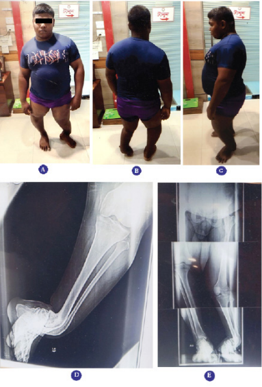
A 21 years old boy sustained motor vehicle injury in the left inferior extremity at the age of 4. He was treated at Combined Military Hospital, Dhaka at that time because father is an Army personnel. Now, he was referred to Bari-Ilizarov Orthopaedic Centre for further management, that is for correction of deformity and L. L. D. Plain X-ray and clinical findings showed his right genu varum deformity due to depressed medial tibial plateau and lower (Lt) tibio fibular varus and procurvatum deformity and left knee genu valgum. His father was anxious regarding his deformity correction.4,5 (Figure 1(A-E))

Figure 1(A-E)
(A) Front view of the patient with procurvatum deformity of tibia (Lt) é ankle varum
(B) Back view of the patient
(C) Patient Side view showing procurvatum deformity
(D) Radiograph of lower (Lt) tibio fibular varus and procurvatum deformity.
(E) Bipedal full length stress X-ray.
Problems before surgery
Obesity
Gradually worsening of deformity and skeletal maturity Right sided genu varum, left sided valgum and Lt ankle varus
Treatment plan
Two steps of surgeries were calculated.
What we hope to achieve
Our aim is to achieve
Waund gait
Eliminating any preexisting LLD
Allow full weight bearing é minimal stiffness of joints
Increase quality of life
Why Ilizarov method?
Ilizarov compression-distraction device provides flexibility and adaptability
Advantages of Ilizarov
Minimally invasive
Allow weight bearing from 1st POD
Early mobilization
Other advantages of Ilizarov
Modular
Customizable
Can treat multilevel, multiplanar and multi-directional deformities
Can articulate across the joints
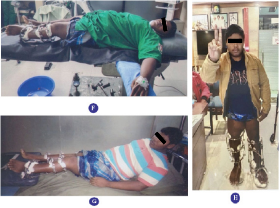
During treatment images, radiographs and follow up (Figure 1(F-H))

Figure 1(F-H)
(F) Patient lying on operation table during application of Ilizarov apparatus
(G) Patient with Ilizarov apparatus
(H) Correction of deformity with Ilizarov apparatus in stu
Technical pearls
Post-operative management and rehabilitation
Exercises were encouraged just day after the pain permitted.
Cleansing the wire sites at regular interval.
Discharged with follow-up every weeks until radiological signs of union.
Apparatus removed with outdoor or indoor basis. (Figure 1(I-L))
None.
The authors declare no conflicts of interest.

©2023 Bari, et al. This is an open access article distributed under the terms of the, which permits unrestricted use, distribution, and build upon your work non-commercially.