MOJ
eISSN: 2374-6939


Research Article Volume 6 Issue 7
Department of orthopedics, South Valley University-Qena, Egypt
Correspondence: Ahmd Khairy, Department of orthopedics, South Valley University-Qena, Egypt
Received: January 08, 2016 | Published: December 28, 2016
Citation: Khairy A (2016) Fixation of Supracondylar Humeral Fracture in Children By Medial and Lateral Pinning Versus Lateral Pinning. MOJ Orthop Rheumatol 6(7): 00248. DOI: 10.15406/mojor.2016.06.00248
Background: Supracondylar fracture of humerus is one of the most common fractures in the first decade of life. There are various modalities of treatment adviced for the management of type III supracondylar fracture of humerus. At present closed reduction and percutaneous pin fixation is most widely accepted treatment method for displaced supracondylar fracture but controversy persists regarding the optimal pin fixation technique. The purpose of this study was to assess results and complications of fixation of supracondylar humeral fracture (type III) by crossing pinning versus two parallel lateral pinning in children. As regard intra operative and postoperative stability, elbow flexion, extension, supination and pronation. Vascular status pre and post operative, risk of nerve injury. Incidence of deformity of the elbow ( Cubitus varus), elbow function.
Method: This prospective randomized controlled study of children with displaced surpracondylar fracture (type III) admitted in Orthopedic Department of Sohag university hospital after taking an informed consent from child's fathers or near relatives and approval from the institute ethical committee in the period between June 2012 and May 2013. There were totally 44 children selected for the study. We lost 4 children for follow up. They were allocated to Group-A (two parallel Lateral wires) 20 children, and Group - B (Medial and lateral wires) 20 children.
Primary assessment was done for major loss of reduction and iatrogenic ulnar nerve injury. Secondary assessment was done for clinical alignment, elbow range of motion, radiographic measurements, Flynn grade, functions and complications.
Results: The two groups were evaluated for pre-fracture characteristics and post reduction evaluation at first week, third week, fourth week, two months, three months and four months. The mean follow up in group A was 3.6 months and group B was 3.8 months. Both groups were also similar in sex distribution, pre-operative displacement, mode of injury and neurovascular status. No major loss of reduction was observed in both groups where as there was no significant difference between mild loss of reduction (3 cases in group-A), change in Baumann angle, change in Humerocapitellar angle , Flynn grade, elbow extension and flexion, carrying angle, total range of motion (p>0.05). But there was only one case of ulnar nerve injury in group B that had completely improved after three months follow up.
Conclusion: This study concluded that there was no significant difference between medial and lateral crossing wires and 2 parallel lateral wires as regard postoperative clinical results, complications and incidence of iatrogenic ulnar nerve injury. But medial and lateral crossing wires give more biomechanical stability than 2 parallel lateral wires in fixation of supracondylar humeral fracture (type lll) in children.
Supracondylar fracture of humerus is a common injury in children, comprising 3-18% of all pediatric fractures and 60% of pediatric fractures around the elbow.1 More than 97.7% of the fractures are of extension type and only 2.2% are of flexion type. most occurred in males, especially between the age of 5 and 8 years.2 Posterior displacement suggests a hyperextension injury, usually due to a fall on the out-stretched hand, while anterior displacement is thought to be due to direct violence (e.g. a fall on the point of the elbow) with the joint in flexion.3
Gartland classification
This classification of supracondylar fractures is based on the radiological appearance of fracture displacement.4
Various treatment options has been discovered for type III supracondylar fracture such as closed reduction and long arm cast or slab, Dunlop skin traction, olecranon traction, but all of these methods had significantly large complication rate.6 The standard current treatment for displaced supracondylar fracture has been close reduction and percutaneous pin fixation. This method has consistently given excellent results reported by various authors.7,8 However, controversy persists regarding whether medial and lateral pin fixation or lateral pin fixation is satisfactory technique in terms of stability and iatrogenic ulnar nerve injury.9 Ideally medial and lateral pin fixation engage medial and lateral column at fracture site whereas lateral pin stabilizes lateral and central column. Medial and lateral pin fixation has been presumed to be more stable but it can cause iatrogenic ulnar nerve injury. Therefore, we conducted this prospective study to compare whether lateral pin construct, if placed properly, can provide the same stability like medial and lateral pin fixation, at the same time avoiding the possibility of iatrogenic ulnar nerve injury.10,11Current method of treatment of supracondylar fracture of humerus in children is based on Gartland classification. Flynn et al. reported the incidence of cubitus varus deformity after treatment was 5% where as Arino et al. reported that it was almost 21%, ulnar nerve deficit was found in 15% of patients who were treated with medial and lateral pin as per the report of Chai.2-4
It is a prospective randomized study of children with displaced surpracondylar fracture (type III) admitted in Orthopedic Department of Sohag university hospital to assess the result of fixation of displaced supracondylar humeral fractures in children by two parallel lateral pins versus crossing medial and lateral pins after taking an informed consent from child's fathers or near relatives and approval from the institute ethical committee in the period between June 2012 and May 2013.
There were 44 children in our study 4 were lost for follow up. They were divided into two groups; group-A (two parallel lateral wires) and group-B (two crossing medial and lateral wires). The average age in group-A was 5.4±3.1 and in group-B was 6.2±2.3 There were 13 boys (65%) and 7 girls (35%) in group-A and 15 boys (75%) and 5 girls (25%) in group-B. the mode of trauma was falling from height (tree, donkey, wall. . . . . .etc) in 10 children in group-A and 8children in group-B, and while playing in 7 children in group-A and 10 children in group-B and due to road traffic accident in 2 children in group-A and group-B, and due to other causes in only 1 child in group-A. The affected side was right side in 7 children of group-A and 4 children of group-B and left side in 13 children of group-A and 16 children of group-B.The neurovascular status was pulseless viable limb in only on child of group-A and weak pulse in 2 children of group-A and 3 children of group-B and median n. injury in 3 children of group-A and 1 child o group-B and there were no cases of radial n. injury. Preoperative 19 children of group-A and 18 children of group-B were splinted with above elbow slab in 70-90 degree flexion of the elbow. Anteroposterior and lateral few x-ray was done on the elbow and the displacement was posteromedial in 14children of group-A and 13 children of group-B. Posterolateral in 4 children of group-A and 6 children of group-B and posterior in 2 children of group-A and one child of group-B. The average hospital stays in hrs was 15±10 hrs. In group-A and 20±8 hrs. In group-B (only one case with impalpable pulse was admitted for three days) (Table 1).
|
Result |
Rating |
Carrying Angle Loss(o) |
Flexion Loss(o) |
Extension Loss(o) |
|
Satisfactory |
Excellent |
0-4.9 |
0-4.9 |
0-4.9 |
|
Good |
5.9.9 |
5.9.9 |
5.9.9 |
|
|
Fair |
10-14.9 |
10-14.9 |
10-14.9 |
|
|
Unsatisfactory |
Poor |
≥15 |
≥15 |
≥15 |
Table 1 Modified Flynn’s criteria and overall rating.
All children with suspected supracondylar fracture of elbow were seen either at orthopedic emergency room or orthopedic outpatient department by orthopedic resident doctor and the orthopedic senior surgeon. They were assessed for general evaluation of the general condition, associated other injuries and assessment of the vascular and neurological status of the affected limb. Anteroposterior and lateral radiographs of the elbow were done. All displaced supracondylar fractures were admitted and injured elbow was immobilized in splint with elbow in 70 to 90 degree of flexion according to the vascular condition of the affected limb with elevation. Pulseless viable limb and nerve injuries were also included for the study. Patients were reassessed in the ward for neurovascular injuries. Surgery was planned on the same day after obtaining written informed consent from child's parents or near relatives. Preoperative investigations (blood picture and prothrombine time & concentration) were done for all cases in our study. Patients were randomly selected by drawing lots with even number included in group A (two parallel lateral wires) and odd number in group B (medial and lateral wires). Surgical techniques were standardized in terms of pin location, the pin size (weight less than 20.kg size 1.5mm; more than 20 kg 2mm.), stability on table, position of elbow for medial and lateral pin placement and the post operative course.
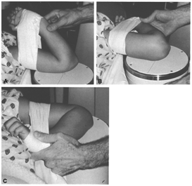
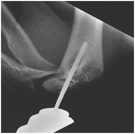
Surgery was performed by senior orthopedic surgeon who is well trained for this technique. General anesthesia was used for all patients with the injured upper limb at the side of the table. The injured elbow was placed on the plate of image intensifier which was adequate for the surgery due to the small size of the elbow. Closed reduction was done and confirmed by image intensifier. If acceptable, assistant would clean and drape the limb along with image intensifier and surgeon goes for scrub. Fracture would be reduced again and fixed under image intensifier according to the selected configuration (Figures 1 & 2).

Figure 1 Position of the patient on the operating table.17

Figure 2 Placement of the lateral pin17
For the lateral fixation technique two pins were inserted from lateral aspect of elbow across the lateral cortex to engage the medial cortex keeping the elbow in hyper flexion. For the pin construct to be acceptable and biomechanically stable one pin had to be placed in lateral column and another in central column. Pins were placed in parallel configuration with the adequate separation at fracture site.
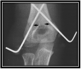
For the medial and lateral fixation technique, first the lateral pin was inserted from lateral cortex across the lateral cortex to engage the medial cortex keeping the elbow in hyper flexion. Then the elbow was extended to less than 90 degree and about 2cms, of medial incision was made over the medial epicondyle (not in all cases; in some cases we identified medial epicodyle manually with elbow flexed at 70-90 degree and the wire was putted manually then use the drill to introduce the wire carefully). Blunt dissection was done to locate the medial epicondyle and ulnar nerve rolled back with opposite thumb and the medial pin was inserted from the medial cortex to engage the lateral cortex with the elbow in less than 90 degree of flexion. The pin configuration was considered to be acceptable if one pin was placed in lateral column and another pin in medial column. Pins were cutted short bove the skin with good sterilized dressing to avoid the pin site local infection. Elbow was immobilized with posterior slab with elbow in 70 to 90 degree of flexion depending upon the swelling and neurovascular status. All patients were given single dose of broad spectrum antibiotics followed by oral antibiotics for five to seven days. Neurovascular examination was performed preoperatively and immediate post operatively and at one week follows up. All the patients were evaluated clinically and radiografically at one week, three weeks, four weeks, two months, three months and four months. In both groups the K wires were removed in three to four weeks and active assisted mobilization started Figure 3.

Figure 3 Supracondylar fracture humerus fixed with two crossed pins.17
A long arm posterior plaster splint is worn for 3 weeks. Ulnar, radial, and median nerve function should be checked after anesthesia. The pins are removed at 3 weeks, and another posterior splint is applied. At 4 weeks, intermittent active range of motion exercises are started at home; they should be taught by physical therapist to the child and the parent, explaining that the child is to carry out his own active range of motion program. Passive motion or forceful manipulative motion must be avoided in children because they will decrease the range of motion and frighten the child. Clinical evaluation was done by senior orthopaedic surgeon who includes passive range of motion, measurement of carrying angle, neurovascular status, superficial and deep infection and necessity to re-operate. Clinical evaluation was graded according to carrying angle and elbow range of motion using the criteria of Flynn et al.12 Radiographic evaluation was performed by anteroposterior and lateral radiographs of the elbow. Satisfactory fixation was confirmed intra operatively under image intensifier and radiograph taken. Follow up radiographs were taken at one week, three weeks, four weeks, two months, three months and four months. Baumann angle and Humerocapitellar angle were calculated on the immediate radiographs and after three months for any loss of Baumann angle and Humerocapitellar angle. At the three months and four months follow up child were evaluated for full function, minor limitation of function and major loss of function. Iatrogenic ulnar nerve injury was evaluated immediate postoperatively who had normal ulnar nerve function on the preoperative examination. Any patient with immediate post operative ulnar nerve deficit was putted under intensive follow up.
Methods of Follow Up
*Clinically; neurological and vascular assessment, range of motion, deformity and stiffness.
*Radiological; follow up x-rays using antero-posterior and lateral views.
Both group A and group B were comparable in terms of pre-fracture characteristics, fracture patterns, post reduction radiographic measurements showing satisfactory randomization. During this study period 44 children were treated for completely displaced type III supracondylar fracture in humerus. 4 children were excluded from the study due to loss for follow up.
The group-A comprised twenty patients. The mean age was 5.4 years. Among which 13 patients (65%) were boys and 7 patients (35%) were girls. In 10 patients injury occurred due to fall from height, 7patients were injured while playing whereas 2 due to road traffic accident and one due to some other cause. 7 patients (35%) had right elbow and 13 (65%) had left elbow fracture. One patient had pulseless viable hand, 2 patients had weak pulse 3 pts, had median nerve palsy and no one had radial nerve palsy. In majority of patients (95%) primary splintage was done in posterior splint and elevation given. Displacement was posteromedial in 14 patients, 4 had posterolateral and two had direct posterior displacement. No iatrogenic ulnar nerve injury was found in this group. The mean Baumann angle loss, Capitohumeral angle loss and carrying angle loss was 5.10, 5.80 and 3.300 respectively.3 patients had mild loss of reduction(rotational instability) in follow up x-rays.the mean loss of elbow flexion and extension were 8.40, 7.90 respectively, Total range of motion was 1350. Flynn grade showed excellent result in 15 patients, good in 3 and fair in 2 patients. 2 patients had superficial pin tract infection. No re-operation was needed in this group. The mean hospital- treatment duration was 6.5 hours. Finally 18 patients had full return to function and only 2 had minor limitation.
The group-B comprised 20 patients. The mean age was 6.2 years. Among which 15 patients (75%) were boys and 5 patients (25%) were girls. In 8 patients injury occurred due to fall from height, 10 patients were injured while playing whereas 2 due to road traffic accident. 4 patients (20%) had right elbow and 16 (80%) had left elbow fracture. no patient had pulseless viable hand, 3 patients had weak pulse, one had median nerve palsy and no one had radial nerve palsy .In majority of patients (90%) primary splintage was done in poserior splint and elevation given. Displacement was posteromedial in 13 patients, 6 had posterolateral and one had direct posterior displacement. One child had postoperative iatrogenic ulnar nerve injury in this group but completely improved after four months postoperative follow up. The mean Baumann angle loss, Capitohumeral angle loss and carrying angle loss was 4.80, 6.000 and 3.170 degree respectively. The mean loss of elbow flexion and extension were 7.6, 8.2 degree respectively. Total range of motion was 1400. Flynn grade showed excellent result in 16 patients, good in 3 and fair in 1 patient. There were 3 patient had superficial pin tract infection. No patient needed immediate re-exploration. The mean hospital- treatment duration was 6.9 hours. Finally 19 patients had full return to function and only one had minor limitation. Both groups were compared in terms and pameters given in the Table 2 and Table 3 there were no significant differences (p> 0.05) between groups with regard to any of these variables except one case had iatrogenic ulnar nerve palsy which not needed re-operation and this case completely improved after four months follow up (Figure 4).

Figure 4 Show mild loss of reduction (rotational instability) in using two parallel lat. Wires (s. univ. hospital).
|
Data of the patients |
Group – A |
Group – B |
P-value |
|
No of patients |
20 |
20 |
|
|
Age* (yrs) |
5.4 |
6.2 |
0.451 |
|
Sex @ |
13 |
15 |
0.234 |
|
Male |
|||
|
Female |
7 |
5 |
|
|
Mode of trauma@ Fall from height While Playing |
10 |
8 |
0.312 |
|
Road Traffic accident |
7 |
10 |
|
|
Other |
2 |
2 |
|
|
1 |
0 |
||
|
Affected side @ |
7 |
4 |
0.642 |
|
Right |
|||
|
Left |
13 |
16 |
|
|
Neurovascular Status @ |
12 |
0 |
0.211 |
|
Pulseless viable hand |
3 |
||
|
Weak pulse |
|||
|
Median nerve injury |
3 |
1 |
|
|
Radial nerve injury |
0 |
0 |
|
|
Primary spintage @ Yes |
19 |
18 |
0.521 |
|
No |
1 |
2 |
|
|
Displacement @ |
14 |
13 |
0.318 |
|
Posteromedial |
|||
|
Posterolateral |
4 |
6 |
|
|
Posterior |
2 |
1 |
|
|
Injury-Hospital Duration hr.* |
15±10 |
20±8 |
0.303 |
Table 2 Patients data.
*The values are given as the mean and standard deviation. @The values are given as the number of patients.
|
Group-A |
Group-B |
P-value |
|
|
Loss of reduction |
|||
|
Major |
0 |
0 |
|
|
Mild |
3 |
0 |
0.072 |
|
None |
17 |
20 |
0.411 |
|
Iatrogenic Ulnar nerve injury @ |
0 |
1 |
0.632 |
|
Bauman angle loss*(deg) |
5.10±5.0 |
4.8±5.2 |
0.478 |
|
Humerocapitellar angle loss*(deg) |
5.8±5.2 |
6.0±5.1 |
0.267 |
|
Carrying angle loss*(deg) |
3.30±4.25 |
3.17±4.15 |
0.698 |
|
Elbow flexion loss*(deg) |
8.4 |
7.6 |
|
|
Elbow extention loss*(deg) |
7.9 |
8.2 |
|
|
Range of motion*(deg) |
|||
|
Extension |
-2 |
0 |
|
|
Flexion |
137 |
140 |
0.217 |
|
Total motion |
135 |
140 |
|
|
Flynn grade@ |
|||
|
Excellent |
15 |
16 |
0.321 |
|
Good |
3 |
3 |
0.521 |
|
Fair |
2 |
1 |
0.421 |
|
Poor |
0 |
0 |
|
|
Superficial Infection@ |
2 |
3 |
0.459 |
|
Re-operation@ |
0 |
0 |
|
|
Hospital-Treatment Duration hrs.* |
6.5±2.7 |
6.9±2.6 |
0.218 |
|
Return to function@ |
|||
|
Full |
18 |
19 |
0.421 |
|
Minor limitation |
2 |
1 |
0.321 |
|
Major limitation |
0 |
0 |
Table 3 Patients results.
*The values are given as the mean and standard deviation. @The values are given as the number of patients.
Supracondylar fracture of the humerus in children is one of the most common fractures seen in orthopaedic outpatient department all over the world accounting for 60% of all elbow fracture in children in the first decade of life.1 Traditionally this type of fracture is associated with high rate of malunion, nerve injuy, and vascular complications.2 Supracondylar fracture of the humerus is a condition that needs a most important skill that the orthopaedic surgeon must develop. Namely, the ability to choose from a number of treatment modalities the best treatment for a given condition in a given patient. To suggest that all supracondylar fractures are best treated by one treatment method ignores the variability of this fracture. The treatment of supracondylar fractures aims to restore anatomical or near anatomical reduction, early restoring elbow function with good ROM, avoid complications like neurovascular, deformity, elbow stiffness………etc. Decrease physical and psychological impairment of the fracture on the children and their parents.
This prospective study aims to assess and compare between using two parallel lateral wires and two crossing medial & lateral wires as regard stability, safety, complication, postoperative ROM and incidence of iatrogenic ulnar nerve injury. This study includes 44 children. 4 were lost for follow up. They were divided into two groups. Group-A two parallel lateral wires. Group-B two crossing medial and lateral wires. 20 children in each group. In group-A the mean age was 5.4 yrs. Among which 13 patients (65%) were boys and 7 patients (35%) were girls. In group-B the mean age was 6.2 years. Among which 15 patients (75%) were boys and 5 patients (25%) were girls. As regard Flynn criteria for grading the results were excellent in 15 children in group-A and in 16 children in group-B. Good in 3 children in both groups. 3 children in group-A had postoperative mild loss of reduction (rotational instability) in follow up x-rays but no one in group-B and this mean that medial and lateral wires give more stability. We had only one case of iatrogenic ulnar nerve injury in group-B that shows complete recovery in the follow up and no cases need re-exploration. It mostly due to neuropraxia or axonotemesis due irritation or compression of the ulnar nerve by the medial wire. No cases of ulnar nerve injury occurred in group-A. There were loss of carrying angle more than 10º in two cases of group-A and one case of group-B. There were two cases of superficial pin tract infection in group-A and three cases in group-B and all improved on first generation cephalosporin antibiotic. The average total ROM of the elbow was 135º in group-A and 140º in group-B. There was mild limitation of function in two cases of group-A and one case of group-B. 18 children had return to full function of the elbow in group-A and 19 in group-B. We compared our results to previous studies as Foead et al. & others.11,13-16 regard results, complications and follow up duration. The results were compared to the results of these studies with less complication rates. Less incidence of iatrogenic ulnar nerve injury in crossing wires with spontaneous recovery without exploration. No iatrogenic ulnar or radial nerve injury in two parallel lateral wires and with more stability in cases of two crossing medial and lateral wires. Lower incidence of pin tract infection, no cases of postoperative vascular injury and the duration of the study shorter than the others.
Foead et al.13 reported a prospective study of 66 children. 11 were lost for follow up. With mean period of follow up 8.9 months, the average loss of carrying angle was 3.57 and 3.7 degree in medial & lateral wires and 2 lateral wires respectively. The average loss of elbow extension and flexion was 7.14 and 8.68 respectively in medial & lateral wires and 7.11 and 11.26 respectively in 2 lateral wires. The average loss of baumann’s angle was 5.96 and 5.3 in medial & lateral wires and 2 lateral wires respectively and there were 5 cases of iatrogenic ulnar nerve injury in medial and lateral wires and 2 cases of iatrogenic ulnar nerve injury and one radial nerve injury in 2 laterl wires cases.
Vaidya et al.11 reported a prospective study of 66 children. 6 were lost for follow up. With mean period of follow up 6 months. No major loss of reduction was observed in both the groups where as there was no significant difference between mild loss of reduction. Change in Baumann angle, change in Humerocapitellar angle, flynn grade, elbow extension and flexion, carrying angle, total range of motion. But there was three ulnar nerve injury in group B. three cases of ulnar nerve need immediate post operative re-exploration among which two had tenting of ulnar nerve over the pin and in one case pin was causing constriction of cubital tunnel since no direct compression over the nerve was found.
Chakraborty et al.15 reported a retrospective study of 92 children. 56 were fixed by medial and lateral crossing wires and 36 were fixed by 2 lateral wires, there 4 cases of iatrogenic ulnar nerve injury in crossing wires and 4 cases of radial nerve injury in 2 lateral wires. There were 4 cases of cubitus varus in crossing wires and 10 cases of cubitus varus in 2 lateral wires. 4 cases of ulnar nerve injury were explored.
Anwar et al.14 reported a prospective study of 50 children, 25 were fixed by medial and lateral crossing wires and 25 were fixed by 2 lateral wires, as regard carrying angle loss according to Flynn,s criteria the results were excellent in 72% and good in 28% in both methods, the mean loss of elbow flexion and extension were 8.36 and 7.26 respectively. There was one case of iatrogenic ulnar nerve injury in crossing wires.
Maity et al.16 reported a prospective study which was long term study between October 2007 and October 2010 of 160 children, 80 in each group. The follow up duration was 3 months. 30 of 160 children did not complete the follow up visits. Abhijan.16 reported that there was no significant difference between the two methods as regard results and complication.
This study is a prospective study not a retrospective study like that of Chakraborty et al.15 short term not like that of Maity et al.16 with excellent results rate according to Flynn's criteria higher than that of Anwar et al.14 with incidence of iatrogenic ulnar nerve injury in crossing wires less than that of Chakraborty et al. and others.11,13,15 were no cases of ulnar nerve injury or radial nerve injury as that of Chakraborty et al. & Foead et al.13,15 in cases 2 lateral wires. We had low incidence than that of Chakraborty et al.15
In this study the number of cases is lower than the other studies. This study included cases of two parallel lateral wires and not the divergent lateral wires which may take place in other study in our department. The duration of this study was shorter than other studies.18-22
From this prospective study we concluded that there was no significant difference between medial and lateral crossing wires and 2 lateral wires as regard postoperative clinical results , complications and incidence of iatrogenic ulnar nerve injury, but medial and lateral crossing wires give more biomechanical stability than 2 parallal lateral wires in fixation of supracondylar humeral fracture (type lll) in children.
Prof, Daniel Altaha. Critical review.
None.

©2016 Khairy. This is an open access article distributed under the terms of the, which permits unrestricted use, distribution, and build upon your work non-commercially.