MOJ
eISSN: 2374-6939


Review Article Volume 14 Issue 5
1Department of Musculoskeletal Physiotherapy and Kinesiology, TPCT’s Terna Physiotherapy College, Navi Mumbai
1Department of Musculoskeletal Physiotherapy and Kinesiology, TPCT’s Terna Physiotherapy College, Navi Mumbai
Correspondence: Dr. Medha Deo, Department of Musculoskeletal Physiotherapy and Kinesiology, TPCT’s Terna Physiotherapy College, Navi Mumbai
Received: August 08, 2022 | Published: September 23, 2022
Citation: Deo M, Jain S. Effect of toe - out gait modification on pain and quality of life in patients with knee osteoarthritis: a randomised controlled pilot trial. MOJ Orthop Rheumatol. 2022;14(5):139-145. DOI: 10.15406/mojor.2022.14.00595
Osteoarthritis (OA), a degenerative joint disease (DJD), is one of the most common musculoskeletal impairment and one of the leading causes of physical disability in adults. The disease most commonly affects the knee joint. OA is extremely common after 40 year of age, although it may not always be symptomatic when present. Worldwide OA is estimated to be the fourth leading cause of physical disability.1 OA is the most frequent joint disease with a prevalence of 22 - 39% in India.2,3 Prevalence of OA is increasing due to population ageing and increase in the related risk factors such as obesity. The medial compartment is affected 10 times more often than the lateral compartment,4 which is likely due to greater medial compartment loading during gait.5 A study found that the medial tibiofemoral compartment was most likely to be involved in knees with OA (72%). The external knee adduction moment (KAM) has been associated with increased loading in the medial tibiofemoral compartment.6,7
In OA Knee, there is rubbing and gradual loss of articular cartilage, osteophyte formation, narrowing of the joint spaces causing changes in the biomechanical alignment, leading to unequal and abnormal force distribution at the knee joint surfaces. Common Activities of Daily Living (ADL) affected due to OA - pain during movement, weight - bearing, and gait that may interfere with ADLs, difficulty controlling weight bearing activities like walking, stairs ascent and descent, squatting, sitting down/ rising from a chair, reduced participation in ADLs.8–10
Walking, a dynamic functional activity is commonly affected in patients with knee OA. Normally during walking, the GRF (Ground Reaction Force) passes medial to the knee joint, creating a varus torque at the knee with each step. In patients with knee OA, there is a tendency of increased knee adduction moment while walking, coupled with increase in GRF-Moment Arm(MA) resulting from various impairments, such a pain, muscle imbalances, thus creating more compressive load on the medial compartment of the knee joint. This alteration leads to more and more varus angulation that progresses the medial compartment knee OA. Abnormal mechanical loading is the main culprit in the development of OA, and therefore, no drug can feasibly encourage healing until the underlying biomechanical malalignment issues are addressed.11
As the severity of pain and disease increases, total joint replacement (TKR) is generally preferred so as to improve the functional status and Quality of Life (QoL) of the patient. Physical therapy plays a vital role, in the form of conservative treatment of OA Knee, in improving the patient’s physical, functional abilities thereby improving the QOL.
Conventional treatments for knee OA traditionally has been impairment-based, aiming to reduce pain, restore mobility and improve muscle strength and it generally involves a combination of exercises, electrotherapeutic modalities and lifestyle modification. They may be effective in treating the impairments but may not provide with much improvement in the routine ADLs and functional mobility tasks as this requires combined effects of muscle strength, pain-free movement, and balance along with proper biomechanics and force distribution at the joint. Hence, the exercise programs should include strategies which addressed these problems. Some strategies to reduce the medial knee joint loading are - reduced walking velocity, trunkal sway, use of assistive devices and knee braces, footwear modifications, and gait modification (one of which is out-toeing while walking).10
Gait modification is a relatively new approach which represents a simple and cost-effective treatment strategy that may be employed to reduce medial knee load.11 Improving the walking ability of the patient is clinically important as it is related to the maintenance of functional independence. Toe-out is postulated to reduce the knee adduction moment and thereby reduce the abnormal loading of the knee joint.12 Hence, out-toeing while walking could be an effective method to reduce overall forces acting at the knee joint and redistribute the forces evenly over the articular surfaces by correcting the pathomechanics, thereby preventing the progression of Knee OA.
Hence, combining the effects of conventional treatment with that of gait modification may be helpful in reducing the dynamic medial knee joint load, thus enhancing the functional outcomes and improving the patient’s functional status. This could also be beneficial in slowing, preventing or delaying the progression of Knee OA. This study was thereby undertaken to find out the effectiveness of toe-out gait modification in addition to the conventional treatment of Knee OA.
Effects of 10-week toe-out gait modification intervention in people with medial knee osteoarthritis: a pilot, feasibility study.
Summary: This study was conducted to find out the feasibility of toe-out gait modification program in medial tibio - femoral OA and to assess the changes in the clinical and biomechanical outcomes. In this study, 15 participants underwent 10 - weeks of toe - out gait modification wherein a real-time biofeedback was used to measure the toe - out angle during walking - via the motion analysis system. Results showed improvement in the outcomes Numerical Rating Scale (NRS), Western Ontario and McMaster Universities Osteoarthritis Index (WOMAC) and Knee Adduction Moment (KAM). But the implementation of this program in clinical setup is not feasible as it requires sophisticated laboratory setup. Though, being a new approach in the treatment of OA knee, there is a need for conducting relatively larger scale studies, which could be delivered using less resource - intensive machines.
- KOOS - A systemic review and meta - analysis of measurement properties.
Summary: This study was performed to evaluate the measurement properties of Knee Injury and Osteoarthritis Outcome Score (KOOS). Results showed that KOOS has adequate internal consistency, responsiveness for age-and condition - relevant subscales, test - retest reliability and construct validity in adults with knee injuries and / or osteoarthritis.
The reliability and minimal detectable change of timed up and go test in individuals with grade 1 - 3 knee OA.
Summary: The purpose of this study was to estimate the reliability and Minimal Detectable Change (MDC) of Timed Up and Go (TUG) test in individuals with doubtful to moderate knee OA. Two clinical physiotherapists, experienced and trained in TUG test administration (for the purpose of standardisation) performed inter - rater reliability testing at different times of the same day in an alternating order. For intra - rater reliability testing, the same physiotherapists performed TUG test on 2 consecutive visits with 2 - day interval. Results showed excellent intra- and inter - rater reliability (ICC 0.97, 0.96 respectively). Also, in addition, the OARSI has recommended the use of TUG test as a performance-based test of physical function in individuals with hip or knee OA.
Conservative management of symptomatic knee OA: a flawed strategy?
Summary: The purpose of this paper was to summarise the clinical evidence on conservative management for knee OA and to identify the attributes of the ideal treatment regimen in this patient population. This review comprised of the current standard of care recommendations for conservative management of medial compartment knee OA.
Conservative biomechanical strategies for knee OA
Summary: This study reviewed strategies that directly affect the gait. Knee OA is closely associated with the development of a high external knee adduction moment, which reflects compression of the medial compartment of the knee. The nature of biomechanical loading at the knee joint can be altered by a number of conservative intervention strategies, which are potentially capable of slowing the progression of the disease. Using lateral wedge insoles or thin-soled, flexible shoes and strategies that directly modify gait characteristics, like a toe-out gait, lateral trunk lean and the use of a walking stick can reduce the knee adduction moment and thus contribute to retarding the progression of OA. Intervention strategies that act either directly or indirectly upon the knee joint, such as valgus knee braces and muscle strengthening, can effectively decrease the knee adduction moment.
To assess the effectiveness of 4 week toe- out gait modification in subacute and chronic bilateral knee OA.
To assess the effectiveness of conventional treatment along with toe-out gait modification in subacute and chronic bilateral Knee OA on pain as measured by NRS. To assess the effectiveness of conventional treatment along with toe-out gait modification in subacute and chronic bilateral Knee OA on functional disability as measured by KOOS and TUG Test.
Hypothesis
Null Hypothesis: There will be no additional effects seen in the reduction of pain and improvement of physical function in patients with sub - acute and chronic bilateral knee OA, with by toe-out gait modification.
Experimental Hypothesis: There will be reduction of pain and improvement of the physical functions in patients with sub - acute and chronic, bilateral knee OA by toe-out gait modification, when given along with the conventional treatment.
Study design: Experimental, Intervention-based Randomised Controlled Trial.
Population setting.
Study setup: Tertiary health care centres
Sample size: 30 subjects with primary bilateral knee OA.
Materials required: Baseline protractor (Figure 1), Interferential Therapy (IFT) device
Inclusion criteria
Exclusion criteria
Outcome measures
Study procedure and intervention
A total of 30 Subjects, both males and females, of age group 45 - 70 years with the clinical diagnosis and radiographic evidence of sub - acute and chronic knee OA were selected in the study. The individuals with OA knee were screened for exclusion criteria. Individuals who met the inclusion criteria were included in the study. A prior consent was taken and they were explained about the procedure, purpose and duration of the study in their understandable language. The required assessment was done and documented. The subjects were then randomly divided into 2 groups. Group A was the experimental group and Group B was the control group. Both groups were assessed on the basis of the outcome measures of the study and documentation was done. The outcome measures were assessed pre intervention i.e. the first day and post intervention i.e. after 4 weeks of the study for both the groups. The pain was measured prior to intervention using NRS and functional ability was assessed using TUG test and KOOS.
Treatment protocols: 1 week of supervised physiotherapy session was given to both the groups. After which, patients of both groups were given a treatment session of 1 day /week for 3 consecutive weeks. Home exercise program was given to the patients of both the groups.
Experimental Group (A): Toe - out gait modification along with Conventional treatment.
A baseline protractor was used to teach the patient the range of toe-out angle (15 - 20 degrees), which was to be maintained while walking. The patient was made to learn the toe-out angle range every day during the supervised treatment session, in order to promote learning. 2 sessions of increased toe - out walking on a flat surface at a comfortable self-selected pace (similar to their over ground walking speed) for 20 minutes/session for 4 weeks, along with conventional treatment was given. The out - toeing was performed on one limb at a time while the gait training sessions of 20 minutes. (10 minutes for each limb). Patients were made to practice and were instructed to maintain the increased toe-out angle while walking in their daily livings.
Control Group (B): Conventional treatment and walking in the patient’s own gait pattern with no modification or alterations, for 10 minutes/session.
Conventional treatment was given to both the groups. This included - IFT along with conventional treatment inclusive of Static and Dynamic Quadriceps, Vastus Medialis Oblique (VMO) muscle strengthening, Hamstrings and Tendo-Achilles (TA) stretching, Side - lying hip abductors 4 strengthening, Prone - Hip extensors strengthening (hip extension with flexed knee), Forward lunges, Mini - squats were given.
Home exercises for group A included the conventional exercises and walking with out-toeing (range 15 to 20 degrees). These exercises were prescribed on a daily basis. Home exercises for group B included conventional exercises along with walking in the patient’s own gait pattern with no modification or alterations or feedback, for 10 minutes/session. Follow - up testing was done after the successful completion of the intervention. 4 weeks duration was chosen as an appropriate intervention length to provide sufficient time to establish motor learning of the new gait pattern.
For intra - group analysis of parametric tests -Paired t test
For intra - group analysis of non-parametric tests - Wilcoxon test
For inter - group analysis of parametric tests - Unpaired t test
For inter - group analysis of non- parametric tests - Mann - Whitney U test
MS Excel 2007 and Graphpad Prism 8.0 (GPS-0876226-TDOA-1FA44) were used to analyse the data
Graph 1-Graph 10
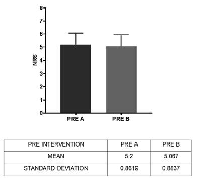
Graph 1 PRE NRS of Group A and Group B.
Inference Test of Normality was performed. P value = 0.7471 (not significant).

Graph 2 POST NRS of Group A and Group B.
Inference- Pain had reduced significantly in the experimental group when compared with the control group following treatment. P value= 0.0197.
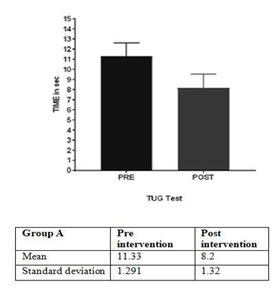
Graph 3 TUG test of Group A.
Inference - Time required to complete the TUG Test reduced significantly in the experimental group following intervention. This implies that the speed of walking improved significantly in the experimental group following intervention. P value < 0.0001
.
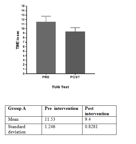
Graph 4 TUG test of Group B.
Inference – Time required to complete the TUG Test reduced significantly in the control group following intervention. This implies that the speed of walking improved significantly in the control group following intervention. P value < 0.0001.
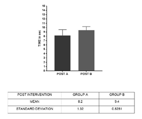
Graph 5 Comparison - Post Intervention TUG Test Of Group A and Group B.
Inference - Time required to complete the TUG Test was significantly reduced in the experimental group when compared with the control group following intervention. P value = 0.0059
.
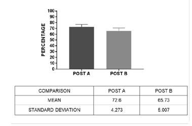
Graph 6 Comparison of pain (Subscale of KOOS).
Inference - ADLs improved significantly in the experimental group when compared with the control group, following intervention. P value = 0.0002.

Graph 7 Comparison of symptoms (Subscale of KOOS).
Inference - Symptoms had improved significantly in the experimental group when compared with the control group, following intervention. P value <0.0001
.

Graph 8 Comparison of oral (Subscale of KOOS).
Inference – QoL improved significantly in the experimental group when compared with the control group, following intervention. P value = 0.0327
The findings of this study could be due to the following
Reduction in pain: The probable reasons for this pain reduction could be as follows:
Based on Pain subscale of KOOS and NRS: The study showed that there was reduction of pain in both the groups. Significant improvement was seen in the experimental group (group A) when compared with the control group (group B).
Findings could be attributed to the following -
Based on symptom and ADL subscale of koos and tug test - Significant improvement was seen in the experimental group (group A) when compared with the control group (group B). Improvement in the symptoms and ADLs in the patients of both the groups could be due to
Function, sports and recreational activities of the koos subscale
The intra- group analysis of the scores of the function, sports and recreational activities of the KOOS subscale for both the groups significantly improved (p value < 0.0001), but the inter group analysis did not show a significant result (p value = 0.0565). This could be attributed to the fact that in our country, we have a culture which is different than the western countries, here the individuals are not as much indulged in sports and recreational activities as theirs. This could be a probable reason why the inter - group analysis did not show significant results.
QOL subscale of koos
Intra-group analysis of both the groups showed significant improvement in the QoL (p value < 0.0001). Inter - group analysis also showed significant difference between scores of both the groups (p value <0.0001). The experimental group showed a significant improvement in the QoL, when compared with the control group. Improvement in QoL of both the groups could be due to the following:
Also, based on the analysis of demographic data, according to BMI of all the subjects, it was observed that OA knee is more common in over weight and obese individuals than in individuals with normal BMI. Studies in the past have shown a linear correlation between BMI and OA Knee.20
The gender predominance was also noted, females being more affected with OA knee than males. Studies in the past have also reported that women are more likely than men to suffer from osteoarthritis, and women experience more severe arthritis in the knee.21 The results were similar to the results of this present study. The prevalence of knee OA is higher in women than men, and the incidence increases around menopause.
After analyzing the results of this study it could be concluded that toe - out gait modification, when given in addition to the conventional treatment, would be more effective, than conventional treatment alone, in reduction of pain and improvement of functional abilities in patients with sub acute and chronic knee OA. It could be incorporated as a part of conventional treatment protocol.
The maintenance of toe-out angle (as a part of the home program) after the supervised sessions was difficult.
Long term follow-up of the subjects could be taken so as to assess the motor learning of the toe-out gait pattern. Use of visual feedback as a part of home program in various forms for example the use of baseline protractors or could be helpful.
The goal of rehabilitation is to decrease pain and improve physical function of OA Knee individuals. The exercise protocol of the experimental group had a beneficial effect on both pain and physical function. The result showed significant improvement in the outcome measures. Hence, the toe-out gait modification can be used in addition to the conventional treatment of OA Knee.
None.
The authors declare no conflict of interest.

©2022 Deo, et al. This is an open access article distributed under the terms of the, which permits unrestricted use, distribution, and build upon your work non-commercially.