MOJ
eISSN: 2374-6939


Research Article Volume 5 Issue 6
1Chief Consultant, Bari-Ilizarov Orthopaedic Centre, Visiting and Honored Prof., Russian Ilizarov Scientific Centre, Russia
2Bari-Ilizarov Orthopaedic Centre, Bangladesh
Correspondence: Mofakhkharul Bari, Chief Consultant, Bari-Ilizarov Orthopaedic Centre, Visiting and Honored Professor, Russian Ilizarov Scientific Centre, Kurgan, Tel +88 01819 211595
Received: July 13, 2016 | Published: October 4, 2016
Citation: Bari MM, Islam S, Shetu NH,Rahman M (2016) Distraction Osteogenesis Technique in the Treatment of Acquired and Congenital Shortening ofHand Bones Using Mini-Ilizarov Fixator. MOJ Orthop Rheumatol 5(6): 00202. DOI: 10.15406/mojor.2016.05.00202
Distraction osteogenesis is the most used method for bone lengthening and deformity correction of the hand bones. The lengthening technique for congenital and post traumatic hand bone pathology started at our Bari-Ilizarov Orthopaedic Centre in 1995.1 The application of mini-Iliarov fixator to short tubular bones is low traumatic and the performance of the operation is not so difficult. The set of mini-Ilizarov fixator parts and instrumentation for surgeries is neither complex nor costly. The lengthening of hand bone segments is realized by creating regeneration in the osteotomized bone or stump. Bone grafting is not necessary. The mini-Ilizarov fixator can be applied to several bones simultaneous for creating distraction osteogenesis into or more bones in order to reduce a number of interventions, shorten the treatment and rehabilitation time. Lengthening procedures the combined with traditional methods of phalangization, web-plasty and bone deformity correction. We are using this mini-Ilizarov fixator for the last 20 years for various hand pathologies.
Keywords: Mini-Ilizarov fixator, Acquired andcongenital shortening, Phalanges, Metacarpal bones, Distraction osteogenesis
Post traumatic stumps and congenital shorts segments in adults and children of 3 years and older.
Instrumentation and techniques
Lengthening techniques are perform the use of Ilizarov mini external fixator for the management of hand pathology supplied with additional parts such as plates, hinge, post, bushings and threaded rods.
Thin 1 mm or 1.5 mm Kirschners wires which as suitable for paediatric or adult hand bones have 3 sharpened end facets. The mini-Ilizarov fixator is composed of fixation units (Figure 1) for attaching the wires in to the slots (7) of slotted washers (3) and their fastening with a fixation nut (8) (Figure 2). (A) composed of slotted washers (3,7), space washer (4), bolt (5) with an axial hole and perpendicular threaded hole in its head, locking screw (6), fixation nut (8).
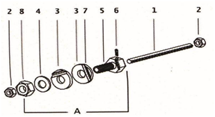
Figure 2 Threaded rod (1), nuts (2) and wire-fixation unit. (A) Composed of slotted washers (3,7), space washer (4), bolt (5) with an axial hole and perpendicular threaded hole in its head, locking screw (6), fixation nut (8).
Distraction or compression along a flat- side threaded rod (1) is produced by turning the nuts (2) on both sides of the bolt (5) that has a threaded hole in the head for a locking screw (6) that rests on a flat of the rod.
Placement of mini-Ilizarov fixator and osteotomy Step 1: Two or three proximal wires are drilled at 90° to each other from the dorsolateral bone surfaces and should pass both cortices but exit only to one or 2 mm from the cortex opposite to the direction of insertion. They are bent so that the support (wire fixation unit) is located 3 or 4 mm over the skin and are attached one by one in to the slots of slotted washers (Figure 1). Next the wires are fixed in the proximal fixation unit which must lie parallel to the bone axis. The flat-side rod for distraction is inserted in to the proximal wires fixation unit.
Step 2 Distal wires are drilled the same way as described above and are fixed in the distal wire support after bending. The locking screws of both units must be position at 90° to each other.
Step 3 The rod is turned so that the flat is perpendicular to the threaded hole of the bolt head in the distal support and the screw locks the distal assembly on the flat. Next, the locking screw of the proximal support strongly fixes the threaded rod with the bolt.
Step 4 Before osteotomy the locking screw of the distal part that connects with the flat must be released. Percutaneous osteotomy of the segment is performed through a 0.5 cm dorsolateral skin incision so that the soft tissues are minimally injured. The accuracy of its completion each checked by c-arm following an acute distraction to 3 or 4 mm. The bolt screw in the distal unit must be locked again once the contact between the fragments ends has been achieved by compression. The wound is closed aseptically.
Lengthening technique with mini-Ilizarov fixator depends upon the length of a post traumatic bone stump or a bone segment that is short due to congenital disorders. Lengthening of stump or a segment that is shorter than 30 mm requires bridging of the joint that is located proximally to the segment under lengthening while a segment which is longer than 30 mm does not require joint fixation.
Bone segment longer than 30 mm
Diagram 3 shows the placement of fixation units on phalangeal stumps or hand bone segments that are longer than 30 mm (Figure 3). The lengthening runs under favorable functional conditions for joints as physical exercises are possible after the operation.
Distraction started on the 5th postoperative day, continued for 35 days to lengthen 2.5 cm, d. smiling patient with mini-Ilizarov fixator, e. 2.5 cm lengthening of the thumb is visible (Figure 4).

Figure 3 Diagram of the lengthening technique for a phalanx or stump with mini-Ilizarov fixator that is longer than 30 mm.
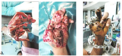
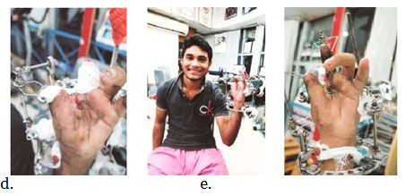
Figure 4 a. Severe machinery crush injury of the left hand. b. 1 month follow up after the treatment of Ilizarov fixator. c. Posttraumatic stump at the level of the left proximal thumb phalanx in a 17 years old boy was osteotomized, distraction started on the 5th postoperative day, continued for 35 days to lengthen 2.5 cm. d. Smiling patient with mini-Ilizarov fixator. e. 2.5 cm lengthening of the thumb is visible.
Bone segments of 30 mm or shorter
When the length of a bone segment or a stump is 30 mm or shorter the proximal fixation unit lies over the segment which is proximal relative to the segment under lengthening (Figure 5) and has two wires. One or two wires pass in the base of the segment under lengthening, and they are also fixed in the proximal support. Three or two wires are inserted in the distal part of the segment under lengthening and should be fastened into the distal fixation unit.
Lengthening of the middle phalanx in a congenital anomaly requires adjacent distal and proximal joints bridging (Figure 6). The technique foresees gradual stretching of the bridged joints by a total amount of 6 or 8 mm performed two weeks prior to removal of the fixator. The wires that are in the lengthened segment must be cut and taken off in order not to hinder to stretch the joint (Figure 7).
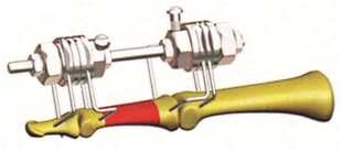
Figure 6 Variant of the mini-Ilizarov fixator mounting for lengthening the middle phalanx in a congenital anomaly.
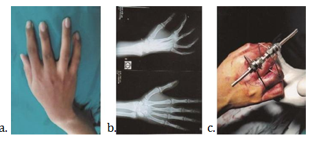
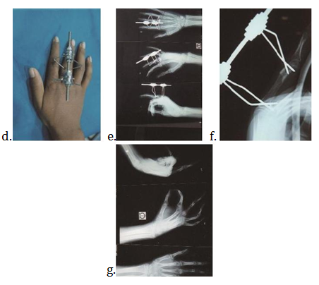
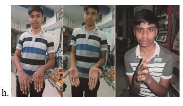
Figure 7 a. Posttraumatic shortening of proximal phalanx of right middle finger. b. Radiograph of right hand, shortening of proximal phalanx of right middle finger. c. After the surgery with mini-Ilizarov fixator in OR, d. External view of the mini-Ilizarov fixator. e. Radiographic view of osteotomized and lengthening part of proximal phalanx with ongoing callus formation. f. Radiographic view of lengthening proximal phalanx of right middle finger with mini-Ilizarov fixator. g. Final radiographic follows up after 3 months and h. Clinical appearance of the patient after 4 months.
Technique for lengthening of the 1st metacarpal bone and it’s phalangization
This lengthening technique is used for amputation stumps at the level of the first metacarpal and is aimed at improving the gripping function in the 1st web space. Two wire fixation units are placed on the second metacarpal bone and the medial transport along the transverse threaded rod is performed in order to grow skin for subsequent web-plasty (Figure 8). Double osteotomy of the metacarpal bone is possible if there is enough soft tissue on the stump end is possible if there is enough soft tissue of the stump end (Figure 9). The technique completion requires metacarpal joint stretching by 4 or 5 mm during distraction and fixation periods.
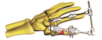
Figure 8 Diagram of the 1st metacarpal bone lengthening by mini-Ilizarov fixator and skin stock growing in the 1st web space.
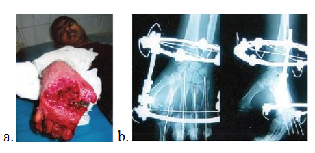
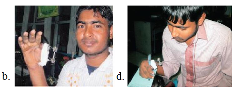
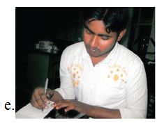
Figure 9 a. 18 years old boy- Machinery injury of right hand, with auto amputed thumb, hanging metacarpals and fractures of 2nd, 3rd, 4th, 5th metacarpals, b. Radiograph of right hand with Ilizarov in situ. Fixation of 2nd, 3rd, 4th, 5th metacarpals with k/wires, c. External view of mini Ilizarov, d. Patient can hold the pen with the help of mini Ilizarov fixator and e. Now the 18 years old patient can write with his hand.
Distraction is started within 5 to 6 days after the operation with an increment of 0.25 mm made three times a day to reach 0.75 mm of daily lengthening. To produce the distraction increment, the locking screw of the distal fixation unit is released so that there is some contact between the screw end and the flat of the threaded rod for moving the distal unit that should not rotate. The screw must be locked upon completion of the nut turning.
Callotasis is checked with regular x-ray taken every 10 days. Once an uneven regeneration density is seen in the radiographs or distraction callus is shaped like an hour-glass, the distraction rate must be decreased or even stopped. The amount lengthened at a single osteotomy level is within 1 or 2 cm on average.
Fixation of the lengthening achieved lasts from 4 to 8 weeks and depends on the quality of regeneration and lengthening amount. The mini-Ilizarov fixator can be removed once the density of the regenerated bone approximates the density of the bone areas adjacent to regeneration and when the cortex on the radiographs is uninterrupted. Clinical test for mechanical stiffness of the regenerated bone is mandatory.
Dressings must be changed on the first postoperative day and then every 7 to 10 days, or when it is necessary.
The rehabilitative procedures should be undertaken during the course of treatment and it includes exercise therapy, massage of the hand joints and wrist if possible. Physical therapy procedures improve blood circulation and joint motion.
Complications are a fact of life that every orthopaedic surgeon should face. The most frequent one is cutting of the skin with a wire at its insertion point during distraction. So, it is highly recommend to fold the skin in the area between the proximal and distal wires during their drilling. The skin unfolds during distraction. Antiseptic dressing is obligatory while changing dressings.
Wire tract infection is possible when the wire persists to cut the skin. A wire must be taken out if it fails to stop infection. An excessive skin tension happens due to distraction forces. Distraction efforts should be decreased or even stopped if the skin turns pale. Once the blood flow restores, distraction is continued. The scars caused by the incision for the osteotomy or wire entrance points turn almost invisible over time.
Distraction osteogenesis is an authentic and reliable method for lengthening of short tubular bones using mini-Ilizarov fixation both in cases of amputation stumps or congenital malformation.1-6 Lengthening procedures and web plasty improve the esthetical appearance and functioning of the hand.2,7-9 Callotasis of the proximal phalanx of the thumb or the first metacarpal bone in posttraumatic stumps is an effective reconstruction method to compensate for the loss of functions.9
Summary of the technical solutions for the last 20 years of long practical experience has been presented here.
Minor complications during the course of treatment neither affected final outcomes nor prolonged treatment time. Wires cut the skin during distraction in 10% of patients. It resulted in wire tract infection that happened in 7% of our cases. 5% of the cases treated had premature dismounting of the mini-Ilizarov fixator due to infection developed and plaster casts were applied. There was no functionally limiting loss of joint motion in any patient. Joint subluxation or dislocation, regenerate fracture, non-union, arthritis or nerve injury was not observed.
Our series of the cases (95) with an acquired and congenital pathology in hand bones that required lengthening. Posttraumatic cases wire 35, aged from 5 to 50 years in who 25 hand segment were lengthen (10 metacarpals, 5 proximal phalanx and 10 middle phalanx). Congenital hand bone shortening was managed in 60 individual aged from 4 to 40 years. A total of 32 segments were lengthening (10 metacarpal bones, 10 proximal phalanx and 12 middle phalanx). Web-plasty was a rational procedure in 12 cases.10 We are using mini-Ilizarov construct that incorporates biocompatible thin wires instated of half- pins that results in a lesser invasiveness during the intervention and distraction. The thin wires are drilled 90° to each other and relative to the longitudinal bone axis and retain the bone from both sides thus excluding deformities. Distraction rate has great importance and must not be accelerated to allow the regeneration process run in a steady pace suitable for a short tubular bone to exclude non-union but could decreased in the circumstances mentioned earlier. The principle characteristics of mini-Ilizarov fixator are its versatility which provides assemblies for lengthening of both hand phalanges and metacarpal bones. One of the best advantages of the techniques are the possibility to apply a mini-Ilizarov fixators to several bones simultaneously and its ability to be adjusted to grow the skin bulk in the interphalangeal space both in congenital and acquired anomalies. Dermatoplasty for web space skin division can be done easily.2 Mini-Ilizarov fixator is the best choice for lengthening the hand bones which provides good solutions on the basis of specific medical condition and patient desire.
None.
None.

©2016 Bari, et al. This is an open access article distributed under the terms of the, which permits unrestricted use, distribution, and build upon your work non-commercially.