MOJ
eISSN: 2374-6939


Research Article Volume 6 Issue 2
1Professor PhD, Chief Consultant, Bari-Ilizarov Orthopaedic Centre, Visiting and Honored Professor, Russian Ilizarov Scientific Centre, Kurgan, Russia
2Bari-Ilizarov Orthopedic Centre, Bangladesh
3Jessore Medical College Hospital, Bangladesh
Correspondence: Bari-Ilizarov Orthopaedic Centre, 72, Satmasjid Road, Nizams Shankar Plaza, Dhanmondi, Dhaka, Bangladesh, Tel 8800000000000
Received: October 18, 2016 | Published: October 18, 2016
Citation: Bari MM, Islam S, Shetu NH, Rouf AHMA, Rahman M (2016) Distraction Osteogenesis by Ilizarov Technique for Infected Gap Non-Union of the Femur. MOJ Orthop Rheumatol 6(2): 00212. DOI: 10.15406/mojor.2016.06.00212
Introduction: Purpose- To review records of 36 patients who underwent distraction osteogenesis or bone transport by Ilizarov fixator for infected gap non-union of femoral shaft.
Materials and Methods: 30 men and 6 women aged 20 to 57 years underwent adequate debridement and resection of the nonviable bone, followed by bone transport using Ilizarov fixator for infected gap non-union of the femoral shaft. All patients had a bone defect of >4 cm. The lengthening index, x-ray consolidation index, functional status, bone healing and complications encountered during the treatment were assessed.
Results: The patients had undergone a mean of 3 (range 1-6) surgical procedures before presentation. The mean duration from trauma to presentation was 6.5 (range 5-16) months. The mean bone defect after adequate resection was 8.9 (range 4.5-13) cm. The mean treatment time was 8.5 (range 6-16) months. The mean lengthening index was 12.5 (range 10.5-14) days/cm. The mean treatment index was 25.5 (range 25-45) days/cm. The mean follow up time was 18.5 (range 15-45) months. Functional outcome was excellent in 20, good in 10 and fair in 6 patients. All patients had solid union and eradication of infection. None had neurovascular complications and refracture of the regenerated bone, but encountered knee stiffness (n=7) i.e, knee extension contracture which was managed by Judet’s Quadriceps Plasty later on, we did not do any bone grafting for any cases.
Conclusion: Bone transport by Ilizarov fixator is safe, stable, and effective for treating infected gap non-union of the femoral shaft.
Keywords, Ilizarov, Distraction osteogenesis, Femur
Management of infected gap non-union of the femur include debridement and bone transport by Ilizarov technique.1-3 by using Ilizarov fixator we can correct deformities, regenerate new bone without bone grafting, correct LLD and patient can weight bear during the course of treatment.4 We retrospectively reviewed records of 36 consecutive patients who underwent distraction osteogenesis using Ilizarov compression-distraction device for infected gap non-union of the femoral shaft.
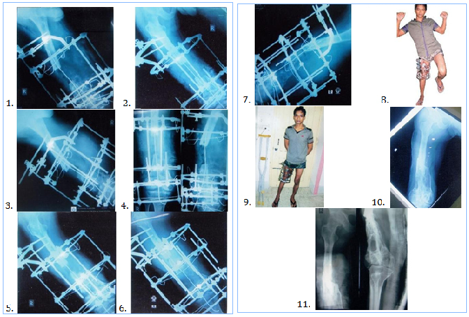
Between 2000 and 2015 30 man and 6 women aged 20 to 57 years (mean 32) were underwent adequate debridement and resection of nonviable bone followed by bone transport using Ilizarov fixator for infected gap non-union of the femoral shaft (Figure 1 to 11).

Figure 1 Big gap non-union of right femur with Ilizarov frame. Intramedullary guide wire in situ to maintain the axis of the limb.
Figure 2 Visible corticotomy in the lower metaphysis of right femur.
Figure 3 Radiograph of right femur and knee with Ilizarov fixator, 10 days after corticotomy.
Figure 4 Radiograph of right femur with Ilizarov in situ after 20 days of ascending distraction.
Figure 5 Radiograph of right femur with Ilizarov in situ after 40 days of ascending distraction.
Figure 6 Radiograph of right femur with Ilizarov in situ after 2 months of ascending distraction.
Figure 7 Good regenerate is visible in the ascending distraction region.
Figure 8 The patient is advised to stand on the affected limb with the frame.
Figure 9 Patient is on Ilizarov frame in the right thigh and knee.
Figure 10 Final radiograph of good union is seen in the right femur (AP view).
Figure 11 Final radiograph of right femur with good consolidation after 11 months.
All patients had a bone defect of >4 cm, complete blood counts, ESR and C-reactive position level were measured. Quantity of patients, presenting symptoms and duration, anamnesis, sinus, skin status, shortening, and deformity and neurovascular status of the knee joint were recorded.
Under spinal anaesthesia patients were positioned supine. The dead bone was resected, debridement were done adequately. Cortical bleeding known as paprika sign was considered the end point of bone resection.1,5,6 Ilizarov was applied accordingly. Monofocal bone transport was performed either the ascending technique (with a distal femoral metaphyseal corticotomy) or the descending technique (with a proximal subtrochanteric femoral corticotomy) through healthy tissue.
Post-operative care is essential and was standardized by pain management active and passive joint mobilization and strengthening exercises for the hip and knees. Antibiotics were continued for a minimum of 6 weeks or until the ESR and C-reactive protein level had returned to normal.6 Rubber stopper should be used in the wire and pin sites for proper application of dressing gauge. Distraction was started on the post-operative days 5 at a rate of 1 mm/day in 4 equal increments.7 X-ray was obtained every 2 weeks during bone transport and every month during the consolidation period, at the time of Ilizarov frame removal.
The lengthening index was defined as the duration to bone transport/lengthening per cm gain in length. The treatment index or x-ray consolidation index was defined as the time in days to the appearance of consolidation of at least 3 cortices on the anteroposterior and lateral x-rays divided by the total amount of bone transported and the amount of lengthening in cm.
Function of the limb was evaluated on the basis of 5 criteria of adversity:
Functional outcome was excellent if the patient was active and able to perform daily activities and the other 4 criteria were absent, good when one or 2 of the other criteria were present, fair when 3 or 4 of the other criteria were present and poor when the patient was inactive, regardless of the other criteria.1,8
Union, infection, deformity, LLD and mechanical insufficiencies at the docking site were assessed. The outcome excellent when the bone was united without infection, <7° deformity, <2.0 cm LLD, good when the bone was united and with 2 of the above criteria, fair when the bone was united with one of these criteria, poor when the bone was un-united, with bone of the criteria fulfilled5.
All intraoperative injuries and difficulties during limb lengthening that were not resolved before the end of treatment we considered that the true complications.
The patients had undergone a mean of 3 (range 1-6) surgical procedures before consulting us. The mean duration from trauma to consultation was 6.5 (range 5-16) months. The mode of injuries were RTI (n=25), fall from a height (n=6); gunshot (n=5). 18 patients had discharging sinuses. 14 patients had a segmental bone defect along with shortening and deformity, whereas 4 patients had shortening only.
The mean bone defect after debridement was 8.9 (range 4.5-13) cm. For bone transport, the ascending technique was used in 28 cases and descending technique in 8. The mean treatment duration was 8.5 (range 6-16) months. The mean lengthening index was 12.5 (range 10.5-14) days/cm. The mean treatment index was 25.5 (range 25-45) days/cm. The mean follow up time was 18.5 (range 15-45) months. Functional outcome was excellent in 20, good in 10 and fair in 6 patients. All patients had a good bone union and eradication of infection.
None had neurovascular complications and refracture of regenerated bone. The problems were pain during the distraction period (n=20), wire site infection (n= 10), soft tissue interposition at docking site (n=3). True complications encountered knee stiffness in 7 cases.
Management of infected gap non-union of femur is a challenge for orthopedic and reconstructive surgeon. The associated problems with bone defect, shortening and deformity may complicate the treatment procedure. The law of tension stress that was pioneered by Academician Ilizarov.4 with distraction osteogenesis based on the biology of bone and soft tissue regeneration. The fundamental and basic principles of the Ilizarov method includes elastic stable fixation, a low energy corticotomy with gradual controlled coordinated distraction and osteogenesis by intramembranous ossification are the same for bone transport and compression distraction.9 Compression distraction method is simpler than bone transport and should be used to close the bone defect directly.1 but transport is essential when gap is very big.10 The long duration of bone transport in femur is sometimes uncomfortable for patients. Proper application of Ilizarov frame and tensioning of wires in the femur gives excellent results. Patients with infected non-union in femur have undergone several multiple interventions. To eliminate infection vascularization of the infected bone is increased by the biological stimulation of corticotomy. We did not apply any autogenous bone grafts after freshening sclerotic bone ends. Distraction is a excellent potent stimulus for osteogenesis. Every successful treatment with Ilizarov patients always depends on proper wound care with meticulous intelligent follow up and monitoring. Fundamental principles of Ilizarov methodology is load and motion, which can be achieved by the active participation of physiotherapist.
Distraction osteogenesis using Ilizarov technique in the femur is a safe and effective tool for treating infected gap non-union of femur.
None.
None.

©2016 Bari, et al. This is an open access article distributed under the terms of the, which permits unrestricted use, distribution, and build upon your work non-commercially.