MOJ
eISSN: 2374-6939


Case Report Volume 18 Issue 2
 Jesús García Álvarez, María del Sol Gómez Aparicio, David Segura Sánchez, Carlos Guillermo Puig Abbs
Hospital Intermutual de Levante, San Antonio de Benagéber, Valencia, Spain
Correspondence: Sergio Pérez Ortiz, Hospital Intermutual de Levante, Autovía CV-35, Km 11,7, 46184 San Antonio de Benagéber, Valencia, Spain
Received: March 10, 2026 | Published: March 24, 2026
Citation: Pérez Ortiz S, García Álvarez J, Gómez Aparicio MS, et al. Delayed posterior interosseous nerve palsy following olecranon fracture fixation with complete recovery: a case report and discussion of inflammatory neuropathy as a non-iatrogenic aetiology. MOJ Orthop Rheumatol. 2026;18(2):46-50. DOI: 10.15406/mojor.2026.18.00739
Background: Posterior interosseous nerve (PIN) palsy following elbow surgery is uncommon and most frequently attributed to iatrogenic injury during radial head procedures requiring lateral exposure. Delayed PIN palsy arising after isolated olecranon fixation via a strictly posterior approach—without lateral dissection—is exceedingly rare. This presentation may reflect postsurgical inflammatory neuropathy (neuralgic amyotrophy, NA) rather than a direct surgical insult.
Case presentation: A 26-year-old male truck mechanic sustained a work-related intra-articular comminuted displaced olecranon fracture (AO/OTA 21-B1.3) with an intact coronoid process. Preoperative neurovascular examination was normal. Open reduction and internal fixation was performed on 11 July via a posterior approach using a precontoured olecranon locking plate (Acumed). Neurological examination was normal on postoperative day 1 and at week 2. At postoperative week 3, the patient developed sudden complete inability to extend his fingers (MRC 0/5) with preserved wrist extension and finger flexion (5/5), without compartment syndrome. The patient had undergone prolonged immersion of the left forearm in very hot water the day before symptom onset.
Results: Electromyography at postoperative week 6 confirmed very severe axonotmesis of the left PIN. Conservative management was instituted: dorsal extension orthosis and oral prednisolone 60 mg/day for two weeks with progressive tapering. Initial clinical motor recovery was recorded at week 18. Serial electromyography demonstrated proximal reinnervation polyphasia at week 23 and active global reinnervation at week 32. Complete motor recovery was achieved by week 48. Elective plate removal was subsequently performed without recurrence.
Conclusion: Delayed PIN palsy after olecranon fixation with a normal early postoperative course is inconsistent with iatrogenic nerve injury and should prompt consideration of postsurgical inflammatory neuropathy. Recognition of this entity supports conservative management, avoids unnecessary surgical re-exploration, and enables timely initiation of corticosteroid therapy.
Keywords: posterior interosseous nerve, olecranon fractures, neuralgic amyotrophy, parsonage–turner síndrome, axonotmesis, electromyography, peripheral nerve injuries, corticosteroids
The posterior interosseous nerve (PIN) is the deep motor branch of the radial nerve. After separating from the superficial radial nerve at the level of the lateral epicondyle, it traverses the radial tunnel and pierces the supinator muscle at the arcade of Fröhse, subsequently innervating the extensor musculature of the wrist and fingers with the exception of the extensor carpi radialis longus and brevis.1 Clinically, PIN palsy manifests as finger drop without complete wrist drop, given the proximal innervation of the extensor carpi radialis longus; sensory function is preserved, as the PIN is an exclusively motor branch distal to its origin.1
PIN injury in the context of elbow surgery has been reported primarily after radial head fracture fixation or arthroplasty, where lateral exposure in proximity to the radial neck places the nerve at risk.² Carbonell-Escobar et al., in a series of 62 patients undergoing radial head arthroplasty, identified a PIN palsy incidence of 3.2%, attributing the injuries to excessive anterior retraction at the radial neck level and prolonged tourniquet time.2 Critically, both palsies in that series presented in the immediate postoperative period and resolved within eight weeks - a temporal pattern fundamentally inconsistent with the presentation described in this report.2
Neuralgic amyotrophy (NA), or Parsonage–Turner syndrome, is an inflammatory peripheral neuropathy characterised by immune-mediated focal axonal damage within the brachial plexus and its branches.3,4 Its incidence is estimated at approximately 1 per 1,000 per year, though this figure is likely underestimated owing to systematic diagnostic delay and frequent misclassification.4,5 The classic phenotype- sudden severe pain followed by patchy paresis - is present in approximately 70% of patients; however, isolated involvement of distal motor branches such as the PIN may occur without a prominent painful prodrome, as demonstrated by Akane et al. in a series where fewer than 53% of patients with AIN/PIN involvement reported pain preceding paresis.6 The PIN and the anterior interosseous nerve (AIN) are among the most commonly isolated distal targets in NA,6–8 and surgical procedures constitute well-recognised precipitating triggers, acting through immunological mechanisms involving disruption of the blood–nerve barrier and T-cell–mediated endoneurial inflammation.3,9 In a landmark series by Malamut et al., six patients developed idiopathic brachial neuritis between one and thirteen days after surgical procedures entirely remote from the brachial plexus, establishing the concept of postsurgical inflammatory brachial neuropathy as a distinct and underrecognised clinical entity.9,7
We present the case of a 26-year-old male who developed isolated severe axonotmetic PIN palsy at postoperative week 3 following technically uneventful olecranon plate fixation via a posterior approach. The clinical, electrodiagnostic, and temporal features are analysed to support a non-iatrogenic, inflammatory aetiology and to discuss the role of corticosteroid therapy in this context. Informed consent was obtained from the patient for publication of this case report and accompanying radiographs.
A 26-year-old male truck mechanic with no relevant medical history sustained a work-related fall onto his outstretched left upper limb. Radiographic evaluation revealed an intra-articular comminuted displaced fracture of the olecranon with an intact coronoid process (Figure 1), classified as AO/OTA 21-B1.3. Neurovascular examination on admission was entirely normal.
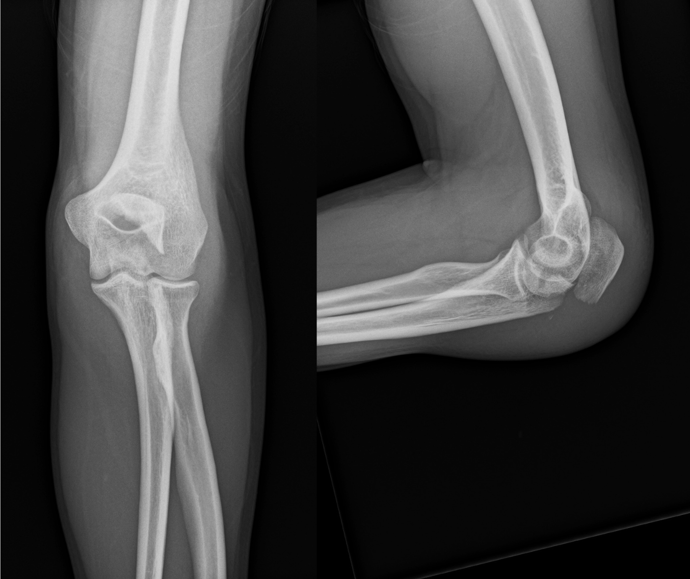
Figure 1 Preoperative radiographs. Anteroposterior and lateral radiographs of the elbow showing an olecranon fracture classified as AO/OTA 2U1C.
On postoperative day 0, the patient underwent open reduction and internal fixation through a standard posterior approach to the elbow. Following identification and neurolysis of the ulnar nerve, fracture reduction was achieved with pointed fracture reduction clamps and provisional Kirschner wire fixation. Definitive fixation was accomplished using a precontoured olecranon locking plate (Acumed, Hillsboro, OR, USA). Intraoperative fluoroscopic assessment confirmed satisfactory articular reduction and implant positioning (Figure 2). Full elbow range of motion was demonstrated at the conclusion of the procedure. No retraction or dissection was performed in the lateral or anterolateral compartments, and the PIN was entirely outside the operative field.
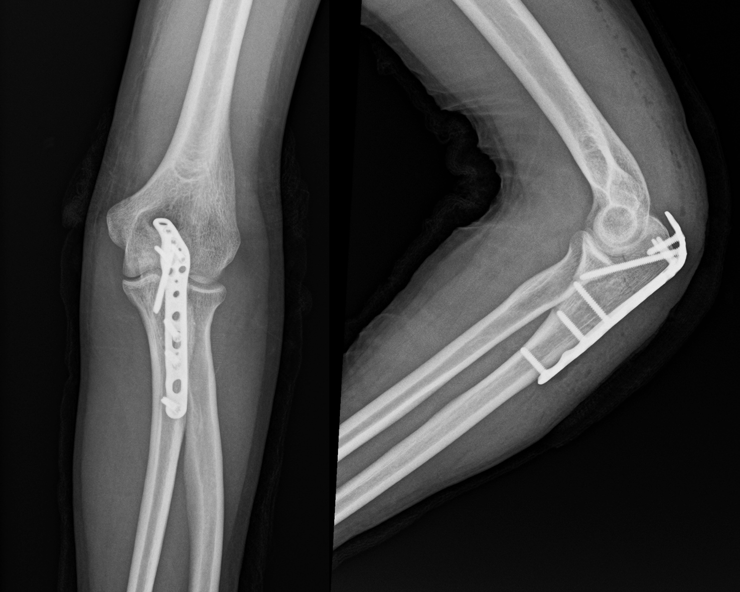
Figure 2 Postoperative radiographs. Anteroposterior and lateral radiographs of the elbow after open reduction and internal fixation of the olecranon fracture using a precontoured olecranon plate (Acumed, Hillsboro, OR, USA).
Neurological examination of the left upper extremity was entirely normal on postoperative day 1. At the two-week follow-up review on 26 July, the patient demonstrated full elbow, wrist, and digital range of motion without motor or sensory deficit. At postoperative week 3, the patient reported sudden inability to extend his fingers. Assessment on 2 August confirmed complete absence of finger extension (MRC grade 0/5), with fully preserved wrist extension (5/5) and finger flexion (5/5). No forearm swelling, haematoma, skin changes, or clinical signs of compartment syndrome were detected; all forearm compartments were soft on palpation. The patient reported a persistent oppressive aching pain in the left forearm. He disclosed that on the day immediately preceding the onset of the neurological deficit he had immersed his left forearm in very hot water for a prolonged period in the course of his occupational duties.
Nerve conduction studies and needle electromyography were performed at postoperative week 6 - three weeks after onset of the deficit. Electrodiagnostic findings demonstrated very severe axonotmesis of the left PIN, with markedly reduced or absent voluntary motor unit recruitment and abundant fibrillation potentials and positive sharp waves in PIN-innervated muscles consistent with acute denervation. No abnormalities were identified in the ulnar nerve, median nerve, or other radial nerve branches (Figure 3).
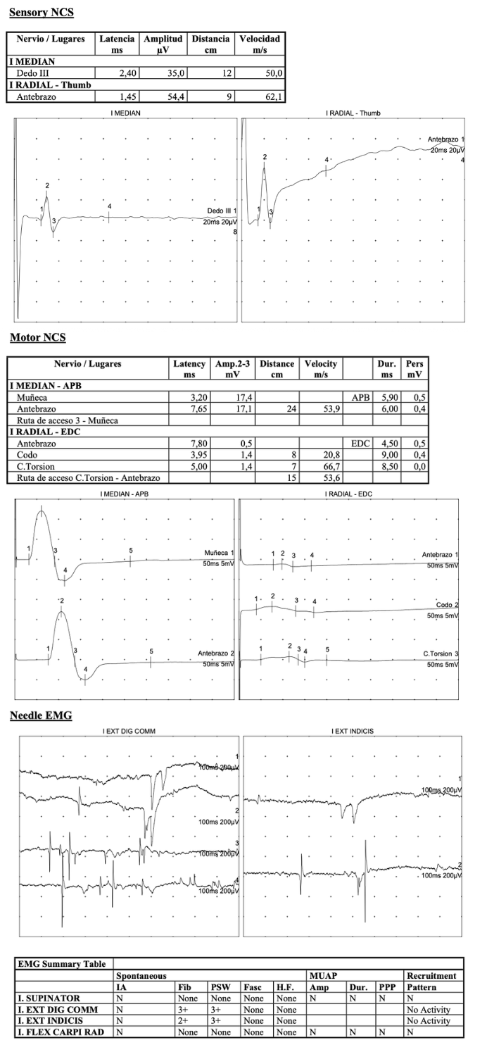
Figure 3 Electromyographic findings. Electrodiagnostic study showing selective motor involvement of the posterior interosseous nerve with preserved sensory conduction and denervation in PIN-innervated muscles, consistent with posterior interosseous neuropathy.
Conservative management was instituted. A custom-fabricated static dorsal extension orthosis was applied to maintain the metacarpophalangeal joints in functional extension and prevent flexion contracture. In addition, oral prednisolone was prescribed at 60 mg/day for two weeks, followed by progressive dose tapering over a further two weeks, in accordance with evidence-based recommendations for acute inflammatory neuropathy.10,11
Initial clinical signs of motor recovery in the PIN territory were first documented at postoperative week 18. Repeat needle EMG at week 23 demonstrated onset of reinnervation polyphasia in the most proximally innervated PIN muscles, consistent with early axonal regeneration. A further EMG at week 32 confirmed active global reinnervation with marked electrophysiological improvement. By postoperative week 48, the patient had achieved complete motor recovery with MRC grade 5/5 in all previously affected muscles. Elective removal of the olecranon plate was subsequently performed without neurological recurrence, and the patient returned to his occupational duties without restriction (Figure 4).
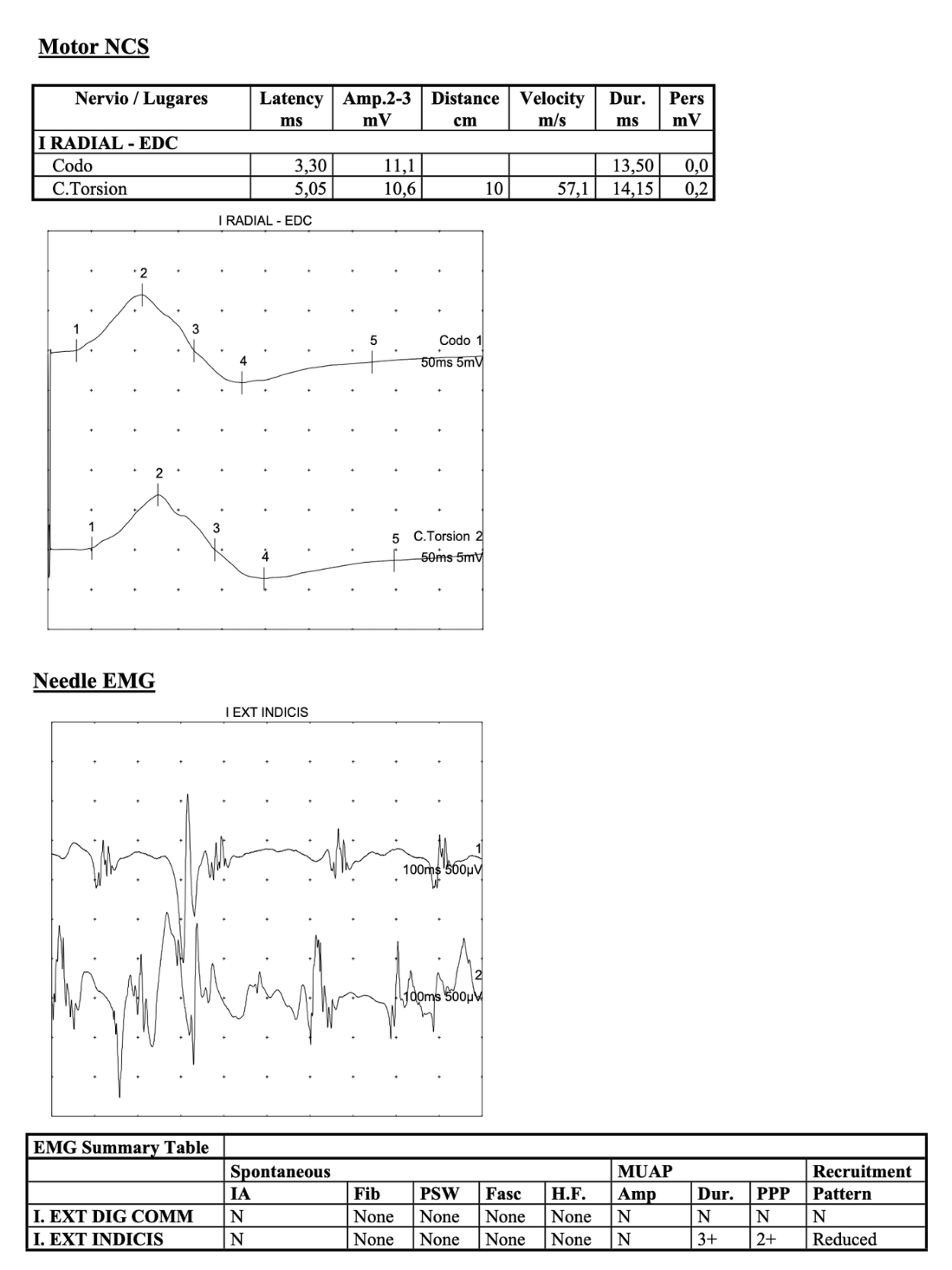
Figure 4 Follow-up electromyographic study. Follow-up EMG demonstrating improvement in radial motor conduction and chronic neurogenic changes with reinnervation in posterior interosseous nerve–innervated muscles, compatible with recovery after idiopathic neuralgic amyotrophy.
The present case raises a fundamental diagnostic question that has direct therapeutic consequences: does the PIN palsy observed three weeks after olecranon fixation represent an iatrogenic surgical injury, or does it reflect a postsurgical inflammatory neuropathy triggered by the operative event? The answer, we argue, lies in a careful integration of the anatomical, temporal, electrodiagnostic, and clinical evidence.
From an anatomical standpoint, direct surgical injury to the PIN in the context of a posterior approach to the olecranon is implausible. The PIN courses through the radial tunnel in the anterolateral compartment of the forearm, entering the supinator at the arcade of Fröhse—a region entirely outside the operative field of a posterior olecranon approach, which involves no lateral or anterolateral dissection whatsoever.1 In contrast, PIN palsy has been documented after radial head arthroplasty precisely because the lateral Kocher approach requires dissection in proximity to the radial neck; even in that setting, Carbonell-Escobar et al. reported an incidence of only 3.2% in 62 patients, and both affected cases presented immediately postoperatively, not with a latency of three weeks.2 The fact that our patient underwent a thorough neurological examination on postoperative day 1 and again at week 2 - both entirely normal - categorically excludes any mechanism of intraoperative nerve injury, whether from direct trauma, excessive retraction, thermal damage, or haematoma compression.
The temporal profile of the deficit, emerging suddenly at week 3 with concurrent oppressive forearm pain and no associated swelling or compartment syndrome, is instead highly characteristic of the acute inflammatory phase of neuralgic amyotrophy (NA) involving the PIN. NA is a multifactorial inflammatory disorder in which genetic susceptibility, immunological activation, and focal mechanical vulnerability of nerve fascicles interact to produce sudden, often severe, peripheral nerve injury.3,4 Its reported incidence of approximately 1 per 1,000 per year is widely considered an underestimate, as the condition is systematically misdiagnosed or attributed to mechanical causes.4,5 Van Alfen and van Engelen, in a landmark series of 246 patients, provided a comprehensive characterisation of the clinical spectrum of NA, noting that while the classic phenotype involves proximal brachial plexus branches with severe shoulder pain, isolated distal motor branch involvement - including the PIN - is a well-established variant.5 Akane et al. further demonstrated that in cases of AIN/PIN involvement, fewer than 53% of patients reported pain preceding the onset of paresis, meaning the absence of a prominent shoulder pain prodrome, as in our case, does not argue against this diagnosis.7
The role of surgery as a precipitating trigger for NA is well established and provides the most plausible primary mechanism in this patient. Malamut et al. described six patients who developed idiopathic brachial neuritis between one and thirteen days after surgical procedures entirely remote from the brachial plexus, and concluded that the delayed postoperative onset, together with the multifocal electrodiagnostic pattern, were the principal arguments against a mechanical perioperative cause.9 The pathophysiological substrate involves disruption of the blood–nerve barrier under surgical stress, enabling cellular and humoral immune effectors to access the endoneurial space and initiate a focal inflammatory cascade.3 The present patient's deficit appeared at week 3- within the temporal window described by Malamut et al. for postsurgical inflammatory brachial neuropathy - and its sudden onset with pain is entirely consistent with this mechanism.9
The patient additionally reported prolonged hot water immersion of the left forearm on the day before symptom onset. While thermal stimuli sufficient to produce local tissue inflammation could theoretically act as a secondary trigger in an immunologically sensitised nerve, direct causal evidence linking thermal exposure to NA is absent from the literature. The surgical procedure itself remains the most parsimonious primary explanation, with the thermal event possibly representing a coincidental or additive factor. The absence of compartment syndrome clinically excludes thermally induced pressure elevation as an independent mechanical cause of nerve dysfunction.
The electrodiagnostic findings are entirely coherent with this interpretation. Axonotmesis—Sunderland grade 2 nerve injury - denotes disruption of the axon and its myelin with preservation of the endoneurial, perineurial, and epineurial connective tissue framework.1 Wallerian degeneration proceeds distally from the site of injury and becomes electrophysiologically detectable as fibrillation potentials and positive sharp waves typically two to three weeks after the insult, which is precisely why the first EMG, performed three weeks after deficit onset (week 6 postoperatively), demonstrated the pattern of very severe acute denervation with absent voluntary motor unit activity.1 Bäumer et al., using high-resolution MR neurography in 19 patients with posterior interosseous neuropathy syndrome, found that 84% harboured proximal fascicular lesions at the upper arm level rather than focal entrapment at the arcade of Fröhse, implicating an inflammatory mechanism in the majority and providing the structural correlate of what is detected electrophysiologically as axonotmesis.7 Maldonado et al. corroborated this in a retrospective reanalysis of 15 patients with nontraumatic isolated PIN palsy, showing that 73% had electrodiagnostic and MRI findings incompatible with simple radial tunnel compression.8 In our patient, the absence of any electrodiagnostic abnormality in other nerve territories further supports a focal inflammatory lesion of the PIN rather than a positional or systemic mechanism.
Axonal regeneration in axonotmetic injuries proceeds at approximately 1–3 mm per day along intact endoneurial tubes, with the earliest electrophysiological sign of recovery being nascent polyphasic motor unit potentials in the most proximally innervated target muscles.1 The serial EMG findings in this case - proximal reinnervation polyphasia at week 23, active global reinnervation at week 32, and complete clinical recovery at week 48 - follow precisely this expected progression. Feinberg et al., in a prospective observational study of 29 affected nerves in Parsonage–Turner syndrome, reported initial electrodiagnostic recovery at a mean of 5.8 months and complete electrodiagnostic recovery in 52.9% of patients followed beyond one year, a timeline closely mirroring our case and providing important prognostic benchmarks.12
The decision to manage this patient conservatively rather than surgically was supported by multiple converging lines of evidence. First, the implausibility of an iatrogenic mechanism removed any indication for re-exploration of the surgical site. Second, the progressive electrodiagnostic evidence of spontaneous reinnervation - already present at week 23 - provided objective confirmation that axonal regeneration was underway along intact endoneurial tubes, rendering surgical intervention unnecessary. Third, the natural history of NA involving distal motor branches is generally favourable with conservative management: Akane et al. reported motor recovery in 71.4% of conservatively treated patients,7 and the algorithm proposed by Gstoettner et al. reserves neurolysis exclusively for cases with documented fascicular constrictions on high-resolution imaging and absent spontaneous recovery after a minimum of three months.3 The dorsal extension orthosis maintained metacarpophalangeal joint position during the reinnervation period, preventing fixed flexion contracture and facilitating functional rehabilitation.
Regarding pharmacological management, oral corticosteroids administered in the acute phase of NA have been shown in observational studies to accelerate both pain relief and early motor recovery. Van Eijk et al., in a retrospective study of 50 prednisolone-treated patients compared with 203 historical controls, demonstrated that the median time to initial pain relief was significantly shorter in the treated group (12.5 vs. 20.5 days) and that the proportion achieving measurable strength recovery at one month was significantly higher (18% vs. 6.3%; p = 0.011).10 A Cochrane systematic review by van Alfen et al. found no randomised controlled trials in this area but concluded that available open-label evidence supports a role for oral prednisolone in shortening the acute pain phase and facilitating early recovery.11,12 Current clinical guidelines recommend prednisolone 1 mg/kg/day for seven days with progressive tapering when patients are seen within the first four weeks of symptom onset.13 The 60 mg/day regimen prescribed in our patient for two weeks with subsequent tapering is consistent with these recommendations and with the dosing used in the observational study by van Eijk et al.,10 targeting both the acute inflammatory insult and the oppressive forearm pain reported by the patient.
This report has important limitations that must be acknowledged. As a single case, it constitutes Level IV evidence, and no definitive causal relationship between the surgical trigger, the thermal exposure, and the inflammatory neuropathy episode can be established from temporal association alone. High-resolution MR neurography or nerve ultrasound - which may have identified hourglass fascicular constrictions pathognomonic of NA3,6- was not performed, representing a diagnostic gap. Hepatitis E virus serology and other targeted inflammatory biomarkers, implicated in 10–15% of NA cases,13 were not obtained, limiting aetiological characterisation. These limitations do not diminish the educational value of the case but underscore the importance of incorporating systematic neuroimaging and serological evaluation in future similar presentations to enable more precise mechanistic characterisation and to build a more robust evidence base for this rare clinical scenario.
Delayed PIN palsy emerging at postoperative week 3 following olecranon fracture fixation via a posterior approach, with a documented normal neurological examination in the preceding two weeks, is an exceedingly rare presentation that is anatomically and temporally incompatible with iatrogenic nerve injury. The clinical and electrodiagnostic pattern in this case - isolated very severe axonotmesis of the PIN, progressive serial EMG reinnervation, and complete motor and sensory recovery at 48 weeks with a conservative regimen comprising short-course oral prednisolone and dorsal extension orthosis - is entirely consistent with postsurgical inflammatory brachial neuropathy involving the PIN as the isolated target nerve. Clinicians encountering a delayed postoperative peripheral nerve deficit should consider this diagnosis before attributing the deficit to the surgical procedure, as accurate recognition avoids unnecessary re-exploration, enables timely corticosteroid therapy, and supports informed prognostic counselling regarding the protracted but favourable natural history. Future cases should incorporate high-resolution nerve imaging and targeted serological investigation to further characterise the inflammatory mechanism and contribute to a growing evidence base for this underrecognised entity.
The Gamma distribution is parameterized in this paper in terms of the mean and the standard deviation. The probability density function of a Gamma distributed variable x is
None.
The authors declare that there are no conflicts of interest.

©2026 Pérez, et al. This is an open access article distributed under the terms of the, which permits unrestricted use, distribution, and build upon your work non-commercially.
