MOJ
eISSN: 2374-6939


Case Report Volume 7 Issue 4
Resident, Grant Medical Center, USA
Correspondence: Rebecca A Sundling DPM, 313 Olentangy River Road, Columbus, Ohio, Tel (740) 507-6739
Received: February 05, 2017 | Published: February 24, 2017
Citation: Sundling RA, Cheney NA (2017) Danis-Weber a Non-Union in a Teenager: A Case Report and Review of Literature. MOJ Orthop Rheumatol 7(4): 00278. DOI: 10.15406/mojor.2017.07.00278
Case: This case focuses on the occurrence and treatment of a non-union of a Danis-Weber A fracture in a teenage girl. After 5 months of protected weight bearing and the diagnosis of a non-union, a bone stimulator was added to the treatment plan. Two months later without noted healing, the patient was taken to the operating room for open reduction internal fixation with both allograft and autograft.
Conclusion: Even though unexpected at times, non-unions are possible in any age group for many possible reasons.
Ankle fractures are a common injury, occurring in 187 of 100,000 people each year in the United States.1 When a fibula is fractured, 75-85% of the time it occurs concomitantly with a tibia fracture, often due to a rotational injury.2-5 Stability of the fracture is assessed and when it is stable and non-displaced, conservative treatment is recommended, with satisfactory results.2,6 This includes 6-8 weeks of protected or non-weight bearing in an immobilizing boot or below knee cast.
Fibula fracture non-unions are relatively rare and infrequently reported, found only in 0.3% to 5.4% of fibular fractures in literature.2 Many of these are asymptomatic and require no treatment. The vast majority of literature discussing fibular non-unions pertains to fractures at or above the level of the tibio-talar joint.2-9 To our knowledge, only one case of a Danis-Weber A (DWA) non-union has been discussed in literature at this time, which was treated with avulsion fragment excision.2 We present a case of a teenage female who developed an atrophic non-union of a more proximal DWA fracture.
A conversation was had with both the patient and the patient’s parents regarding possible publication of case material. Verbal consent was obtained from the patient’s parents.
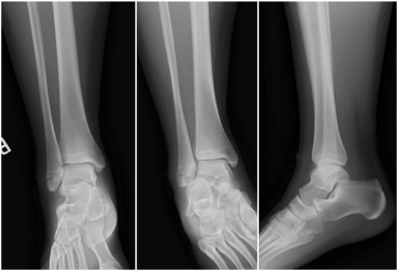
The patient is a 15-year-old skeletally mature female who sustained an inversion injury to her right ankle in January 2015, resulting in a Danis-Weber a fracture (Figure 1). The fracture was non-displaced, complete and intra-articular. At this time, she was seen by a partner of the attending author, placed in an immobilizing boot and allowed to bear weight. The patient was followed with serial radiographs. At 5 months, it was determined that the patient had a delayed union and was instructed to use a bone stimulator daily.

Figure 1 Initial radiographs. Anterior-posterior, Mortise and Lateral views of the right ankle. January 2015.
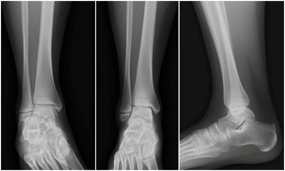
In August 2015, the fracture was re-evaluated, showing no improvement from previous radiographs (Figure 2). At this point, the patient was referred to the attending author for a second opinion. The fracture site remained swollen and painful. It was determined that the best treatment for the patient would be open reduction internal fixation of the fracture (ORIF) with bone grafting, as she had a non-union. Surgery was scheduled for September 2015.

Figure 2 Radiographs after 2 months of bone stimulator use. Anterior-posterior, Mortise and Lateral views of the right ankle. August 2015.
The patient was taken to the operating room and placed in a supine position. A lateral linear incision was made overlying the fibula, centered over the fracture. Dissection was carried deep utilizing blunt technique until the non-union was identified. The non-union was atrophic in nature, which resulted in bone loss at the fracture site with significant fibrous tissue present. After debridement of the fracture site, a gap, measuring approximately 5 mm, was noted between bone segments. The two bone fragments where prepared and a tricortical allograft was fashioned to fit the void, measuring 0.5 cm tall x 1.5 cm deep x 2 cm wide. Autograft from the calcaneus was procured from a lateral incision to fill remaining defects in the graft-bone interface. The correction was assessed with intra-operative fluoroscopy and there was appropriate length and fracture reduction as shown by the presence of the dime sign, which has repeatedly been shown to be important for adequate fracture reduction.10 This was then stabilized utilizing a 6-hole distal fibular hook plate and three locking screws. This particular plate has a device that attaches to the plate for further compression, allowing for better fixation of the fracture site. This was again assessed with fluoroscopy. The wound was then irrigated and closed in layered fashion.
The patient was non-weight bearing in an immobilizing boot for 6 weeks and followed a normal post-operative course, with serial radiographs. The patient continued use of the bone stimulator. At 6 weeks post-operatively, the patient was allowed to begin weight bearing. At final radiographs in December 2015, a complete union was noted (Figure 3). The patient returned to all pre-injury activities without reservation. At one-year out from surgery, the patient is doing well with no complaints or limitations to activity.
Fibula non-unions are very rare and virtually unheard of when the fracture is distal to the tibio-talar joint. When they are reported in the literature, it is most commonly rotational fibular fractures.2-4 Walsh and DiGiovanni discuss one case of a DWA non-union in their retrospective case review.2 In that study, the fracture was an avulsion of the calcaneofibular ligament and was treated with excision of the fragment with addition of a Brostrom procedure.
The cause of delayed and non-unions is well known and documented. Factors that can increase the risk of non-union include increased age, obesity and history of smoking, none of which apply to our patient.2-4,11 The patient is young, active and healthy with no history of substance or tobacco use. Given this, one would expect that she would have healed the non-displaced fracture under traditional conservative measures, such as immobilization and limited weight bearing.
The blood supply to the fibula is known to diminish proximal and distal to the ankle joint, which does put a DWA fracture at higher risk for non-union.2 If blood flow is a concern, bone scans can be utilized to assess blood flow to the fracture fragments.7 Sneppen also showed that supination type injuries, such as in this case, frequently resulted in pseudoarthrosis of fracture fragments.9
Conservative care of non-displaced fractures usually results in satisfactory outcomes for patients, and has lower risk profiles than operative treatment.6 However, non-unions do occur and several ways have been documented on how to treat them.3 In a systematic review of fibula non-unions, Bhadra et al.3 reviewed the treatment options found in the literature.3 These include further conservative management, bone stimulation, external fixation, percutaneous drilling, bone grafting and ORIF with or without bone grafting, arthrodesis, excision and segmental resection.3,8 Segmental resection, however, is not recommended for fractures close to the ankle joint, such as this patient’s.8 Bhadra et al.3 also provided a treatment algorithm that recommends ORIF with bone grafting for atrophic non-unions of the distal third fibula, which is what we did.
This particular case is interesting, as a non-union of a true DWA type fracture has not been noted in the literature. Our patient is young and healthy and underwent traditional conservative care, developing an atrophic non-union. The non-union was unexpected and failed to respond to bone stimulation. Surgical treatment of the non-union with ORIF with calcaneal autograft and tricortical allograft proved to be an acceptable option for Danis-Weber a non-union.
None.
None.

©2017 Sundling, et al. This is an open access article distributed under the terms of the, which permits unrestricted use, distribution, and build upon your work non-commercially.