MOJ
eISSN: 2374-6939


Research Article Volume 4 Issue 5
1Chief Consultant, Bari-Ilizarov Orthopaedic Centre, Visiting and Honored Prof., Russian Ilizarov Scientific Centre, Russia
2Bari-Ilizarov Orthopaedic Centre, Bangladesh
Correspondence: Mofakhkharul Bari, Chief Consultant, Bari-Ilizarov Orthopaedic Centre, Visiting and Honored Professor, Russian Ilizarov Scientific Centre, Kurgan, Tel +88 01819 211595
Received: March 28, 2016 | Published: April 15, 2016
Citation: Bari MM, Shahidul I, Shetu NH, Mahfuzer RM (2016) Correction of Bowing Deformities in Paediatric Femur and Tibia by Ilizarov Technique. MOJ Orthop Rheumatol 4(5): 00153. DOI: 10.15406/mojor.2016.04.00153
Introduction: Correction of multiplanar bone deformities in children is indicated for prevention of secondary orthopaedic complications. Different problems related to surgical intervention were reported: non-union, delayed union, recurrent deformity, refractures, nerve palsy and pin tract infection. The aim of this study was to show the results of children femur and tibia bowing deformities by Ilizarov technique.
Materials and Methods: We analysed 27 cases of children femur and tibia bowing deformities under the age of 13 yrs. Simultaneous deformity correction in femur and tibia was done with Ilizarov device in ipsilateral side. Contralateral side was operated after 14 days.
Results: The duration of Ilizarov fixation was 130 days on an average. The deformity correction was achieved with a proper alignment in all the cases.
Conclusion: Bowing of femur and tibia can be corrected simultaneously by Ilizarov fixation with minimum complications. There were no recurrent deformities in our cases.
Keywords: Bowing femur and tibia, Deformity correction, Ilizarov fixator
Bow legs and bow femur is the disease which occurs more often in girls.1-3 Clinical features become obvious at the beginning of ambulating and are manifested by varus deformity of lower limb, impaired gait, short stature and pathologic features.1,4,5 Traditional medical treatment is phosphate substitution combined with vitamins.2 The conservative treatment is not always successful. In that situation, reconstructive surgical intervention for correction of multiplanar bone deformities is indicated to prevent secondary orthopaedic complications such as pain, degenerative arthrosis and pathologic fractures. Children with deformities are treating by various methods of corrective osteotomy and fixation devices by Kirchner’s wires, plates, epiphysiodesis, Ilizarov devices and intramedullary nailing.5-9,2,3 Multiple problems related to surgical correction were reported: non-union after osteotomies, delayed union, recurrent deformity, deep intramedullary infection, joint stiffness, refracture, leg length discrepancy.8,9
For correcting multiplanar and multiapical deformity nowadays Ilizarov is an ideal and efficient technique. The deformity correction should be done in children in order to prevent early degenerative arthosis. Since 1998 we have been using the Ilizarov method and device, the thin Ilizarov wires are most biocompatible for metabolic character of the disease. The Ilizarov fixation decreases the rate of complications and deformity recurrence in long-term follow up.
We analysed 27 children for correction of bow thigh and legs, surgical intervention done between 1998 to 2014 in Bari-Ilizarov Orthopaedic Centre and NITOR (National Institute of Traumatology & Orthopaedic Rehabilitation).
Inclusion criteria were
Deformities of two segments (femur and tibia) on both sides, age under 13 years, simultaneous surgical intervention in femur and tibia in order to correct a multiplannar deformity of whole lower limb (the second limb is operated in a 14 days) with the Ilizarov device.
Exclusion criteria were
Age over 13 years, reconstructive surgical correction of only one segment (femur or tibia), which did not provide re-alignment of the whole leg.
Diagnosis was done on the typical laboratory findings of hypophosphatemia (the average pre- operative level 2.2mg/dl) elevated alkaline phosphatase activity in combination with clinical features of limb deformities and short stature.
We prescribed adequate oral phosphate and calcium therapy before surgery atleast for 6 months.10
Stress full length anteroposterior x-ray of the entire lower extremities were taken and lateral x-ray of each segment with adjacent joints were obtained. The torsional deformities were assessed by clinical and x-ray findings in all patients. MAD, mMPTA, mLDFA were measured preoperatively, after Ilizarov removal at the beginning of full weight bearing (normally in 2 months after frame removal) and in long-term follow up. Surgical planning is absolutely necessary and we must assess the extent of deformity in the lower limb.
The first step is to introduce the wires and frame assembly followed by percutaneous osteotomies.9,12,13 Multi segmental fixation by Ilizarov method was the main thing of surgical intervention.
Ilizarov frame assembly, placement of olive wires and hinges were dictated by anatomic location and severity of the deformities. If the deformity was located at only one level of the bone segment (one CORA), the single osteotomy was done. If the deformity correction could not be achieved by single approach (e.g. metaphyseal and diaphyseal deformity locations), the double percutaneous osteotomies were done in tibia, the osteotomy of the fibula was performed in the lower third or between the middle-lower thirds of the shaft. In all cases the operation was performed on both segments of the lower limb simultaneously in order to obtain realignment of the whole limb.
The steps of surgery in bowing deformities for application of the Ilizarov device are necessary for correction of osteotomies. Correction of angular and rotational deformities were performed either in the acute or gradual manner, depending on the severity of the deformity. Deformities of 25°-30° and more were corrected partially at once; residual deformities were corrected gradually by controlled coordinated stretching. Gradual correction can be started on the 4th to 5th post-operaive day. In our all cases we strived to eliminate the present deformities and to prevent under or overcorrection. When a homogenous bone regenerate was seen with x-ray evidence of consolidation of at least three of four cortices, the Ilizarov fixator was removed according to being able to walk with partial or full weight bearing without pain.
Table 1 shows basic parameters that concern the age of the first interference, features and location of angulations. Ratio of girls to boys was 20 to 7.
|
Parameters |
Basic Parameters |
|
Age |
11, 8 ± 1,2 |
|
Varus deformity of the lower limb femur in middle and distal third and distal metaphysis |
12 segments |
|
Varus deformity of the whole tibia |
15 segments |
Table 1 Basic parameters that concern the age of the first interference, features and location of angulations
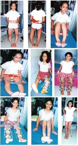
Considering the location of deformities a monofocal bisegmental transosseous osteosynthesis (one osteotomy of femur and one of tibia) was done on 12 limbs and polyfocal (double osteotomy of at least one of the segments) bisegmental transossous osteosynthesis was done in 15 cases (Figure 1).

Figure 1
The following complications that we noted: Pin tract infections in 12 cases, and translation of bone fragments requiring adjust of the Ilizarov fixator under spinal anaesthesia in 6 cases. The results were classified according to Lascombes13 as follows:
Table 2 Present values of MAD, mLDFA mMPTA before surgery, at the beginning of full weight bearing after Ilizarov removal and in the long-term follow up.
|
Initial varus |
Before Treatment |
Before Treatment |
After 10 Years |
||||||
|
MAD |
mLDFA |
mMPTA |
MAD |
mLDFA |
mMPTA |
MAD |
mLDFA |
mMPTA |
|
|
38.8+8.85 |
101±3.5 |
78±2.5 |
0.8±2.0 |
88.1±1.5 |
88.3±1.5 |
20.0±15.0 |
95.0±5.0 |
80.7±5.0 |
|
Table 2 Values of MAD, mLDFA, mMPTA
Deformity correction was achieved with a proper alignment and normal orientation of the knee space according to mechanical axes of segments.
Bow thigh legs due to rickets develop in young age and indications for surgical intervention appear early.4,9,10 Conservative medical treatment consists of oral phosphate substitution and vitamin B.2,4,11 The preoperative average serum phosphate level of 2.2 mg/dl in our series is comparable with 2.0 mg/dl reported by Song et al.8 Deformities of lower limbs should be surgically corrected for biomechanical conditions, and to prevent early arthorosis of hip and knee joints.9
Various treatment modalities are used for fixation of bone fragments after osteotomies: plaster cast, k-wires, plates and screws, Ilizarov fixators and intramedullary nails.4,5,9,14
Ilizarov with parallel connecting rods provides accurate deformity correction.
The problem of Ilizarov is the long period of fixation: from 120 days to 150 days.12,13 Application of locked intramedullary nails is inadvisable in paediatric group.
Song, et al.8 noted in 20 patients 18 major complications (recurrent deformity, fracture after frame removal, peroneal nerve palsy) and 13 minor complications. Fucentese et al.14 describes development of contractures of the ankle.
In our series we observed 2 recurrent deformities due to less correction of the previous deformities and that was overcame by reapplication of Ilizarov after corrective osteotomy
Bow thigh and bow legs deformities in paediatric group should be surgically corrected. Simultaneous correction of femoral and tibial deformities by Ilizarov device is preferable. Ilizarov technique permits achieving correct alignment of the mechanical limb axis from centre of hip to knee and ankle, which is important for growing and formation of catilagenous surfaces of joints. We observed newly formed deformities in distal femoral and proximal tibial metaphysis in two cases.
None.
None.

©2016 Bari, et al. This is an open access article distributed under the terms of the, which permits unrestricted use, distribution, and build upon your work non-commercially.