MOJ
eISSN: 2374-6939


Literature Review Volume 15 Issue 3
1Professor at the department of Stomatology, Faculty of Dentistry, UNICAH, Honduras
2Chief of the Dentistry Service, Hospital Escuela. Professor at the Catholic University of Honduras, Honduras
3Oral and Maxillofacial Surgeon, Teaching Hospital, Honduras
4Dentist, Private practice, Honduras
5Dentistry Student, the Catholic University of Honduras, Honduras
Correspondence: Romero H, Professor at the department of Stomatology, Faculty of Dentistry, UNICAH, Honduras
Received: June 13, 2023 | Published: June 26, 2023
Citation: Romero H, Guifarro JJ, Díaz F, et al. Congenital syngnathia in pediatric patient (virtual surgical planning in a pediatric patient with syngnathia): a case report and literature review. MOJ Orthop Rheumatol. 2023;15(3):118-121. DOI: 10.15406/mojor.2023.15.00630
Congenital craniofacial disorders account for approximately 20% of all birth defects. One of the less common disorders is syngnathia. The case presented includes bony fusion of the alveolar processes of the maxilla and mandible and the ascending ramus with the zygomatic bone. This case report will present details from the gestational birth to the first month of age when the baby underwent the first attempt. to release syngnathia and postoperative control.
Keywords: maxillomandibular fusion, syngnathya, synechiae, congenital
Congenital fusion of the jaws can include soft tissue (synechiae) or bone (syngnathia). 1 The first report of syngnathia was published in 1907 by Kettner, who described a bone-gingival adhesion of the upper and lower jaws, and it can be associated with cleft palate, microglossia, and an abnormal limb. Since then, there have been sporadic reports describing individual cases from around the world.2 A complete review of these combined cases can provide an understanding of how this anomaly presents and sheds light on the relative frequencies of the several clinical features that have been associated with syngnathia. So far the largest number of cases reviewed was published by Naikmasur et al. in 2012, which included 39 cases derived from previously published articles.2
The pathogenesis of the syngnathia is unknown. The persistence of the oropharyngeal membrane is the most common mentioned hypothesis.3 Oral adhesions may represent remaining of the oropharyngeal membrane, which usually disappears during the 4th week of intrauterine life. Other suggested mechanisms are abnormal vascularization by the Stapedial artery, early loss or failure of nerve migration ridge cells resulting in incomplete separation of the first branchial arch in maxilla and mandible, lack of tongue protrusion during development and trauma.3 Teratogens represent a possible etiology of syngnathia, since Meclozine and large doses of vitamin A interfere the jaw development in rats. However, these theories and experimental findings are not supported by clinical data.3
Patients with congenital fusion of the jaws may present with other malformations, in a review of reported cases, it was found that the fusion of the maxillae was exclusively associated with the underdevelopment of the facial structures on the affected side, micrognathia, aglossia and hypoplasia of the branch.4 Other alterations that have been described are: colobomas of the eyelids, low-set ears and other ear deformities hypertelorism and cleft palate.4
On radiological examination of syngnathia, maxilla and mandible continuity can be seen at the alveolar leve. The bony fusion can be extended up to the ramus and the zygomatic bone. Involvement of adjacent structures such as zygomatic bone and TMJ can also be avaluated.5 Separation in cases of synechiae and involvement or cases involving zygomatic bone and TMJ might require planned extensive surgery.5
The role of radiographic examination is very important in differentiating synechiae from syngnathia. It also helps define the extent of bone fusion, as well as the involvement of other structures such as the TMJ and the zygomatic bone.5 The use of three-dimensional computed tomography is helpful in managing the condition4. In our case, a 3D CT the scan was done. Bone fusion of the maxilla with the mandible occurred involving the zygomatic bone bilaterally.5
The complex three-dimensional (3D) anatomy of the facial skeleton adds to the complexity of reconstructing this area and creates a real challenge when it comes to achieving aesthetically pleasing results.6 Traditionally, reconstructive surgery for conditions such as syngnathia and other facial malformations has been based on the subjective evaluation of the surgeon, on preoperative and intraoperative aesthetic criteria, with decision-making based on factors such as: the location of the bone cuts and the shape of the bone segments for facial reconstruction.6
Virtual surgical planning though computer-aided design and computer-aided manufacturing have offered an alternative workflow for more accurate preoperative planning and lees need for intraoperative trial and error. This useful tool offers fabrication of cutting guides and bone models using stereolithography techniques.6
Congenital synechiae are widely reported and reviewed in the Literature1 Dowson proposed the following classification:1
Type 1: simple syngnathia, without Other congenital abnormalities in the head and neck.
Type 2: complex syngnathia, with subgroups:
Type 2a: Syngnathia coexisting with aglossia.
Type 2b: Syngnathia coexisting with proximal agenesis or hipoplasia mandible.1
In 2001, Laster et al. classified the types of syngnathia according to the degree of the mandible to the zygomatic complex and maxillary tuberosity:4
1a Simple anterior
Syngnathia: Fusion of the anterior region without other malformations in face or neck.
1b Complex anterior
Syngnathia: Fusion of the anterior region with malformations in face and neck.
2a Simple mandibulozygomatic: Syngnathia Mandibular and zygomatic fusion causing mandibular micrognathia.
2b Complex mandibulozygomatic: Syngnathia Fusion of the mandibular and zygomatic bones associated with cleft palate or ankylos.4
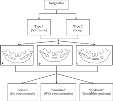
In 2019, Olusanya and Akadiri proposed this classification: (Figure 1)

Figure 1 Proposed classification. (a) Subtype a: midline (anterior); (b) subtype b: bilateral (posterior); (c) subtype c: unilateral (zygomatic-mandibular) 2.
The management of syngnathia requires a multidisciplinary team. The findings of severe micrognathia and polyhydramnios on fetal ultrasounds should raise a suspicion of syngnathia. Prenatal diagnosis allows the planning of delivery in tertiary center.3
Securing the airway is the priority at birth because of the lack of oral access. Blind nasal intubation, fiber optic intubation, and tracheostomy have been used to stabilize the airway of patients with syngnathia, but these approaches have been associated with technical difficulties and/or complications.3
Surgical management division of adhesions is necessary for normal nutrition, eliminating Obstruction of the airways and allowing mandibular function and growth, so must be carried out as soon as possible. Delay in surgical treatment increases the possibility of TMJ ankylosis leading to lack of jaw growth and facial deformities4 nutrition can be challenging in patients with syngnathia. Owing to the wide range of severity, the timing and planning of the surgery has to be adapted to the situation and the general condition of the patient. Very little information exists on the long-term outcome of these patients.7
A newborn referred from the department of neonatology was a product of a 32 weeks on February 4 of 2021 through spontaneous vaginal delivery without any complication, by a 21-year-old mother with no history of systemic diseases or any smoke or alcoholic habits, the baby was referred to the oral and maxillofacial surgery department of Hospital Escuela on February 25 of 2021 (Figure 2).
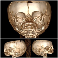
At the clinical examination the maxillomandibular complex where completely fusionated, so in order to give a determined diagnosis, we requested a Computerized Tomography (CT) that illustrated and confirmed the fusion of the gums and bone of both maxilars (Figure 3).

Figure 3 3d reconstruction of computerized axial tomography showing the maxillomandibular fusion and the fusion of the mandibular ramus with the zygomatic complex.
We decided to do a virtual surgical planning to determine the limits of the fusion, precise analysis of the fusion, surgical limits, and to conserve anatomical structures, the fabrication of stereolithographic (SLA) model for the precise treatment planning. The use 3D models have proven to greatly decrease surgical operating time, decrease time under general anesthesia and this model allowed minimize postoperative morbidity (Figure 4).
Cause the baby was a born with low-mass we decided to wait until the newborn get an ideal weight of 10 pounds, and a full count of hemoglobin of 10 g/dl and at least after 10 weeks from birth. In the meantime, the newborn was feeded through a gastric tube. Once the requirements were accomplished we prepared the newborn for surgery under general anesthesia through tracheostomy. Surgery was done by a simple procedure, one incision and a periosteal elevator no separate the fibrous union of the gingivae and the sygnathia was liberate by using a fine Osteotome, the surgery was carried out with no complication, following post-operatory indications with a subsequent use of a mouth gauze to achieve mouth opening and avoiding the relapse of the syngnathia and through physiotherapy by suture threads attached to the tongue and to the alveolar gingivae. This therapy management was done for 15 days (Figure 5).
Syngnathia is an extremely rare condition involving an abnormal development of the splanchnocranium, which is the part of the skull that is derived from the gill arches. Syngnathia can occur with a wide range of severity from a single mucous bands (synechiae) to complete bone fusion (synostosis).7 Our patient presented a bilateral syngnathia of both maxillae which is the least common and most severe form of syngnathia. This presentation can be classified as Type 2b according to Laster et al.1
Olusanya et al.2 proposed a new classification, taking the tissues involved as a reference. It adds whether it is an isolated maxillomandibular fusion, associated with another anomaly or associated with a syndrome. This classification can be very useful to learn more about the patient with the malformation.2
Treatment should focus on securing the airway and placing a nasogastric tube to provide nutrition from birth. This is followed by early removal of the bony fusion with interposition of material to cover the bony ends according to Dawson et al. and followed by active physical therapy. Early treatment has growth retardation and resulting facial deformaties.8–13
Syngnathia is a very rare malformation that can be associated with other anomalies, syndromes or it can be totally isolated. The proper prenatal study and good management in the birth of the newborn can drastically change the patient's prognosis. Good management of the airway, good nutrition, hospital care and a good surgical technique can treat this pathology.
For research articles with several authors, a short paragraph specifying their individual contributions must be provided. The following statements should be used “Conceptualization, Hugo Romero,Mario Gabrie and Henry Valladares; methodology, Juan José Guifarro; validation, Hugo Romero, Juan José Guifarro and Francisco Díaz; formal analysis, Francisco Díaz; investigation, Mario Gabrie, Francisco Díaz, Henry Valladares. data curation, Fabricio Garay; writing-original draft preparation, Hugo Romero and Mario Gabrie; writing-review and editing, Mario Gabrie and Eduardo Osorto; visualization, Francisco Díaz and Juan José Guifarro; supervision, Hugo Romero.Juan José Guifarro; project administration, Eduardo Osorto. All authors have read and agreed to the published version of the manuscript”. Please turn to the CRediT taxonomy for the term explanation. Authorship must be limited to those who have contributed substantially to the reported work.
None.
The author declare that there are no conflicts of interest.

©2023 Romero, et al. This is an open access article distributed under the terms of the, which permits unrestricted use, distribution, and build upon your work non-commercially.