MOJ
eISSN: 2374-6939


Case Report Volume 17 Issue 6
1Assistant Professor, Department of Anatomy, JN Medical College, India
2Professor, Department of Orthopaedic Surgery, JN Medical College, India
3Junior Resident, Department of Orthopaedic Surgery, JN Medical College, India
4Assistant Professor, Department of Orthopaedic Surgery, India
Correspondence: Yasir Salam Siddiqui, Assistant professor, Dept. of Orthopaedic Surgery, J. N. Medical College, Faculty of Medicine, A.M.U., Aligarh, Uttar Pradesh, India, Tel +919837343400
Received: September 09, 2025 | Published: November 19, 2025
Citation: Rahman N, Abbas M, Siddiquee S, et al. Congenital radioulnar synostosis – brief anatomical & functional appraisal. MOJ Orthop Rheumatol. 2025;17(5):151-153. DOI: 10.15406/mojor.2025.17.00725
Congenital radioulnar synostosis is an infrequent malformation characterized by anomalous connection between the forearm bones. The patient characteristically presents with loss of forearm rotation movements with forearm fixed in pronation hindering their day to day activities requiring supination. The shoulder joint compensates to some extent for loss of rotatory movements of forearm, especially in subtle deformities. As far as the treatment is concerned, it ranges from merely observation to surgical intervention for restoring forearm rotations for optimal rehabilitation of the children. The case study presented here deserves reporting not only because of the rarity of the condition but also due to its tricky diagnosis at presentation owing to compensatory mechanism for forearm rotations. An attempt is also made to do briefly discuss the anatomical developmental defect during intra-uterine life leading to the congenital radioulnar synostosis.
Keywords: congenital, radius, ulna, synostosis, pronation, supination
Congenital radioulnar synostosis (CRUS) is an infrequent malformation characterized by anomalous connection between the two long forearm bones (radius and ulna) due to an embryological failure of longitudinal segmentation or separation.1 The condition was first reported by Sandifort in 1793.1,2 It frequently involves both forearms as compared to single one. It is on average an isolated condition without any associated lesions or deformities.2–4 However, its associations with other musculoskeletal conditions and syndromes have been noted in literature, such as arthrogryposis multiplex congenita, developmental dysplasia of hip, clubfoot, osteogenesis imperfecta and tarsal coalitions.5,6 The patient characteristically presents with loss of forearm rotation movements with forearm fixed in pronation hindering their day to day activities requiring supination. The condition is by and large painless except in few cases with snapping elbow and locking elbow, which may be painful.4,7 The shoulder joint compensates to some extent for loss of rotatory movements of forearm, especially in subtle deformities. Therefore the abnormality is missed by the caregivers in initial years of birth and also by young paediatric orthopaedicians, as it is an atypical paediatric pathology often missed unless supported by a good history and clinical examination to raise a diagnostic suspicion of the lesion. Hence the compensatory mechanisms and low index of suspicion can holdup the diagnosis pending the school age.8 As far as the treatment is concerned, it ranges from merely observation to surgical intervention for restoring forearm rotations for optimal rehabilitation of the children. The nature of surgery ranges from excision of synostosis with interposition of soft tissue to derotational osteotomies.9
In literature case studies are reported, highlighting the various aspects of congenital radioulnar synostosis including clinical presentation and management. We believe that the case presented here deserves reporting not only because of the rarity of the condition but also due to its tricky diagnosis at presentation owing to compensatory mechanism for forearm rotations. An attempt is also made to do briefly discuss the anatomical developmental defect during intra-uterine life.
Four years old female was brought to our paediatric orthopaedic clinic by his parents with complaints of restriction of the rotatory movements of both forearms. They further added that the child is having some difficulty in washing face, eating and playing. However the condition was painless. There was no history of any associated trauma, or similar complaints in the family. There was no history of consanguinity. The baby was full term, delivered at hospital without any difficulty, through vaginal route. There was no history suggestive of any maternal infection or any exposure to teratogenic agents throughout the antenatal period. There was no history suggestive of delayed crying or any developmental delay (delayed milestones). No history of any other joint involvement or other systemic illness. Clinical examination revealed both forearms were fixed in approximately 30 degrees of pronation (Figure 1). There was no restriction of motion at the shoulder and elbow joint. There was no swelling, undue bony prominence and varus or valgus instability at elbow. No definite syndromic features were noted. Systemic examination was unremarkable. Radiological examination of both forearms with elbow and wrist revealed bilateral proximal radioulnar synostosis (Figure 2 & 3). Radiologically, it was classified as type 3 CRUS as per Cleary and Omer’s classification. A final diagnosis of bilateral CRUS, Cleary and Omer’s type 3 was made.
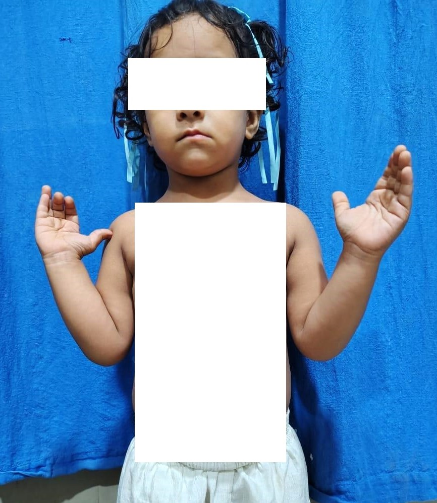
Figure 1 Clinical photograph of patient showing both forearms in pronation.
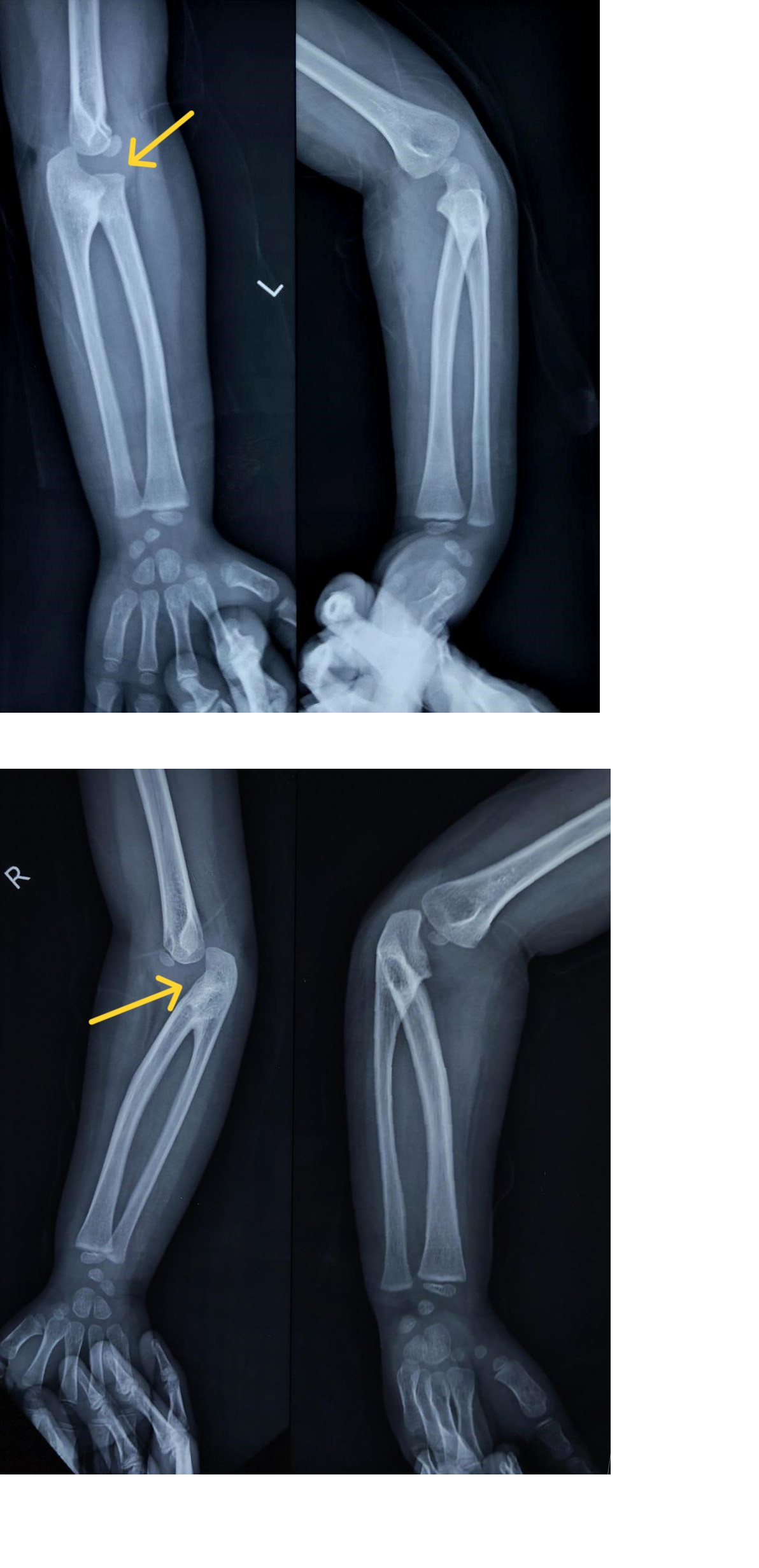
Figure 2 & 3 Radiograph of both forearms including elbow showing bony fusion of proximal radio-ulnar joint with radial head lying posterior in relation to capitulum (Cleary and Omer’s Type 3 CRUS).
In the present circumstance, as the patient was able to carry out her day to day activities, conservative management was advised with regular follow-up. Parents were communicated about the need of regular follow-up for proper treatment, rehabilitation and to monitor progression of the disease.
Congenital radio-ulnar synostosis represents a relatively unusual pathology, frequently diagnosed tardily due to an early low suspicion index. A thorough clinico-radiological examination of every patient is vital for making a diagnosis. CRUS malformation is characterized by an anomalous connection between the forearm bones due to an embryological failure of longitudinal segmentation.1 During embryological development, the upper extremity bud arises from the non-segmented body wall around 4th week of intra-uterine life, with the elbow joint starts developing around 5th week. The cartilaginous analages of the upper extremity long bones (humerus, radius & ulna) are connected before segmentation. Consequently, for a concise period, the radius and ulna shares a common perichondrium.10 Thus forearm begins as a solitary cartilaginous anlage and differentiates into radius and ulna from distal to proximal direction around the 7th week of intra uterine life.2 Thus the lesion results from the failure of longitudinal separation and the persistence of cartilaginous anchoring of the forearm’s perichondrium.11–13 This connection frequently ossifies into a bony synostosis but can also remain unossified as fibrous connection.14,15
The patient characteristically presents with loss of rotatory movements of forearm, with forearm fixed in varying degrees of pronation depending upon the severity and time of insult, thus hindering their day to day activities requiring supination. Functional restrictions at early ages are tolerated due to the flexibility of patients in generating compensatory mechanisms, as was seen in our patient. The Cleary and Omer’s classification divides the congenital radioulnar synostosis into four types based on the radiological manifestation of the synostosis and the position of the radial head in relation to capitulum.15
The patients are offered conservative treatment with minimal to no limitation of performing basic activities of daily living, else surgical intervention is required when patients experiences limitations in essential daily activities. The nature of surgery ranges from excision of synostosis with interposition of soft tissue to derotational osteotomies.9 Determination of the ultimate limb position, achieved through osteotomy and derotation is controversial and is dictated by the functional demands as well as socio-cultural needs of the patient.15
To conclude, congenital radio-ulnar synostosis represents a relatively unusual pathology, frequently diagnosed tardily due to low index of suspicion. The lesion results from the failure of longitudinal separation and the persistence of cartilaginous anchoring of the forearm’s perichondrium. Functionally, patient presents with difficulties in activities of daily living. The nature of surgery ranges from excision of synostosis with interposition of soft tissue to derotational osteotomies with varying results and risk of recurrence. Hence, the future research should be focused on in-utero diagnosis of the lesion, measures to prevent this congenital developmental deformity and surgical procedures to rehabilitate the patient without much risk of recurrence.
None.
The authors declare that there are no conflicts of interest.

©2025 Rahman, et al. This is an open access article distributed under the terms of the, which permits unrestricted use, distribution, and build upon your work non-commercially.
