MOJ
eISSN: 2374-6939


Research Article Volume 7 Issue 2
1Associate Professor of Orthopaedic Surgery, Bone, Joint and Related Tissues Research Center, Akhtar Hospital, Iran
2Assistance Professor of Orthopaedic Surgery, Bone, Joint and Related Tissues Research Center, Akhtar Hospital, Iran
3Professor of Orthopaedic Surgery, Bone, Joint and Related Tissues Research Center, Akhtar Hospital, Iran
4Orthopaedic Surgery Resident, Bone, Joint and Related Tissues Research Center, Akhtar Hospital, Iran
5Medical Student, Faculty of Medicine, ShahidBeheshti University of Medical Sciences, Iran
6Orthotist and Prosthetist, Bone, Joint and Related Tissues Research Center, Akhtar Hospital, Iran
Correspondence: Amir Hossein Keshavarz, MD, Department of orthopaedics, Akhtar Hospital, SharifiManesh St., Elahiye, Tehran, Iran, Tel 912-2486544
Received: September 20, 2016 | Published: January 17, 2017
Citation: Kazemi SM, Esmailijah AA, Abbasian MR, Keshavarz AH, Zafari A, et al. (2017) Comparison of Clinical Outcomes between Different Femoral Tunnel Positions after Anterior Cruciate Ligament Reconstruction Surgery. MOJ Orthop Rheumatol 7(2): 00262. DOI: 10.15406/mojor.2017.07.00262
There is no robust consensus on whether the more oblique femoral tunnel position offers better results than standard surgical technique in term of operative outcomes. Thus, it is important to determine the thorough position of the femoral tunnels. This study investigated whether a change in the femoral tunnel position in both axial and coronal planes can significantly alter the postoperative functional and clinical outcomes of the patients. Method: This comparative, retrospective, single-center study was performed on 44 patients who had underwent single-bundle Anterior Cruciate Ligament Reconstruction (ACLR). To evaluate the tunnel position in coronal and axial planes, radiographic assessments were done. Based on radiographic data, the patients were classified into 4 groups. The time interval between surgery and last visit averaged 23.6 ± 2.2 months (18-30 mos.). Lysholm knee score, and Cincinnati score were completed for all of the patients. Furthermore, the Lachman, anterior drawer and pivot-shift tests were performed. Results: Of the 44 patients included in the study, 9 patients (20.4%) were classified as the low-anterior group, 17(38.6%) were classified as the low-posterior group and 18(40.9%) were classified as the high-posterior group. None of the patients were included in high-anterior group. A greater mean Lysholm score (96±3) in low-posterior group was the only significant difference between the three groups. Conclusion: Findings of the current study show that with the low-posterior placement of the femoral tunnel, based on both anteroposterior (AP) and tunnel-view x-rays, the better clinical outcomes can be achieved in short-term than the routine tunnel placements
Arthroscopic techniques in knee surgeries have been introduced in recent decades and consequently our views onanterior cruciate ligament (ACL) reconstructions has been changed a lot over the years.
Thefavorable placement of the ACL graft is now under investigation. {Williams, 2004 #139}{Williams, 2004 #139}{Williams, 2004 #139}{Williams, 2004 #139}There are some factors that may affect achieving acceptable ACL reconstruction (ACLR) results. Incorrect placement of femoral and tibial tunnels seems to be a reasonable cause of failure in ACL reconstruction outcomes and it has been reported to be 4%-63% by several recent studies [1,2].
The evidences show that anatomic ACL graft positioning can restore rotational stability, resulting in better functional outcomes [3-6].
Recently, the more important role of femoral tunnel than the tibial tunnel has been emphasized [7]. Technically it is difficult to assess femoral tunnel position especially when the double-bundle ACLR is considered [8-10]. Femoral tunnel misplacement may result in a loss of flexion and an elongated graft and knee joint instability as a result of the substantial forces applied on the reconstructed tissue [11-16].
There is no robust consensus on whether the more oblique femoral tunnel position offers better results than standard surgical technique in term of postoperative knee laxity. Moreover, there are few studies concerning the impact of femoral tunnel position in both coronal and axial planes.
In this regard, it is important to determine the correct position of the tibial and femoral tunnels. The purpose of this study was to attempt to investigate whether a change in the femoral tunnel position in both axial and coronal planes could change the postoperative knee joint laxity include anterior, lateral or rotational instability in addition to functional outcomes of the patients.
This comparative, retrospective, single-center study was performed on 60 patients who had underwent single-bundle ACLR using semitendinosus autografts in 2013 by a expert surgeon. The trans-portal technique was used for the all cases.
All of the 60 patients were recalled to further evaluations. The patients had to be aged over 18 years, who did not have a history of multi-ligament injury, inflammatory arthritis and osteoarthritis. Also patients with non-anatomic femoral tunnel position which was recognized by postoperative CT-Scanning and lateral view plain radiographs of the knee were excluded from study. The criteria for acceptable placement of the femoral tunnel based on the CT images were as follows:
The correct position was defined as placing posteromedial surface of the lateral condyle on axial plane. Also, the origin of femoral tunnel should be at 10 o’clock on the right and 2 o’clock on the left knee. The insertion of the tunnel should be on anterolateral, lateral or posterolateral of femur with 3-4 cm distant from lateral condyle [17,18]. The thickness of posterior cortex should be 1-2mm in axial slice.
Also using quadrant method on lateral view plain radiographs, we defined anatomic tunnel positions in sagittal plane. Thus, 44 patients (37male - 7male) aged 27.2±5.6 years were eligible to be included in the study and 16 patients were excluded from the study.
To evaluate the femoral tunnel position in both coronal and axial planes, anteroposterior (AP) - and tunnel-view plain radiographs of the knee were taken. For tunnel view, the knee was placed in 60° flexion.
The tunnels were divided into two groups regarding their location on coronal plane according the AP x-rays.
However, in many of the cases, the tunnel was located between these values. Thus the low-position and high position were considered between 30° to 45° and 45° to 60°, respectively.
Furthermore, we determined the tunnels position based on the tunnel-view radiographs and patients were assigned to two groups:

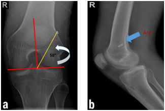
Figure 1: Correlation between the clock-face reference and the tunnel position in plain radiographs. (a) One o’clock position (high-position tunnel) in tunnel-view x-ray of the left knee. (b) The more anterior placement of the high-position femoral tunnel in comparison with low-position tunnel. (Note the endobutton insertion site.)

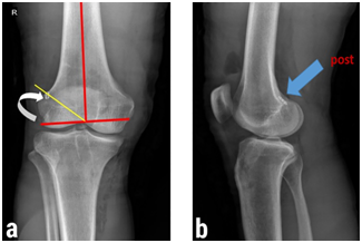
Figure 2: Correlation between the clock-face reference and the tunnel position in plain radiographs. (a) Ten o’clock position (low-position tunnel) in tunnel-view x-ray of the right knee. (b) The more posterior placement of the low-position femoral tunnel in comparison with high-position tunnel. (Note the endobutton insertion site.)
Similar to AP x-rays, in many of the cases, the tunnel was located between these values in tunnel-view x-rays. Thus the low-position and high position were considered between 30° to 45° and 45° to 60°, respectively.
Finally, the patients were classified into 4 groups included: low-anterior, low-posterior, high-anterior and high-posterior.
The time interval between surgery and last visit averaged 23.6 ± 2.2 months (18-30 mos.). Lysholm knee score [19], and Cincinnati score were completed for all of the patients. Furthermore, the Lachman, anterior drawer and pivot-shift tests were performed. Results of the Lachman and anterior drawer tests were considered positive if there was an anterior tibial translation >5mm comparing the normal knee. Pivot-shift test results were graded as follows: 0 (absent), grade I (gentle slide), grade II (definite subluxation), and grade III (subluxation and momentary locking) [20]. All tests were performed by an expert orthopedist who was not a part of the investigation team and was blind to the group assignment. Also the intraobserver reliability of the examiner, based on a pilot study, was 0.9.
In addition, anterior tibial translation was assessed using the KT-1000 knee arthrometer for both operated and normalknees [21-23]. The maximum score for Lysholm knee score was 100 points, while higher scores indicated the better outcomes.
The Cincinnati score was categorized in 4 groups: excellent (80-100 points), good (55-79), fair (30-54), and poor (fewer than 30).
Statistical analysis was performed using SPSS statistical software (version 15.0; SPSS, Chicago, IL). One-way ANOVA and post hoc tests were utilized to compare quantitative data. Besides for comparing qualitative data, the chi-square test was employed. P value < 0.05 was considered significant.
Of the 44 patients, 9 patients (20.4%) were classified as the low-anterior group, 17 patients (38.6%) as the low-posterior group and 18 patients (40.9%) as the high-posterior group. None of the patients were included in high-anterior group. Table 1 show that there was no significant difference between 3 groups in term of age and gender. The mean Lysholm score was significantly higher in the low-posterior group (p<0.001) (Table 2). However, the mean of Cincinnati score was the same in three groups. (Table 2) Of interest, only one patient in high posterior categorized as fair based on Cincinnati score, while all of the other patients were classified as good or excellent (Table 2). Additionally, anterior tibial translation did not differed significantly between the three groups (Table 3). Lachman test was negative in all of the patients. Anterior drawer test was negative in the low-posterior group. However, anterior drawer test was positive in 1 patient in the low-anterior (11.2%) and 1 patient in the high-posterior group (5.5%). Pivot shift test was graded IV in none of the patients (Table 3).
LA Group |
LP Group |
HP Group |
p value |
|
Age, yr |
25.7±4.2 |
28.4±4.4 |
27.5±8.3 |
0.411 |
Gender, n |
0.353 |
|||
Male |
7 |
16 |
14 |
|
-77.80% |
-94.10% |
-77.80% |
||
Female |
2 |
1 |
4 |
|
-22.20% |
-5.90% |
-22.20% |
Table 1: Age-Sex distribution of the patients.
LA Group |
LP Group |
HP Group |
p value |
||
n=9 |
n=17 |
n=18 |
|||
Mean Lysholm Score |
89±5 |
96±3 |
87±4 |
<0.001 |
|
(82-100) |
(88-100) |
(84-94) |
|||
Lysholm Score Grading |
Excellent |
7(77.8%) |
9(53%) |
14(77.8%) |
0.296 |
Good |
1(11.1%) |
8(47%) |
4(22.2%) |
||
Fair |
1(11.1%) |
0 |
0 |
||
Poor |
0 |
0 |
0 |
||
Mean Cincinnati Score |
87±4 |
91±7 |
90±8 |
0.859 |
|
Cincinnati Score Grading |
Excellent |
8(88.8%) |
16(94.2%) |
12(66.7%) |
0.275 |
Good |
1(11.2%) |
1(5.8%) |
5(27.7%) |
||
Fair |
0 |
0 |
1(5.6%) |
||
Poor |
0 |
0 |
0 |
||
Table 2: Comparison of Lysholm and Cincinnati scores in different tunnel positions.
LA Group |
LP Group |
HP Group |
p value |
||
n=9 |
n=17 |
n=18 |
|||
Lachman test |
Positive |
0 |
0 |
0 |
- |
Negative |
9(100%) |
17(100%) |
18(100%) |
||
Anterior drawer test |
Positive |
1(11.2%) |
0 |
1(5.5%) |
0.418 |
Negative |
8(88.8%) |
17(100%) |
17(94.5%) |
||
Pivot shift test |
I |
5(55.5%) |
8(47.5%) |
7(38.8%) |
0.939 |
II |
3(33.3%) |
7(41.2%) |
9(50%) |
||
III |
1(11.2%) |
2(11.8%) |
2(11.2%) |
||
IV |
0 |
0 |
0 |
||
Anterior tibial translation* (mm) |
2.2±0.4 |
2.5±0.7 |
2.4±0.5 |
0.444 |
|
Table 3: Comparison of clinical tests in different tunnel positions.
The main goal of the study was to see whether analteration in the femoral tunnel position in both axial and coronal planes could change the postoperative joint laxity include anterior, lateral or rotational instability in addition to functional outcomes of the patients.
As stated by literature it is possible that patients reconstructed with a higher tunnel positions have an increased laxity as a result of misplaced femoral graft that does not mimic the positioning of the intact or normal ACL. In contrast, graft placement can restore normal knee motion if we perform it in the anatomic fashion [24-28].
Our findings demonstrated a greater mean Lysholm score in the low-posterior group in term of functional outcomes comparing the other groups of the patients. However, we did not find a significant difference in the remaining clinical evaluations include Cincinnati score, Lachman test, pivot shift test and anterior drawer test. A study by Lee et al. [29] showed a lower Lysholm score and higher femoral tunnel positioning in the knees with positive pivot shift test than in the knees without pivot shift.
This is not in accordance with the results of Tsudaet al. [30] who found that the difference between the low- and high-positionsis not enough to convince them that different tunnel positions canbe associated with clinical and functional outcomes. Beside, Markolf reported on a method for the impact of linear regression slopes for the femoral tunnels on postoperative results. He concluded that the slope difference between the above-mentioned positions was not so significant as to be a reason for any advantage of the oblique (or low) tunnel over standard (or high) tunnel positions [31]. It seems that femoral tunnel obliquity may result in marked clinical outcomes if there be a great difference between tunnels linear slope.
Practically the other findings in our study are not clarifying in favor of which tunnel position is preferable. In this regard, the harvested data from anterior drawer and Lachman tests support that all the 3 tunnel positions are quite enough for stopping the anterior tibial translation. This maybe because of the ACLR surgery and its outcome, in which almost all anatomic reconstructed ACLs can control anterior tibial translation [31].
It has been common to place the femoral graft at 11o’clock position to recover the function of the AM bundle of the ACL [14,32].Once an ACL reconstructed knee is subjected to rotatory loads, the high-position tunnel for graft placement will not avoid rotational instability anymore [6,33,34]. Moreover, It has been revealed that the 10o’clock position resembles the PL bundle attachment and can be more sufficient at rotatory loads and limiting anterior tibial translation [35] which was confirmed by other biomechanical studies [14,36]. So it can answer this question that why the mean Lysholm score in the low-posterior group could be greater comparing two other groups and consequently justify the fact that how our remaining non-biomechanical evaluations have the same results.
We did not found a detectable difference in the pivot test between three groups. This test is the most widely used dynamic test, which correlates with instability symptoms [37]. However, our results could be due to the low sensitivity of the pivot shift test [38]. This is in accordance with the result of Jepsen et al. who found no difference between the high- and low-position tunnels regarding anterior laxity and pivot shift test [39].
There are a number of potential limitations that warrant consideration. The first is that in our view the postoperative radiologic assessment of tunnel positions can be somehow challenging with routine radiography, as this is 2-Dimensional illustration of a 3-Dimensional situation [40]. This is a reason why we should investigate tunnel positions in both axial and coronal planes concurrently. Furthermore, it is found that the tunnel-view radiograph is not satisfactory to assess the femoral tunnel placement, because there are variations in radiographic projection at the different phases of postoperative evaluation of the same patient [39]. The second limitation concerns that the mean follow-up time was about 2 years, and thus we cannot debate about the long-term surgical outcomes associated with clinical and radiologic developments. According to the literature Lysholm score was not sensitive to detect changes over time, and then it cannot be a precise scoring scale for long-term postoperative follow-up [41].
The final limitation was that like most related studies [39,42,43] we did not consider the tibial tunnel position which can be one of the important factors on clinical results.
The methodological pitfalls that we encountered with them include: first, the number of patients in this study should be more due to the study type. The second is the fact that this retrospective study investigates the tunnel positions and ACL grafts in patients with previous ACLR surgeries and consequently we had no role in their tunnel positioning.
Although the anatomic ACLR can sufficiently restore the knee stability and be associated with considerable functional improvement, the current study showed that low-posterior tunnel placement resulted in significantly higher knee scores. It is important to consider the femoral tunnel position in different planes and further investigations are required.
The authors report no conflict of interest concerning the materials or methods used in this study or the findings specified in this paper.

©2017 Kazemi, et al. This is an open access article distributed under the terms of the, which permits unrestricted use, distribution, and build upon your work non-commercially.