MOJ
eISSN: 2374-6939


Research Article Volume 15 Issue 6
1Department of Trauma & Emergency (Orthopaedics), AIIMS, India
2Department of Orthopaedics, SCB Medical college, India
3Department of Orthopaedics, S.L.N Medical College, India
Correspondence: Dr Sandeep Velagada, Department of Orthopedics, SLN Medical College Koraput, Odisha, India
Received: October 25, 2023 | Published: November 8, 2023
Citation: Behera S, Rana R, Velagada S, et al. Comparison between primary total elbow arthroplasty versus open reduction and internal fixation in elderly patients with distal humerus fractures: a prospective study. MOJ Orthop Rheumatol. 2023;15(6):204-208. DOI: 10.15406/mojor.2023.15.00646
Objective: It aims to assess the efficacy of these treatments, specifically in the presence of osteoporotic bone conditions. The findings of this study offer insights into the suitability of TEA as an alternative treatment option in such cases.
Methods: In this study, sixty patients with distal humerus fractures were divided into two groups. The first group received ORIF for fracture fixation, while the second group underwent primary TEA treatment. The study evaluated various outcomes, including elbow range of movement, elbow stability, and comparison of Mayo Elbow Performance Scores (MEPS) between the two groups at three- and twelve-month follow-up periods.
Results: In the three- and twelve-month follow-up periods, noticeable differences in elbow motion range were observed between the ORIF and TEA groups. The ORIF group experienced more significant restrictions in daily activities due to stiffness and pain, unlike the TEA group. However, both groups showed improvements in elbow function after one year. Furthermore, there were significant differences in the mean MEPS between the two groups at the three- and twelve-month follow-up period. However, no considerable differences were found between these periods within either group.
Conclusion: TEA is emerging as a promising option for elderly patients with comminuted distal humerus fractures, considering the osteoporotic bone condition. The results of this study hold substantial implications for the decision-making process in treatment selection.
Keywords: arthroplasty, replacement, elbow, fractures, comminuted, humeral fractures, osteoporotic fractures
The elbow is a complex joint consisting of three articulations. Around 7% of adult fractures are attributed to elbow injuries, with distal humerus fractures accounting for 33% of these cases. Thus, distal humerus fractures constitute roughly 2% of all adult fractures and 5% of fractures in older individuals with osteoporosis. This type of fracture exhibits a bimodal age distribution, affecting younger males (around 12-19 years) and elderly females above the age of 80.1–6 Studies indicate that the incidence of distal humerus fractures has increased twofold between1970 and 1995, and it is expected to triple by the year 2030.3 This rise can be attributed to two main factors: the aging population and extended life expectancy, which necessitate more surgical interventions and early functional rehabilitation. Additionally, osteoporosis is prevalent in this age group, either as a result of advanced age or other immunological conditions requiring steroid use, further contributing to the occurrence of these fractures.
Treating distal humerus fractures in the elderly presents numerous challenges compared to other types of elbow fractures, primarily due to the following factors: (1) communition difficulties, (2) the complex anatomy of the distal humerus, and (3) the presence of osteoporosis, limiting the available options for internal fixation. These fractures often result in symptoms such as stiffness, pain, and weakness. Thus, it is of utmost importance to attain a painless, stable, and mobile elbow joint in order to achieve optimal functional outcomes.1,2
In 1913, Albin Lambotte introduced the concept of osteosynthesis, which advocated for the significance of anatomical reduction and early functional recovery, claiming comparable results to conservative treatment. However, during that era, the high risk of infection and hardware failure created state of confusion and uncertainty between traditional and surgical management approaches. Subsequent studies by Evans7 demonstrated that reasonable reduction under anesthesia and immobilization yields satisfactory results, indicating that perfect reduction may not always be necessary for successful treatment. In the past, Riseborough and Radin,8 Brown and Morgan,9 and others favored non-surgical management for distal humerus fractures. However, the trend has shifted towards surgical management in the last quarter of the century due to improved outcomes associated with operative interventions. The AO-Association for the Study of Internal Fixation (AO-ASIF) group promoted anatomical reduction of the articular surface and rigid fixation in all cases. In recent decades, not able advancements have been made in fixation devices, surgical approaches, and rehabilitation protocols, which have shown favorable outcomes, particularly in young adults with good bone quality. Nevertheless, these aforementioned approaches may not consistently yield optimal results in the elderly population, mainly due to poor bone stock and comminution (fragmentation).3,10–12
In this regard, total elbow arthroplasty (TEA) has emerged as a viable treatment option for addressing complex osteoporotic fractures in elderly patients. In cases where prolonged immobilization is required due to non-rigid fixations, functional outcomes are often unsatisfactory, leading to increased dependency and the potential need for additional surgeries. However, TEA has demonstrated promising effects in treating comminuted osteoporotic distal humerus fractures among elderly individuals.
It is acknowledged that the majority of studies on this subject are retrospective, and there is a scarcity of literature specifically focusing on the Indian subcontinent. In this study, we aim to compare the outcomes of total elbow arthroplasty with standard internal fixation in elderly patients who experienced comminuted distal humerus fractures. For our study, we included patients aged 50 years and above, considering that physiological age often exceeds the actual age in developing countries like India.1,13 Through a prospective study, we aspire to provide valuable insights into the existing body of literature and assist in decision-making regarding the treatment of distal humerus fractures in the elderly population.
This prospective longitudinal study was conducted at our institution, which is a prominent tertiary institute, spanning from December 2013 to October 2020. The primary objective is to compare the outcomes of TEA with open reduction and internal fixation (ORIF) in the treatment of comminuted distal humerus fractures in elderly patients. A total of sixty patients participated in the study, with thirty patients assigned to each treatment group which were operated by the same surgeon. The patients for this study were carefully selected randomly based on specific inclusion criteria. These criteria encompassed individuals with isolated fresh traumatic close comminuted distal humerus fractures with articular involvement and were aged over 50 years. Moreover, patients with comminuted fractures with joint dislocation were also included. However, certain patients with open fractures, previous infections, severe bone loss, extensor system disruptions, or severe medical co-morbidities were excluded from the study.
In the TEA group, the surgical technique outlined by Baksi et al. was followed, which involved performing the procedure under either general or regional anesthesia.14,15 A posterior approach was utilized to create full-thickness flaps on both the medial and lateral sides. Further, the ulnar nerve was identified and isolated, and the medial and lateral epicondyles were exposed. Then, comminuted bone fragments were removed, and the distal humerus and proximal ulna were incised. During the surgical procedure, special attention was given to preserving the triceps and brachialis insertions on the proximal ulna. The canals of the humerus and ulna were prepared for stem insertion. Additionally, longitudinal grooves were made on the distal humerus to accommodate the flanges of the humeral stem. Following the trial fitting, cement was applied, and the 3rd generaration Bakshi’s sloppy hinge TEA prosthesis was inserted, ensuring immediate stability. The triceps tendon insertion was repaired to the olecranon, no anterior transposition of ulnar nerve, and the wound was subsequently closed. To facilitate healing, the elbow joint was immobilized using a plaster splint, and postoperative rehabilitation was initiated gradually.
In the ORIF group, the patients underwent surgery in a lateral decubitus position. A chevron osteotomy of the olecranon was performed to gain access to the intra-articular portion of the joint. The articular surface was then carefully reduced and secured using provisional K-wires and a cancellous lag screw. The reduced segment was then fixed to the medial and lateral columns of the shaft using K-wires and plates (Figure 1). Throughout the procedure, care was taken to preserve the ulnar nerve, and in 7 cases, anterior transposition was performed as deemed necessary. After closing the incision, a compression bandage was applied around the elbow, which was splinted in a 90-degree flexed position. During the postoperative period, the patients were immobilized in an arm sling, and range of motion exercises were initiated at a later stage.

Figure 1 Clinico-radiological presentation of a patient from the open reduction and internal fixation group A: Pre-operative radiograph B; Intra-operative radiograph showing the perpendicular arrangement of plates C: Post-operative radiograph.
A minimum follow-up period of one year was implemented, involving comprehensive reviews at intervals of 2 weeks, 6 weeks, 3 months, 6 months, and 9 months for all patients. During these follow-up sessions, the functional outcomes of the patients were evaluated using the Mayo Elbow Performance Score (MEPS) at both the 3-month and 1-year marks. Additional parameters, such as range of motion (Figure 2), pain levels, local temperature, and ability to perform routine activities, were also evaluated as part of the assessment process. Radiographic assessments were also performed to examine the positioning of the components and identify any signs of loosening. The data obtained from the study were subjected to statistical analysis using SPSS software and Graph Pad Prism version. Numerical variables were summarized as mean and standard deviation, while categorical variables were presented as counts and percentages. For non-normally distributed numerical variables, the median and interquartile range were provided. Student's independent samples t-test was used to compare normally distributed numerical variables between groups, and unpaired proportions were compared using the Chi-square test or Fisher's exact test, as appropriate. The test statistics and degrees of freedom were determined for each t-test, and p-values were obtained to assess statistical significance. The study was carried out in accordance with the World Medical Association's Declaration of Helsinki on Ethical Principles for Medical Research Involving Human Subjects, and was reviewed by our institution's Institutional Review. Written informed consent from patient was taken to publish the clinical and radiological photograph without disclosing the name and identity.
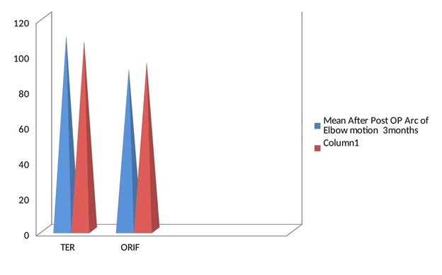
The study conducted at the Orthopedics Department, from December 2013 to October 2020, aimed to compare the outcomes of two treatment approaches for distal humerus fractures in elderly patients. The study involved 60 patients, divided into two groups: one group underwent open reduction and internal fixation (ORIF), while the other group received primary total elbow arthroplasty (TEA). All patients were followed for at least 12 months post-operation. The average age of the study population was 61.37±6.2 years, with a majority of female patients (60%). There were no statistically significant differences in age distribution (p=0.9347) or co-morbidities (p=0.7861) between the two groups, indicating age-matched cases and controls were selected for the study. The dominant hand and the affected elbow also did not show a significant association in both the TER and ORIF groups (p=1.0000). The mean interval between injury and operation was not significantly different between the two groups (p=0.2748), suggesting that the timing of the operation did not influence the choice of treatment (Table 1). Triceps weakness was generally minimal in both groups, with no statistically significant difference observed (p=0.5428). The TEA group demonstrated a significantly better mean postoperative arc of elbow motion at three months and twelve months follow-up compared to the ORIF group (p=0.0137) (Figure 3). In terms of complications, the TEA group had no major complications, while the ORIF group experienced a few complications, all of which were successfully resolved with appropriate treatment (Table 2). The functional outcomes were significantly better in the TEA group, with the majority of patients achieving excellent results at twelve months (Figure 4). The ORIF group also showed positive functional outcomes, with the majority achieving good results (Figure 5).

Figure 3 Comparision between the groups of Mean Post-Operative Arc of Elbow motion after 3 and 12 months follow-up.

Figure 4 Comparision between the groups of Mayo Elbow Performance Score (MEPS) after 3 and 12 months follow-up.
|
Co morbidity |
TER |
ORIF |
TOTAL |
|
DM |
4 |
4 |
8 |
|
DM +HTN |
2 |
0 |
2 |
|
HTN |
4 |
8 |
12 |
|
HTN+OSTEOPOROSIS |
2 |
2 |
4 |
|
No |
18 |
16 |
34 |
|
TOTAL |
30 |
30 |
60 |
Table 1 Distribution of dominant in two groups
p=1.0000, Statistically not significant.
|
Complication |
TER |
ORIF |
TOTAL |
|
Fixation |
0 |
22 |
2 |
|
Hematoma |
4 |
2 |
6 |
|
Infection |
2 |
2 |
4 |
|
No |
22 |
20 |
42 |
|
Ulnar Neuropraxia |
2 |
4 |
6 |
|
TOTAL |
30 |
30 |
60 |
Table 2 Distribution of complication in two groups
Our study evaluated the outcomes of 60 patients who had distal humerus fractures and underwent either TEA or ORIF. The average age of the patients was 61.37±6.2 years, with 60%female. It is worth mentioning that the generation and gender distribution in our study were consistent with the findings of previous studies.16,17 Moreover, the overall complication rates, such as infection and ulnar neuropraxia, were comparable to those reported in earlier studies; however, we did not observe any loosening or heterotopic calcification in this study.18–20
The studies conducted by Gambirasio et al.21 and Garcia et al.22 have also reported good functional outcomes and low complication rates in patients treated with TEA. Similarly, our study aligns with these results, as we observed two cases of triceps weakness and four cases of pain caused by hematoma, all of which resolved over time. Meanwhile, the functional activity was more restricted in the ORIF group compared to the TEA group, primarily due to stiffness. Nevertheless, it is worth noting that all patients in this group had stable joints with normal supination and pronation movements. Notably, the ROM was slightly limited in some cases compared to previous studies, possibly ascribed to the late initiation of physiotherapy in patients with osteoporosis, especially in the ORIF group. Triceps weakness was observed in two cases in the TEA group and four cases in the ORIF group, which agrees with the findings of previous studies.
Furthermore, the TEA group had a MEPS of 97, with 28 out of 30 achieving excellent results, whereas the remaining two reached good results. The ORIF group showed a mean MEPS of 91, with 22 patients achieving outstanding results, six patients attaining good results, and two achieving fair results. Interestingly, there were no poor results in either group.
These findings were comparable to earlier studies by Gambirasio et al.,21 Gracia et al.,22 Frankle et al.17 and Kamineni et al.,23 which showed good to excellent outcomes and high patient satisfaction in patients treated with TEA. Moreover, in a randomized control trial conducted by McKee et al.,24 better results were obtained in the TEA group compared to the ORIF group. Additionally, two systematic reviews have indicated comparable outcomes between these two groups.25,26 However, it is relevant to point out that our study had limitations, including a short-term follow-up period and a relatively small sample size. Thus, further randomized control trials with larger sample sizes and more extended follow-up periods are warranted to establish more robust evidence.
It is important to note that our study mainly focused on non-rheumatoid patients, which may have influenced the observed outcomes. Other studies have indicated that TEA may yield better results than ORIF in MEPS but with higher complication rates.17,27–30 Surprisingly, our study did not observe such higher complication rates, which could be attributed to excluding rheumatoid patients from our cohort.
Our study suggested that TEA and ORIF can lead to satisfactory outcomes in treating distal humerus fractures. However, TEA showed slightly superior functional effects and higher MEPS scores than ORIF. Future research should address the limitations of our study and delve into the long-term consequences of these approaches, encompassing the utilization of validated assessment tools, such as the Disabilities of the Arm, Shoulder, and Hand (DASH) score, for better comparability.
Managing distal humerus fractures in elderly patients presents unique challenges due to comminution and osteoporosis. Careful consideration is necessary regarding fixation failure and limitations on heavy weight lifting when considering TEA as a treatment option for elderly patients. It is crucial to provide thorough counseling to elderly patients and their families, discussing the advantages and potential consequences of both arthroplasty and fixation approaches. TEA may be a suitable treatment option for elderly patients with distal humerus comminuted fractures.
None.
There was no financial support from public, commercial, or non-profit sources.
The authors have no conflict of interests to declare.

©2023 Behera, et al. This is an open access article distributed under the terms of the, which permits unrestricted use, distribution, and build upon your work non-commercially.