MOJ
eISSN: 2374-6939


Clinical Report Volume 18 Issue 1
 AM Shayan R Bari,1 Shahidul Islam,1 Tahsin Ferdous,2 Safwanul Aman3
1Department of Orthopaedic and Traumatology, Bari-Ilizarov Orthopaedic Centre, Bangladesh
2Department of Orthopaedic and Traumatology, Bangladesh Institute of Health Science (BIHS), Bangladesh
3Department of Orthopaedic and Traumatology, Omega Hospital, Bangladesh
Correspondence: Md. Mofakhkharul Bari, Department of Orthopaedic and Traumatology, Bari-Ilizarov Orthopaedic Centre, Bangladesh
Received: December 30, 2025 | Published: January 9, 2026
Citation: Bari M, Bari AMSR, Islam S, et al. Clinical case report: bimodal salvage of a distal femoral periprosthetic fracture and subsequent biofilm infection in a high-bmi geriatric patient. MOJ Orthop Rheumatol. 2026;18(1):4-7. DOI: 10.15406/mojor.2026.18.00731
periprosthetic fracture, TKR, ilizarov, DAIR, CLAP
There is an inherent complexity in the clinical management of periprosthetic distal femur fractures (PDFF) in the elderly population due to bone quality and the presence of prosthetic devices. In the presence of significant excess weight in the patient, there is an added complexity of tissue restraints and high mechanical stress loads attributed to destabilization devices.1,2 The following report outlines the successful binary limb-salvage strategy for the patient who suffered a Periprosthetic supracondylar fracture in October 2024 and subsequent periprosthetic joint infection in November 2025. The report also illustrates how high tension circular fixations and CLAP were utilized effectively for preservation of the original prosthesis in 2022.3,4
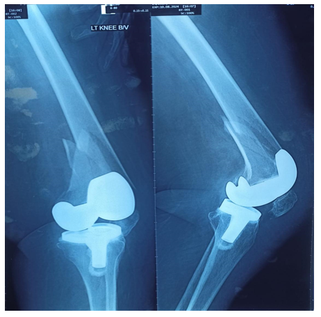
Figure 1 Periprosthetic supracondylar fracture of the femur without dislodgement of prosthesis .
|
Date |
Clinical milestone |
Intervention strategy |
|
2022 |
Primary TKA |
Management of severe osteoarthrosis |
|
Oct 2024 |
Periprosthetic Fracture |
Ilizarov apparatus Fixation |
|
Oct 2024 - Mar 2025 |
Osteosynthesis |
5-month consolidation period |
|
March 2025 |
Fixator Removal |
21-day posterior back slab stabilization |
|
November 2025 |
PJI Presentation |
DAIR with 10-day CLAP therapy |
|
Late 2025 (Dec last week) |
Follow-up |
Complete resolution; TKA component retained |
Table 1 Clinical progress and applied procedure
Classification
In October 2024, the patient presented with a displaced supracondylar fracture proximal to her stable TKA. The injury was classified as:
|
Classification System |
Grade/Type |
Key diagnostic findings |
|
Lewis & Rorabeck |
Type II |
Displaced fracture; Stable femoral component. |
|
Su Classification |
Type II |
Fracture starts at the proximal femoral flange and moves upward. |
|
Kim Classification |
Type IB |
Stable prosthesis; fracture is irreducible (cannot be set manually). |
Table 2 Supracondylar Fracture (Proximal to Stable TKA) classified as below.5
Ilizarov Osteosynthesis in Obese patient
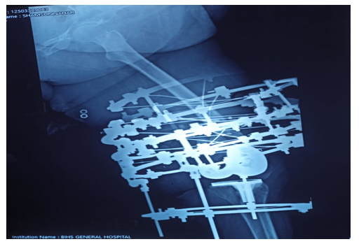
Figure 2 Periprosthetic supracondylar fracture was aligned and fixed with Ilizarov apparatus .
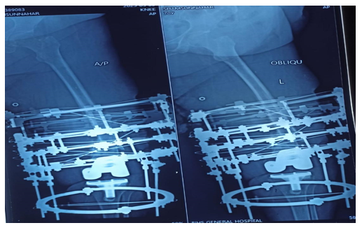
Figure 3 Multiple olive wires inserted to fixing the Periprosthetic fracture .
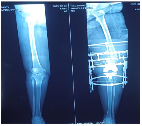
Figure 4 On X-ray stress view no limb length discrepancy .
|
Factor |
Mechanical Risk to Consequence |
Risk to Hardware |
|
Ring Size |
Decreased construct stiffness |
Increased sagging and non-linear displacement |
|
Axial Loading |
Dominance of "cable" behavior |
Yield failure of the wire under heavy mass |
|
Cyclic Loading |
Repetitive stress at bone-stopper interface |
Metal fatigue leading to sudden wire breakage |
The circular fixator was chosen for the purposes of multi-planar stability as well as early mobilization. Nevertheless, the patient’s BMI caused "difficulty in application." The rings had to be large in size (220-240 mm) in order for there to be a 2 cm skin margin, which inherently decreases the axial rigidity.1,6,7
To mitigate these effects, Russian bolts were used for tensioning purposes. The Russian bolts enable manual tensioning of high torque (1600N to 1695N), adequate to provide stiffness to prevent wire fatigue when the patient walks.1,2,8
Justification of the 21-Day Back Slab
After five months of union, the frame was removed in March 2025. A posterior fixation with a back slab was then used for 21 days to offer temporary stabilization. This is an essential step in the fixation of the pin tracks because the cavities created by the 1.8 mm wires are known as “stress risers” of the bone, which tend to increase the chances of secondary fractures during the process of weight-bearing stress.1,6,8
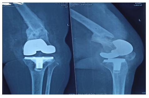
Figure 5 After demounting of the Ilizarov apparatus the Periprosthetic fracture was healed by callus formation.
In November 2025, the patient was in the late stages of PJI. The treatment was DAIR therapy supplemented by CLAP therapy.9,10
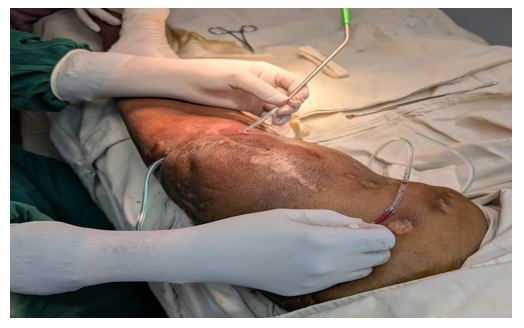
Figure 6 The CLAP therapy was applied to the Prosthetic Joint infection.
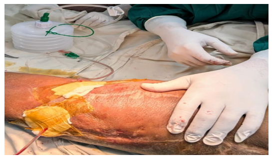
Figure 7 The negative pressure drain was used as outflow tract in CLAP therapy .
CLAP Therapy Dose Calculation
The protocol was developed in order to keep a bactericidal dose of an antibiotic in the biofilm site itself,3,4,10
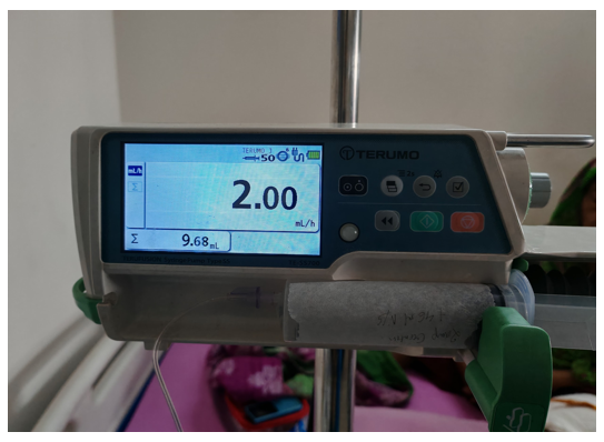
Figure 8 Syringe pump is used for accurate dose calculation in inflow tract of CLAP therapy .
Carrier Solution: Gentamicin 160mg (2×80mg ampules) + 46ml 0.9 % Normal Saline.
Localised Concentration: 160 mg in 50 ml = 3.2 mg/ml = 3200 µg/ml
Perfusion rate: 2 ml/hr via intra-joint dual-lumen tubes
Daily Dose Rule/Formula: 2 mL/hr × 24 hr × 3.2 mg/mL = 153.6 mg/day
Progress and monitoring
The 10-day CLAP course showed rapid biological response:
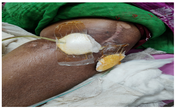
Figure 9 Swelling and inflammation subsided after application of CLAP therapy.
Inflammatory Trends: Serum C-reactive protein (CRP) reached a 'fast response pattern,' lowering to a ratio of < 0.4 of its original value by day 4.
Clinical Findings: The erythema, warmth, and effusion resolved completely by the end of the 10-day perfusion phase.3,4
Safety Profile: Despite the fact that the patient was elderly with significant weight, the serum gentamicin trough levels were kept below values of less than 2.0 μg/mL.10
The success of this case emphasizes the balance of mechanical and biological salvaging techniques that must be used on the high-risk patient.
In Phase 1, The Ilizarov technique was chosen due to the fact that traditional internal plating has a complication rate as high as 35% in obese, osteoporotic populations, often secondary to hardware "cut-out". A circular frame allowed axial load distribution over multiple points, providing controlled micromotion to allow for secondary bone healing.1,2,8
In Phase 2, CLAP therapy overcame the basic inadequacies of systemic use of antibiotics. In clearing mature biofilms from implant surfaces, concentrations 100 to 1000 times the MIC are needed-levels that would be systemically toxic if given intravenously. Locally infusing 153.6 mg/day, the investigators maintained an intramedullary gradient of ≥ 500 μg/ml. In addition, Continuous Passive Motion (CPM) was incorporated during the 10-day perfusion period to allow fluid to circulate throughout the recesses of the joint to ensure complete coverage of the antibiotic on all prosthetic surfaces and to prevent arthrofibrosis.3,9,10
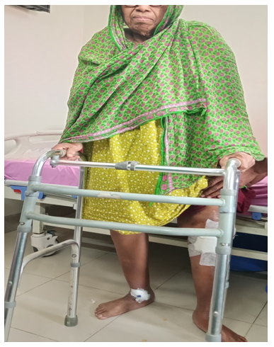
Figure 10 On Follow up patient is able to stand by the support of Walker and there is no limb length discrepancy.
The patient has since made a full recovery, with the primary 2022 TKA functional. This represents that morbid obesity and advanced age are not absolute contraindications to successful limb salvage in complicated periprosthetic cases.1,2,10 Such a resolution was rested on three pillars:
High tension hardware constructs
(> 1600N) to avoid component failure within a high-BMI setting.
Transitional immobilization to protect the bone during consolidation of pin-site stress risers.
Local pharmacological perfusion allows for the safe use of bactericidal concentrations not attainable by systemic methods.
None.
The authors declare that there are no conflicts of interest.

©2026 Bari, et al. This is an open access article distributed under the terms of the, which permits unrestricted use, distribution, and build upon your work non-commercially.
