MOJ
eISSN: 2374-6939


Letter to Editor Volume 17 Issue 5

1Dragonfly Foundation for Research & Development, USA
2PHD Regeneration Technologies, USA
3Division of Basic Medical Sciences, Mercer University School of Medicine, USA
4Department of Surgery, Mercer University School of Medicine, USA
5Department of Pediatrics, Mercer University School of Medicine, USA
6 Department of Obstetrics and Gynecology, Mercer University School of Medicine, USA
7Department of Anesthesiology, Mercer University School of Medicine, USA
Correspondence: Henry E Young, Dragonfly Foundation for Research & Development, Macon, GA 31210 USA
Received: August 18, 2025 | Published: September 9, 2025
Citation: Young HE. Choice of endogenous cells that should be utilized for regenerative medicine. MOJ Orthop Rheumatol. 2025;17(5):77-78. DOI: 10.15406/mojor.2025.17.00713
Clinical studies utilizing mesenchymal stem cells (MSCs) for regenerative medicine have shown that while it is 100% safe to transplant autologous MSCS, the efficacy for restoring neuronal tissue or pancreatic islet tissue has been abysmal. It is inconceivable that a cell that is destined to only form fat, cartilage, and bone, would be chosen for restoring cell types from other lineages, such as ectodermal germ layer lineage nervous tissue and endodermal germ layer lineage pancreatic tissue that are outside the MSCs mesodermal germ layer lineage. One wonders if there is an understanding of normal development for the derivation of these cell types. Included is a lineage map of unidirectional embryonic development from the zygote to differentiated cell types for those attempting to treat individuals in the realm of regenerative medicine.
Keywords: regenerative medicine, stem cells, lineage stem cells, mesenchymal stem cells
Normal embryogenesis
Knowledge of normal embryogenesis and subsequent differentiation potential1 is paramount for anyone attempting to use any type of stem/progenitor cell for regenerative medicine, especially for individuals with neurological, cardiovascular, pulmonary, autoimmune, hepatic, pancreatic, and/or renal chronic diseases, terminal diseases, acute and chronic trauma, and/or those with severe burns.2 Figure 1 is a Lineage Map detailing the pattern of normal developmental embryogenesis from zygote to adult differentiated cell types.3 In the flow diagram, differentiation from a primitive cell type (zygote) to a mature (differentiated) cell type is in a unidirectional direction, from the least primitive cell type to the most differentiated cell type. Dedifferentiation of adult cells followed by their transdifferentiation does not normally occur (# Academia.edu). Rather, it only occurs artificially when embryonic genes, such as the Yamanaka factors (e.g., Oct3/4, Sox2, Klf-4, and c-Myc) are inserted into adult differentiated cells4 to form some variation of induced Pluripotent Stem Cells (iPSCs).5
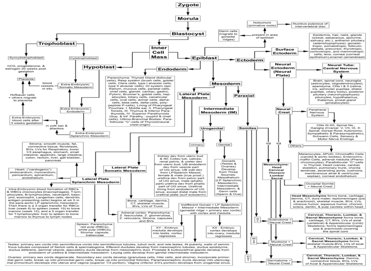
Figure 1 Lineage Map of unidirectional cell differentiation pathway for cells derived from the embryonic zygote. The totipotent zygote divides into two cells (blastomeres), which continue to divide to form a solid of ball of cells, termed morula. With further differentiation, a hollow ball of cells forms, termed the blastocyst. The blastocyst divides into the trophoblast (future syncytiotrophoblast and cytotrophoblast) and inner cell mass (future embryo). The inner cell mass forms the epiblast. Future notochord cells and germ cells split off as individual epiblast cells and continue their development independent of the remaining epiblast. A knot (primitive knot/node) of notochord cells induces the epiblast to divide into three layers: the ectoderm, mesoderm, and endoderm. The ectoderm further differentiates into surface ectoderm, neural ectoderm, and neural crest. The mesoderm further differentiates into the paraxial mesoderm, intermediate mesoderm, and lateral plate mesoderm. The paraxial mesoderm differentiates into somitic mesoderm, which further differentiates into vertebral column and annulus fibrosis of the intervertebral disc. The notochord, an independent entity, forms the nucleus pulposus of the intervertebral disc. The intermediate mesoderm differentiates into the genitourinary system, which further differentiates into the stroma of the genital organs, ovary and testis, and urinary organs. The germ cells (oogonia and spermatogonia), independent separate entities, migrate into the stroma of their respective organs along the lateral plate splanchnic mesoderm. The lateral plate mesoderm differentiates into somatic mesoderm and lateral plate splanchnic mesoderm, and further differentiates into progenitor cells and then into differentiated cell types. The endoderm differentiates into progenitor cells and the adult parenchymal cell types (1,2). Reprinted with permission from Adult Stem Cells, by H.E. Young and A.C. Black, Jr., Fig. 1, Anatomical Record Part A 2004; 267A:75-102. Wiley-Liss, Inc.
Therefore, if one wants to repair and/or restore neuronal tissue one does not choose a cell type within the mesodermal germ layer lineage that will only differentiate into fat, cartilage, and bone (e.g., mesenchymal stem cell).6 But rather one chooses a cell type that will differentiate into ectodermal germ layer lineage neurons and glial cells, such as neural stem cells, or a less differentiated cell, such as an ectodermal stem cell.7 Similarly, if one wanted to repair and/or restore pancreatic islets one does not choose a cell type within the mesodermal germ layer lineage that will only differentiate into fat, cartilage, and bone (e.g., mesenchymal stem cell).6 But rather one chooses a cell type that will form endodermal germ layer lineage pancreatic islet cells, such as endodermal stem cells.7
It is of concern that stem cell clinics in USA and abroad are using MSCs for a myriad of treatments that are outside the differentiation potential of MSCs. For example, a single cell clone of MSCs were derived by repetitive single cell clonogenic analysis. The clone was then examined with human recombinant proteins, morphogenetic proteins, and exosomes derived from differentiated cells. The ONLY cell types the MSCs would form were fat, cartilage, and bone, as discussed previous,6 but more specifically would only form unilocular white adipose tissue (fat), hyaline cartilage (cartilage), and intramembranous bone (bone).7 MSCs did NOT form nervous tissue, nor did they form pancreatic islet tissue.7
One would think that those performing transplants in regenerative medicine would at least be aware of the differentiation potential of the cells they were using. Assuming that is not the case, I have attached a condensed lineage map of embryonic development from the zygote to differentiated cell types (circa, 2004),3 that will hopefully be a reference point for using endogenous naturally-occurring cells for future regenerative medicine endeavors.
None.
The author declares that there are no conflicts of interest.

©2025 Young. This is an open access article distributed under the terms of the, which permits unrestricted use, distribution, and build upon your work non-commercially.
