MOJ
eISSN: 2374-6939


Case Report Volume 4 Issue 5
1Department of Orthopaedic Surgery & Trauma, Ondo State Trauma & Surgical Centre, Nigeria
2Department of Medicine, Kidney Care Centre, Nigeria
3Department of Radiology, Ondo State Trauma & Surgical Centre, Nigeria
4Department of Plastic, Burns & Reconstructive Surgery, Ondo State trauma & Surgical Centre, Nigeria
Correspondence: Adetunji M Toluse, Department of Orthopaedic Surgery & Trauma, National Orthopaedic Hospital, PMB 2009, Igbobi, Yaba Lagos, Nigeria
Received: February 19, 2016 | Published: April 19, 2016
Citation: Toluse AM, Akinbodewa AA, Ogunsemoyin AO, Ezeah I (2016) Cervical Spinal Cord Injury following High-Voltage Electrocution: A Case Report. MOJ Orthop Rheumatol 4(5): 00154. DOI: 10.15406/mojor.2016.04.00154
Aims and Objectives: This case report is aimed at highlighting cervical spinal cord injury with tetraplegia following electrocution which is an uncommon aetiology of spinal injury and to the best of our knowledge, has not been reported in our environment.
Patient and Method: We present a case report of a 47 year old male staff of an electricity company who presented with transient loss of consciousness and tetraplegia following electrocution and fall from height, while at work.
Result: The patient made marginal neurologic improvement on non-operative care with intensive care support and multidisciplinary management.
Conclusion: High voltage electrocution is an uncommon cause of spinal cord injury with potential for immediate, delayed and long term neurologic problems. Multidisciplinary management and long term follow-up is required. Occupational safe practices should be emphasized among electricity workers.
Keywords: Cervical spinal cord injury, Tetraplegia, Electrocution, Occupational safety
The cervical spine is prone to injury and is involved in about one third of all spinal injuries.1 Traumatic tetraplegia is a severe disabling condition with long lasting impact on victim’s quality of life, life expectancy and it also places a huge burden on the family and society with attendant long term dependence on healthcare personnel and resources.2
High voltage electrical injuries are uncommon and may result in mortality or have debilitating neurologic sequelae3. Children and young men are the common victims. Most admissions of adults on account of electrical injury are occupationally related. Almost two thirds of the fatalities occur in people between the ages of 15 and 40 years.3-4 The incidence of spinal cord injury following electrical trauma ranges between 2% and 5%.Electrical injury may produce an immediate or delayed myelopathy. Immediate injury typically produces decreased levels of consciousness, paresthesia, and weakness. Significant or complete recovery is frequently observed. Delayed spinal cord injury is usually incomplete and progressive, and improvement is less common.5
A 47 year old male electricity company staff was electrocuted while working on an 11KVA electric pole, about six meters from the ground and fell to the ground. He sustained partial thickness burn wounds to the forearms and occipital scalp laceration. There was transient loss of consciousness lasting for two hours and inability to move all four limbs. He was initially taken to a private hospital by co-workers, where he received initial resuscitation prior to referral to our centre for expert management. At presentation at our facility, he was fully conscious, had total amnesia about the electrocution, with partial thickness burns in both hands and forearms and sutured 4cm laceration in the occipital region. The heart rate during the first 3-5 days ranged between 44 and 60bpm. The earliest electrocardiogram revealed bradycardia, RSR pattern in the QRS complex, but normal ST-T segment waveform (Figure 1). The respiratory and abdominal systems examination findings were normal. He had intact light touch sensation in all dermatomes, however muscle power was zero globally, tone and deep tendon reflexes were diminished and plantar responses were equivocal.
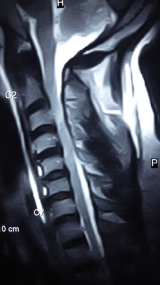
Investigations revealed normal haemogram and serum electrolytes. Plain radiograph of the skull was normal, while spinal radiographs revealed loss of normal cervical lordosis. No fracture or dislocation was seen. Spinal magnetic resonance imaging (MRI) showed generalized loss of disc signal intensities and posterior bulges. Marked canal stenosis at C3 vertebral level. There is also mildly increased parenchymal spinal cord intensity at this level and focal blooming artifacts (Figure 2). Features of hemorrhagic contusion at C3/C4 disc segment in a background Grade III degenerative disc disease and canal stenosis.

Figure 2 Spinal magnetic resonance imaging (MRI) showes generalized loss of disc signal intensities and posterior bulges.
He was commenced on fluid resuscitation, nutritional support, physiotherapy, wound care and pain control. Five days post admission, he was observed to be desaturating, hypotensive and bradycardic. Electrocardiogram done showed persistent right bundle branch block. He was transferred to the intensive care unit where mechanical ventilation was commenced with ionotropic support. He had elective tracheostomy a week after intubation. He subsequently made progressive improvement and mechanical ventilation was discontinued after seventeen days. He regained ability to shrug his shoulders and was mobilized on wheel chair. He was discharged home five weeks post-admission to continue rehabilitation as outpatient and for repeat cervical spine MRI scan and possible posterior decompression and fusion surgery.
Electrical injuries can occur in low-voltage settings, such as with household use, and high-voltage exposures from occupational hazards and lightning strikes.4 Several pathophysiological mechanisms of injury to the nervous system have been proposed, including thermal injury, electroporation, and vascular damage through direct injury as well as indirect injury.3,6 The index patient was a victim of occupational high voltage electrical injury with associated fall from height. The fall from height could also account for cervical spinal cord injury, however, plain radiograph and MRI showed no feature of bony or ligamentous injury. Hence, we inferred that the neurologic deficit is a result of the electrical injury.
The patient showed a combination of features of immediate and delayed onset spinal cord injury. This is similar to report by Johl et al.3 Several other case reports have also documented similar findings.3s-7 Cervical myelopathy with late-onset progressive motor neuron disease following electrical injury has also been described by Ghosh et al.8 In their report, the patient was examined 12 years after the injury, at which time MRI demonstrated cervical cord atrophy. No signal intensity abnormalities were described. Arevalo et al.4 reported two cases of neurologic symptoms immediately following electrical injury in which CT and MR imaging were both normal. However, the MRI done in our patient revealed signal intensity abnormalities.
While electrical injury is commonly associated with fatal cardiac standstill and ventricular fibrillation,9 it has also been associated with other transient cardiac changes with better prognosis. These include bundle branch block, sinus bradycardia, atrial tachycardia, ventricular ectopics and ventricular tachycardia.10 In our index case, evidence of right bundle branch block (bradycardia, Rsr wave pattern) was seen on electrocardiogram (Figure 3).
High voltage electrocution is an uncommon cause of spinal cord injury with potential for immediate, delayed and long term neurologic problems. Multidisciplinary management and long term follow-up is required. Occupational safe practices be emphasized among electricity workers.
None.
None.

©2016 Toluse, et al. This is an open access article distributed under the terms of the, which permits unrestricted use, distribution, and build upon your work non-commercially.