MOJ
eISSN: 2374-6939


Review Article Volume 17 Issue 5

Consultant Orthopaedic Surgeon, Heart Beat Medical Centre & One Day Surgery and Medeor Hospital UAE
Correspondence: Dr. Safaaeldin Abaza, FRCS (Glasgow), Consultant Orthopaedic Surgeon, Heart Beat Medical Centre & One Day Surgery and Medeor Hospital, Abu Dhabi, UAE, Tel +971 55 972 7644
Received: August 20, 2025 | Published: September 9, 2025
Citation: Abaza S. Biologic phenotyping of knee osteoarthritis using doppler ultrasound: a pragmatic algorithm for synovitis, ITB Syndrome, and SCIF. MOJ Orthop Rheumatol. 2025;17(5):80-85. DOI: 10.15406/mojor.2025.17.00714
Background: Knee osteoarthritis (KOA) pain is often misattributed to cartilage structural degeneration. Yet cartilage is aneural, avascular, and alymphatic-incapable of generating pain. Doppler ultrasound enables biologic phenotyping, distinguishing synovitis, iliotibial band syndrome (ITBS), and subchondral insufficiency fracture (SCIF). “KOA is not a picture-it’s a phenotype.” 1
Objective: To classify KOA pain into Wet, Dry, and Equivocal phenotypes using Doppler ultrasound and guide targeted interventions based on biologic activity rather than structural findings.2,3
Methods: Over a decade of Doppler-guided assessments in a single-surgeon orthopedic clinic, patients were phenotyped based on vascular signal and fascial strain. Interventions were tailored to biologic activity.4
Results:Wet KOA shows Doppler-positive synovitis and responds to anti-inflammatory strategies. SCIF presents as acute pain with marrow edema and cytokine activation. Emerging biologics (IL-6, TNF-α, GM-CSF inhibitors) and Arthrosamid® hydrogel support the logic of targeting synovitis directly. Even if unavailable locally, they validate the Doppler framework.5–7
Dry KOA, dominated by ITBS and GTPS, mimics sciatica and is frequently misdiagnosed. MRI often misses fascial strain and early synovitis, leading to unnecessary spinal interventions.8–10
“Dry knees hurt-but not from the joint.”
New Insight: Dry KOA phenotypes frequently present with coexisting ITBS and GTPS, both arising from strain along the iliotibial band and its fascial continuum. GTPS, once considered bursitis, is now recognized as gluteal tendinopathy-primarily involving the gluteus medius and minimus, compressed by ITB tension. (Radiopaedia.org, 2025; European Spine Journal, 2018) “GTPS and ITBS are not neighbors-they’re twins.”
These overlapping conditions are often misdiagnosed as sciatica, leading to ineffective treatments and unnecessary imaging. Doppler ultrasound and clinical palpation redirect therapy toward fascial strain and localized intervention. (TRACE Body Rejuvenation, 2024; Orthobullets, 2025) “Pain in the outer thigh isn’t always spinal-it’s often trochanteric. sterolateral pain is not a nerve-it’s a fascia.”
Conclusion: KOA pain is a biologic signal-not a structural artifact. Doppler-guided phenotyping enables pragmatic, low-cost, targeted treatment. MRI findings must be reframed as contributors-not causes. “MRI sees everything-except the pain.” We don’t chase shadows-we treat the signal.” “KOA is not a picture-it’s a phenotype.”
Abbreviations: KOA, knee osteoarthritis; SCIF, subchondral insufficiency fracture; ITBS, iliotibial band syndrome; GTPS, greater trochanteric pain syndrome; MRI, magnetic resonance imaging; FLS, fibroblast-like synoviocytes; IL-1RA, interleukin-1 receptor antagonist; TNF-α, Tumor necrosis factor alpha; CSF-1, colony-stimulating factor 1; NSAIDs, non-steroidal anti-inflammatory drugs; GM-CSF, granulocyte-macrophage colony-stimulating factor
Knee osteoarthritis (KOA) has long been mischaracterized as a cartilage disease. Yet cartilage is aneural, avascular, and alymphatic-incapable of generating pain. This misconception led to orthopedic disengagement, as pain biology was outsourced to radiologists and cellular biologists. Patients arrived with pain, but imaging revealed cartilage thinning, meniscal tears, osteophytes, and Baker cysts-findings that often failed to explain symptoms. “We chased shadows on X-rays-while biology lit the fire.”
MRI added distractions
Meniscal tears
MRI shows >90% of OA knees have meniscal tears-symptomatic or not.11
McMurray’s, Apley’s, Thessaly, and joint line tenderness tests have low specificity in older adults.12
BMJ guidelines advise against routine arthroscopy for degenerative meniscal changes.13
Cutting the meniscus doesn’t cut the pain
ACL ruptures: 87% develop OA within 10 years despite reconstruction.14
Osteophytes: Reflect remodeling, not pain
Ligamentous changes: Common in aging knees, rarely pain generators
Cartilage thinning: Poor correlation with pain severity
Baker cysts: Indicate effusion, not pathology
MRI sees everything-except the pain.”
“MRI sees aging. Doppler sees inflammation.”
What MRI Misses
Fascial tension (ITBS, GTPS): Not visualized on standard MRI sequences.
Early synovitis: Requires dynamic contrast or Doppler-not routinely performed.
Pain generators: MRI shows structure, not signal.
“MRI sees the cartilage-but misses the cause.”
“Pain isn’t a picture-it’s a phenotype.”
Supporting literature
Radiopaedia: Synovitis Imaging confirms that early enhancement and vascularity are key indicators of active synovitis, best seen on Doppler or dynamic MRI.
RSNA Radiographics 2022 highlights how MRI artifacts distort cartilage lesion depth, leading to overdiagnosis or misclassification.
Radiopaedia: Cartilage Injury Overview explains how tension forces and fascial strain contribute to secondary cartilage injury, often missed on static imaging.
In 2015, the author explored this diagnostic ambiguity in a clinical reflection titled “The Dilemma of KOA&ITBS”, highlighting thedisconnect between radiographic findings and patient-reported pain. By 2020, a second article-“Painful KOA and ITBS: A New Approach”-introduced the concept of Dry KOA, where pain arises from periarticular strain syndromes such as iliotibial band syndrome (ITBS), rather than intra-articular inflammation.
The current manuscript builds on those foundations, integrating both intra-articular and periarticular drivers into a unified framework: the SCIF & Synovitis and Strain cascade.15,16
“KOA is not a picture-it’s a phenotype.” We treat the phenotype-not the picture.”
This manuscript synthesizes over a decade of clinical experience applying Doppler ultrasound to phenotype knee osteoarthritis (KOA) pain. All patients were adults presenting with symptomatic KOA across Kellgren–Lawrence grades, evaluated in a single-surgeon busy orthopedic clinic in Abu Dhabi, UAE.
Diagnostic protocol
Doppler ultrasound
High-frequency linear probe.
Real-time assessment of synovial vascularity, effusion, and soft-tissue strain.
Vascular signal interpreted as biologic activity (Wet phenotype); absence as Dry phenotype.
Phenotype classification:
Wet knees: Doppler-positive effusion and synovial vascularity
Dry knees: Doppler-negative, often with ITBS or posterior chain strain
Equivocal cases: Mixed features
|
Phenotype |
Doppler findings |
Clinical features |
|
Wet Knees |
Doppler-positive effusion and synovial vascularity |
Inflammatory pain, capsula distension |
|
Dry Knees |
Doppler-negative |
ITBS, posterior chain strain |
|
Equivocal Case |
Initial ITB protocol followed by reassessment |
Mixed features |
Intervention pathways
Wet knees
If acute severe pain → MRI to exclude SCIF
If SCIF excluded → aspiration ± intra-articular biologics
Dry knees
ITB palpation and steroid injection if tender
Posterior chain therapy and gait correction
Equivocal
Equivocal knees require staged reassessment. Treat as Dry, ITB protocol & reassess in 3–5 days, and escalate if Doppler signal emerges.
“Start outside. If it fails, go inside.”
“Equivocal isn’t indecision-it’s unfolding biology.”
Reframing pain: from structure to signal
SCIF: from edema to microfracture
Subchondral Insufficiency Fracture (SCIF) is a biologic emergency, not OA. It begins calling it as bone marrow edema then changing name to bone marrow lesion BML and now called sub condylar insufficiency fracture SCIF it is a true microfracture and the trial of the repair in a synovial joint with biological reaction ends in synovitis and effusion.
MRI reveals:
Bone marrow lesions
Subchondral collapse
Effusion and synovitis
SCIF must be excluded in acute severe pain with Doppler-positive effusion. If absent, synovitis is the likely driver.
SCIF triggers cytokine release (IL-1β, TNF-α, VEGF), igniting synovitis. Recent trials show IL-1 inhibition (e.g., Anakinra) can halt this cascade.
“SCIF is the spark. Synovitis is the fire. Doppler is the flashlight.
Capsule Quote: “SCIF is not OA-it’s a fracture in disguise.”
Synovitis: a treatable phenotype
Synovitis is now recognized as a primary pain generator in KOA. It involves:
Macrophage activation
Cytokine release (IL-6, TNF-α, CSF-1)
Doppler-detectable vascularity
Effusion and synovial hypertrophy
Neuronal sensitization
Recent studies confirm:
CSF-1 levels correlate with KOA severity.3
Synovial fluid drives pain and inflammation17
Synovitis correlates more strongly with pain than cartilage damage
Capsule Quote: “Synovitis is no longer silent-it’s a drug target.”
Synovitis magnifies pain biologically. Doppler detects this process in real time.
The Missing link
The failure to explain pain through structural findings led to decades of misdiagnosis and mistreatment. MRI revealed more features, but not more answers. The true pain generators - synovitis and SCIF - remained invisible to static imaging. Doppler ultrasound changed that.
Synovitis is rich in nociceptive innervation and cytokine activity.
SCIF, previously mislabeled as BML, represents microfracture and biologic activation.
These features are vascular, dynamic, and phenotype-specific - and they correlate directly with pain.
“KOA pain is not a picture - it’s a phenotype. Doppler reveals what MRI hides.”
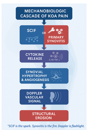
Figure 1 The true pain generators - synovitis and SCIF.
Synovitis-targeted therapies: the new frontier in koa management
Synovitis is no longer a passive bystander-it is a primary pain generator and a drug target. Doppler ultrasound reveals vascular activity that correlates with cytokine release, macrophage activation, and neuronal sensitization. The therapeutic landscape is rapidly evolving, with biologic agents now targeting specific molecular pathways.
Updated therapeutic targets and mechanisms
|
Target |
Mechanism |
Emerging agents |
|
IL-6 |
Suppresses inflammatory cascade |
Tocilizumab, Sarilumab |
|
TNF-α |
Blocks pro-inflammatory signaling |
Adalimumab, Etanercept |
|
GM-CSF–CCL17 Axis |
Modulates macrophage polarization |
Namilumab, Otilimab |
|
CSF-1 |
Regulates synovial macrophage proliferation |
Pexidartinib |
|
IL-1β |
Halts SCIF-triggered synovitis |
Anakinra, Canakinumab |
|
Neurotrophins (NGF) |
Reduces pain sensitization |
Tanezumab (under review) |
|
FLS Subsets |
Targets stromal inflammation |
Anti-CD90, Anti-Podoplanin agents |
|
Hydrogel (Arthrosamid®) |
Fuses with synovium, modulates inflammation |
Non-biologic, but biologically active |
Capsule Quotes:
“Synovitis is no longer silent-it’s a drug target.”
“Hydrogel doesn’t float-it fuses. It treats the tissue, not the fluid.”
“MRI shows anatomy. Doppler reveals pain. Targeted therapy begins with phenotype.”
Clinical implications
These therapies validate the Doppler-guided framework: vascular signal is not incidental-it’s actionable.
Even if biologics are not regionally available, their success in trials reinforces the logic of phenotype-first treatment.
Future directions include topical biologics, intra-articular gene therapy, and FLS-specific modulation-all aimed at reversing synovial inflammation without systemic immunosuppression.
Supporting evidence
Berenbaum et al., Springer, 2025: Synovitis is a viable therapeutic target in OA
Qian et al., Arthritis Res Ther, 2024: FLS subsets are emerging drug targets
Németh et al., Ann Rheum Dis, 2023: Synovial fibroblasts perpetuate inflammation and tissue damage
Frontiers in Immunology, 2022: Synovial biology-directed therapeutics hold promise for OA and RA
The science is ready-the system is not.” “We treat the phenotype-not the postcode.”
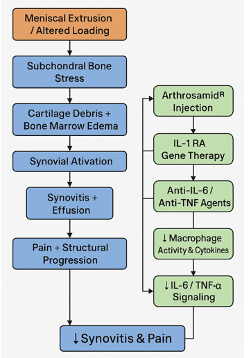
Figure 2 Synovitis-Targeted Therapies: The New Frontier in KOA Management
Hyaluronate: legacy lubrication, limited logic
Hydrophobic nature limits interaction with effusion
No cytokine suppression - ineffective in synovitis
Benefit often attributed to additives like mannitol or sorbitol
FDA reclassified hyaluronate as a drug in 2018
“It’s not the gel - it’s the sorbitol.” “Lubrication is legacy. Inflammation is the enemy.”
The misnomer of “Dry Knee Pain”
Intra-articular injections fail
MRI findings mislead
Doppler clarity redirects therapy
“Dry knees hurt-but not from the joint
ITBS as the dominant pain generator in dry knees
In our clinic, Dry KOA phenotypes outnumber Wet knees by a ratio of approximately 10:1, making iliotibial band syndrome (ITBS) the most frequent cause of painful KOA. This observation is consistent across thousands of cases and has been validated by Doppler ultrasound, clinical palpation, and MRI findings.
Clinical Presentation of ITBS
Posterolateral knee pain, often misdiagnosed as sciatica or radiculopathy
Tenderness over the lateral epicondyle
Negative Doppler signal (no effusion or synovitis)
Dramatic response to local steroid injection
Frequently associated with varus deformity and medial compartment OA
“Dry knees hurt-but not from the joint.” “ITBS is the silent giant-missed on MRI, loud on touch.”
MRI Support for ITBS
MRI often reveals:
Edema and high T2 signal in the fatty tissues between the ITB and lateral femoral condyle
Fibrovascular irritation and thickening of the ITB
Signal abnormalities consistent with strain, not intra-articular pathology
These findings are frequently overlooked or misattributed to degenerative changes. Yet they correlate strongly with clinical pain and Doppler-negative knees.
Supported by:
Vasilevska et al., J Radiol, 2009: ITBS signs in 74% of severe medial OA knees.9
Fairclough et al., Clin Anat, 2006: Histologic evidence of fibrovascular irritation.8
Radiopaedia.org, 2025: MRI shows ill-defined signal abnormalities lateral to the femoral condyle
“Posterolateral pain in medial OA is a diagnostic detour.” “In varus knees, the ITB isn’t tight-it’s tortured.”
ITBS and GTPS: a fascial continuum, not separate syndromes
In our clinical experience, Dry KOA phenotypes-dominated by ITBS-frequently coexist with Greater Trochanteric Pain Syndrome (GTPS). These are not isolated entities but manifestations of a shared fascial pathology. The iliotibial band (ITB) is not merely a tendon inserting at Gerdy’s tubercle-it is a longitudinal fascial sheet that extends distally and posteriorly, enveloping the lateral aspect of the leg and contributing to pain patterns that mimic sciatica.
Anatomical and pathophysiological insights
ITB Insertion: Beyond Gerdy’s tubercle, the ITB continues through the lateral fascia, integrating with the crural fascia and extending down the leg. This explains why pain often radiates to the posterolateral calf, simulating radiculopathy.
Fascial Continuum: The ITB originates from the tensor fascia latae and gluteus maximus, traverses the lateral thigh, and glides over the greater trochanter. Repetitive friction here irritates the trochanteric bursa, leading to GTPS.
Shared Etiology: Both ITBS and GTPS arise from strain, friction, and postural overload along the same fascial line-especially in varus knees, altered gait, or chronic pelvic tilt.
Capsule Quotes:
“Posterolateral pain is not a nerve-it’s a fascia.”
“In varus knees, the ITB isn’t tight-it’s tortured.”
“GTPS and ITBS are not neighbors-they’re twins.”
Clinical and imaging evidence
MRI and Doppler often reveal:
Edema and high T2 signal in the fatty tissues between the ITB and lateral femoral condyle
Thickening and fibrovascular irritation of the ITB
Signal abnormalities lateral to the femoral condyle and greater trochanter
Radiopaedia and Orthobullets confirm overlapping features of GTPS and ITBS, often misdiagnosed as sciatica.
European Spine Journal (2018): 11% of patients referred for sciatica had GTPS; 2.7% had both GTPS and true radiculopathy.
“Pain in the outer thigh isn’t always spinal-it’s often trochanteric.” “Dry knees hurt-but not from the joint. They scream through the fascia.”
Clinical and imaging evidence
GTPS and ITBS are part of a shared diagnostic spectrum, often misinterpreted as radiculopathy or lumbar disc disease.
Radiopaedia and Orthobullets confirm that trochanteric bursitis and ITBS are overlapping conditions, both caused by friction and strain along the ITB.
Springer’s European Spine Journal (2018) reported that 11% of patients referred for sciatica were actually suffering from GTPS, with another 2.7% having coexisting GTPS and true sciatica.
TRACE Body Rejuvenation (2024) emphasized that **misdiagnosing GTPS as sciatica leads to ineffective treatments, unnecessary imaging, and prolonged suffering.
“GTPS mimics sciatica-but it’s a fascia, not a nerve.” “Pain in the outer thigh isn’t always spinal-it’s often trochanteric.”
These missteps drain resources, delay recovery, and obscure the true pain generator.
“We don’t treat the postcode-we treat the phenotype.” “Misdiagnosis costs more than money-it costs clarity.”
KOA is a biologic joint failure. Our Doppler-guided algorithm offers a simple, phenotype-first approach:
Doppler-positive with effusion
If acute severe pain → MRI to exclude SCIF
If not SCIF → treat synovitis with aspiration ± biologics
Doppler-negative
Examine ITB → steroid injection if tender
Equivocal doppler
Start ITB protocol → reassess in 3–5 days → escalate if signal develops
Start outside. If it fails, go inside.”
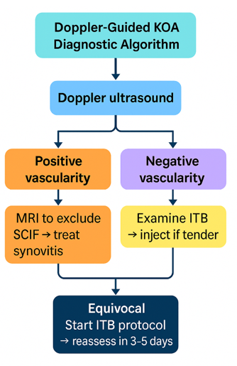
Take-home message &algorithm
Final Capsule: “It’s a simple phenotype way to treat a complex disease-no extra cost, no dilemma, no misdiagnosis.”
Scientific advantages
Precision: Targets biologic pain generators, not incidental findings.
Efficiency: Avoids unnecessary MRI, surgery, and misdirected therapy.
Accessibility: Uses affordable tools (Doppler, steroid, magnesium).
Clarity: Reduces diagnostic confusion and wasted resources.
Impact: Aligns with global push for phenotype-based OA care2.
“We don’t chase shadows-we treat the signal.” We don’t chase shadows
“It’s a simple phenotype way to treat a complex disease-no extra cost, no dilemma, no
Limitations
Single-center scope
Observational design
Limited access to biologics
Operator-dependent Doppler interpretation
Future research and global relevance
Multicenter validation
Phenotype prevalence studies
Cost-effectiveness analysis
Training integration
Biologic access advocacy“
Collective capsule quotes
“SCIF is the spark. Synovitis is the fire. Doppler is the flashlight.”
“Hydrogel doesn’t float - it fuses. It treats the tissue, not the fluid.”
“MRI shows anatomy. Doppler reveals pain. Targeted therapy begins with phenotype.”
“Dry knees hurt - but not from the joint.”
“If Doppler whispers, the ITB is shouting.”
“Cutting the meniscus doesn’t cut the pain.”
“We treat the phenotype - not the picture.”
“In varus knees, the ITB isn’t tight - it’s tortured.”
“Lubrication is legacy. Inflammation is the enemy.”
“Start outside. If it fails, go inside.”
None.
The author declares that there are no conflicts of interest.

©2025 Abaza. This is an open access article distributed under the terms of the, which permits unrestricted use, distribution, and build upon your work non-commercially.
