MOJ
eISSN: 2374-6939


Mini Review Volume 8 Issue 5
1Department of Physical Education, Federal Institute for Education, Science and Technology of Minas Gerais, Brazil
2Department of Physical Education, Physiotherapy, and Occupational Therapy School, Federal University of Minas Gerais, Brazil
3Department of Physical Education, Federal University of Vi?osa, Brazil
4Senior Visiting Researcher, Department of Physical Education, Federal University of Maranhao and CAPES/FAPEMA, Brazil
Correspondence: Alex de Andrade Fernandes, Federal Institute for Education, Sciences and Technology of Minas Gerais, Av. Joao Valentim Pascoal, s/n - Centro - Ipatinga - Minas Gerais, Brazil, Tel 55-31-3899-2076, Fax 55-31-3899-2076
Received: July 15, 2017 | Published: August 8, 2017
Citation: Fernandes AA, Pimenta EM, Moreira DG, Marins JCB, Garcia ES (2017) Application of Infrared Thermography in the Assessment of Muscle Damage in Elite Soccer Athletes. MOJ Orthop Rheumatol 8(5): 00328. DOI: 10.15406/mojor.2017.08.00328
The participation in a soccer match may result in a large number of micro-injuries leading to a number of physiological responses which, in turn, increase skin temperature (Tsk) of the lower limbs. The measurement of Tsk using infrared thermography (IRT) is being used as a tool to evaluate the internal load of the athlete in order to assist the management of training load and to help in the prevention of muscle injuries.
Keywords: Infrared thermography, Muscle damage, Skin temperature, Soccer athletes
Tsk, Skin Temperature; IRT, Infrared Thermography
Participation in a soccer match results in a large number of micro-injuries originated by the eccentric mechanical actions, generating muscle fiber ruptures, cell membrane damage, and sarcomere degeneration.1 Following this process, an acute local inflammatory response is triggered involving the release of different cytokines, migration of neutrophils to the trauma areas, and the release of certain agents into the damaged fibers to attract macrophages that ingest and digest the dead tissue, promoting temperature increase.2 In this sense, studies have demonstrated a significant increase in tsk assessed through IRT after physical effort, which could be an indicator of muscular wear, evidencing the athlete's physical exhaustion.2,3
The acute inflammatory response presented after soccer matches results in the appearance of some cardinal signals produced by the organism such as: heat, redness, pain and swelling.4 The redirection of arterial blood flow to the exercised muscle generates a higher muscle temperature and a greater local cutaneous vasodilation. This process is followed by a sensation of perceived heat, as well as the redness due to the greater number of erythrocytes that transited in the affected area.4 In addition, during the inflammatory process, the blood flow velocity is reduced, which allows the interaction of circulating cells with endothelial cells expressing surface molecules capable of binding to leukocytes.5 It is also important to note that during exercise and post-exercise there is a release of proinflammatory cytokines TNF-α, IL-1β and IL-6 that can remain elevated for a period of 24-48 hours.6 and concomitantly act as endogenous pyrogens, thus contributing to the increase of the internal temperature.5
Considering the factors that trigger the inflammatory process, which consequently alter the skin temperature, it has been proposed the assessment of skin temperature through IRT as an indirect way of measuring muscle damage caused by different types of exercise, especially those with eccentric characteristics. Therefore, the use of IRT can be used as a noninvasive way of measuring body's responses (heat) to the inflammatory process, and can provide valuable information about how the external load was assimilated by the athlete. Therefore, it would be important to study the potential of this strategy for controlling the training load of high performance athletes.7
It is important to emphasize that muscular and cutaneous vasodilatation lead to increased blood flow in the exercised limb. However this is not the only reason for the increase in Tsk. In fact, the highest blood supply during exercise, with arterial blood at a higher temperature in muscle and skin, is the main factor that promotes the increase of Tsk. For example, in resting conditions, the expected blood temperature range is 36.8-37.9 °C8 and consequently the displacement of this blood flow at a higher temperature in exercised subjects will result in a higher muscle and skin temperatures.9
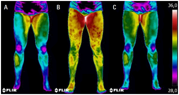
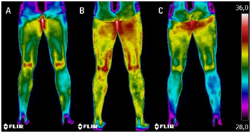
Fernandes et al.2 studied the Tsk changes after a professional football match of the Brazilian championship of the First National Division. The Tsk 24 hours after the game ranged from 2.0 °C to 2.5°C higher in the posterior area of the thighs than before the game. However, no evidence of injury was identified by the medical staff. Therefore, the authors concluded that the increase of Tsk after the match could be attributed to the inflammatory process, since the results of creatine kinase tests were also elevated 24 h after the game. Figures 1 & 2 present the thermal images recorded at the different moments in anterior and posterior views respectively.

Figure 1 Thermograms of the anterior views: (A) 24 hr before the match, (B) 24 hr after starting the match, and (C) 48 hr after starting the match.2

Figure 2 Thermograms of the posterior views: (A) 24 hr before the match, (B) 24 hr after starting the match, and (C) 48 hr after starting the match.2
One of the advantages of using IRT to measure Tsk is the relative low cost of some infra-red camera models. Since it is a non-invasive technique, Tsk monitoring can be performed focusing on a particular body region of interest (local analysis) as well as a broad view of the whole body, enabling a global analysis.10,11
The use of IRT may be a noninvasive alternative to measure Tsk, which can be applied as an indirect measure of the inflammatory process due to muscle damage, and being used as a tool to evaluate the internal load of the athlete in order to assist the management of training load and to help in the prevention of muscle injuries.
The authors of this article are indebted to the following research funding agencies: Federal Institute for Education, Science and Technology of Minas Gerais (IFMG); Coordination for the Improvement of Higher Education Personnel (CAPES); Foundation for Research Support of the Minas Gerais State (FAPEMIG); National Council of Scientific and Technological Development (CNPq).
No potential conflict of interest relevant to this article was reported.

©2017 Fernandes, et al. This is an open access article distributed under the terms of the, which permits unrestricted use, distribution, and build upon your work non-commercially.