MOJ
eISSN: 2374-6939


Mini Review Volume 12 Issue 2
Rehabilitation Service, Hospital Universitario Santa Cristina, Madrid, Spain
Correspondence: Marcos Edgar Fernández-Cuadros, Calle del Maestro Vives 2 y 3, Physical Medicine and Rehabilitation Service, Hospital Universitario Santa Cristina, Madrid, Spain, Tel +34- 915 57 43 00
Received: February 27, 2020 | Published: April 10, 2020
Citation: Fernández-Cuadros ME, Albaladejo-Florín MJ, Álava-Rabasa S, et al. Algorithm of management based on treatment goals for calcifying tendonitis of the shoulder: rigidity, pain and size of calcification. MOJ Orthop Rheumatol. 2020;12(2):26-29. DOI: 10.15406/mojor.2020.12.00511
Purpose: The objective of the present manuscript is to propose a step-by-step algorithm for the management of calcifying tendonitis (CT) of the shoulder based on treatment goals, from conservative to surgical approaches.
Method: A clinical note to present the main treatments for the management of calcifying tendonitis of the shoulder based on pain, rigidity and size of calcification based on the clinical experience and previous publications of the authors has been performed.
Arguments: Treatment is conservative and surgical although there is controversy on the most adequate treatment. Kinesiotherapy is the recommended therapy for shoulder rigidity. For pain management after NSAIDs; microwaves, short waves, TENS, ultrasounds and Interferentials are effective. For the management of size of calcification, Iontophoresis, Electroshock wave therapy, Ultrasound needle guided aspiration and arthroscopic surgeries are the recommended alternatives, in that order.
Conclusions: CT of the shoulder must be treated based on specific goals, mainly pain, rigidity or size of calcification. The proposed step-by-step algorithm of treatment is suggested based on the effectiveness of available techniques. If rigidity is present, kinesiotherapy is the recommended technique. For pain management, physical therapy such as microwaves, short waves, TENS, Ultrasound and Interferentials are effective techniques. For the treatment of calcification, iontophoresis is the most common, safe and inexpensive technique. If all previous conservative techniques failed, advanced techniques such as ESWT, US guided aspiration and arthroscopy are recommended, although they are not exempt of risk factors and complications.
Keywords: shoulder pain, calcifying tendonitis, iontophoresis, ESWT, ultrasound, physical therapy
Calcifying tendonitis (CT) of the shoulder is a common cause of shoulder pain and labor absenteeism.1 It is so frequent that affects 7% of general asymptomatic population, and it is present in 42.5% of symptomatic patients (painful shoulders). Therefore, CT is a common cause of visit to Rehabilitation setting.2 CT affects predominantly middle age women (between 30-60 years of age) and it is related to labor activities such as repetitive maneuvers, sustained positions and loading charges.3
CT is defined as the deposit of calcium carbonate/phosphate over a previously healthy tendon, due to degeneration or chronic inflammation.4 The degeneration and necrosis of the tendon could facilitate the deposit of crystals, in the absent of crystallization inhibitors and favored by chronic inflammation and the presences of nitric oxide.2,4
In the natural history of CT there are three clinical, histological and radiological stages clearly defined: 1) pre-calcification (tenocyte metaplasia and chondrocyte transformation); 2) calcification with a) formative [reservoir of vesicles in matrix], b) resorptive [spontaneous resorption of macrophages by phagocytosis]; 3) post-calcification (collagen remodeling and tendon repair).
CT clinical presentation is highly variable. There are asymptomatic patients in whom CT is only an incidental radiographic/ultra-sonographic finding. On the contrary, pain might be present in patients as acute painful crises or as chronic pain. In patients with long periods of pain, rigidity of the shoulder might be an added complication.3,4
In the natural history of CT, spontaneous pain relief and calcification resorption might be present but with great variability. Bosworth et al and Garner et al have stated in different papers that the reduction of calcification varies from 9-33% in 3 years follow-up; on the contrary, Wagenhauser et al.4 has reported a 27% rate of resorption at 10 years.
Diagnosis of CT is clinical and radiological. Treatment is initially conservative and should be directed towards the main complaint (pain, rigidity or calcification size).1,3,4 Conservative treatment includes NSAIDs (non-steroidal anti-inflammatory drugs), rehabilitation, and physical therapy including different electrotherapy techniques (microwaves, shortwaves, transcutaneous electrical nerve stimulation, ultrasounds, iontophoresis, Interferentials and pulsed electromagnetic therapy). Advanced therapeutic options include to electro shock wave therapy (ESWT), ultrasound guided needle aspiration and arthroscopy.1–4
The objective of the present paper is to propose a step-by-step algorithm for the management of CT of the shoulder based on treatment goals, from conservative to surgical approaches.
Aclinical note of the main treatments for the management of calcifying tendonitis of the shoulder based on pain, rigidity and size of calcification based on the clinical experience and previous publications of the authors has been performed.3,4
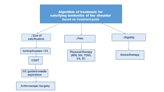
CT is a common, painful and crippling disease that produces great socio economic impact, great use of medical resources and work absenteeism.3 Chronic pain is the main cause of disability because of the loss of function and if prolonged in time, it can cause rigidity.3 Jimenez-Martin5 has reported that patients with painful CT showed limited movement in three spatial axes. Fernandez-Cuadros et al.3 found in their series shoulder rigidity in up to 40% of their patients. However, after 28 sessions of kinesiotherapy, all patients regained mobility in all range of movement (p=0.000). Kinesiotherapy included passive, active and assisted mobilization in order to regain range of movement. In Fernandez-Cuadros series, all patients regained mobility in all range of motion (flexion, abduction, internal and external rotation) from the beginning to the end of kinesiotherapy treatment, and with statistical significance.3 In light of these results, and because of the common of the presentation, for shoulder rigidity, kinesiotherapy must be the first option of treatment (Figure 1).

Figure 1 Algorithm of treatment for calcifying tendonitis of the shoulder based on treatment goals (reduction of pain, rigidity or calcification size).
MW, microwaves; SW, shortwaves; TENS, transcutaneous electrical nerve stimulation; US, Ultrasound; IF, Interferentials; ESWT, electroshock wave therapy
After rigidity, pain is the main cause of disability because of loss of function. Indeed, symptoms can persist up to one year after the first visit in 50% of patients.6 Therefore, pain control must be the second goal of treatment. Conservative treatment includes NSAIDs (non-steroidal anti-inflammatory drugs) and electrotherapy with its diverse modalities such as microwaves, short waves, TENS (transcutaneous electrical nerve stimulation), ultrasounds, Interferentials, pulsed electromagnetic fields and iontophoresis.7 There is still controversy on the ideal treatment of CT; therefore, in daily practice, a combination of treatments is employed.3 Fernandez-Cuadros et al.3,7 has demonstrated that ultrasounds, TENS, microwaves, shortwaves and Interferentials were capable of decreasing pain in patients with CT of the shoulder without statistical difference between them. Physical therapies have demonstrated effectiveness in the management of pain in CT of the shoulder (Figure 1). Unfortunately, neither kinesiotherapy nor electrotherapies were capable of decreasing size of calcification.
For the management of calcification there are many alternatives to date, such as iontophoresis, ESWT (electroshock wave therapy), ultrasound guided needle aspiration and arthroscopic surgery.
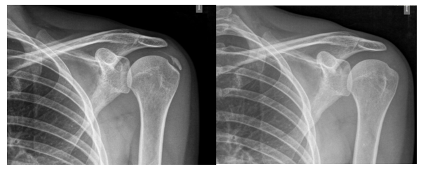
Iontophoresis with 5% acetic acid is a safe, common and cheap treatment and it was the only alternative in Rehabilitation Departments to act over the calcification.1,4,7 Fernandez-Cuadros, Chico-Alvarez and Rioja-Toro have demonstrated that Iontophoresis with 5% acetic acid, at either 20 sessions, 30 sessions and 40 sessions respectively were capable of decreasing significantly pain and size of calcification without any adverse effects on CT of the shoulder.1,4,7 Fernández-Cuadros4,7 has reported a successful rate of 57%, an improvement rate of 25%, and a failure rate of 18% (Figure 2).

Figure 2 Example of a 47 years-old female patient with shoulder pain (Visual analogue scale of 8/10) and calcifying tendonitis of the left shoulder. After 20 sessions of Iontophoresis with acetic acid and Ultrasound (1w/cm2/3MHz/5min) calcification disappeared and pain ameliorated (Visual analogue scale of 2/10).
If iontophoresis is unsuccessful, ESWT, ultrasound (US) guided needle aspiration and arthroscopy is advanced therapeutic options for the management of CT.7 These techniques are as effective as iontophoresis; however, they are painful, expensive, use complex devices (ESWT equipment, US equipment, arthroscopic equipment), and not free of risks or complications.1,7
In the case of ESWT, three sessions are performed with an interval of 1session/week, and the effect is not observed until 6months.2 US guided aspiration is employed if the previous techniques failed, but the effect is not observed until 6 months follow-up.2 Moreover, the effect of treatment is dependent on the type of calcification, that is, resorptive calcification has better prognosis than formative calcification.2 Lanza et al.8 has stated that the effectiveness rate for US guided aspiration is of 50% and the improvement can be observed even at a 2 years follow-up. There is controversy on which technique is superior on CT, ESWT or US guided aspiration. Suzuki et al.9 states that ESWT is as effective as US guided aspiration for the management of CT, but Arirachakaran10 states that US guided aspirations has better results than ESWT on pain, size of calcification and Constant-Murley functional scale. For that reason, Formigo-Couceiro2 suggests that formative calcification must be treated by ESWT and resorptive calcification must be treated by US guided aspiration.
In case all described alternatives failed, arthroscopic surgery is the definitive alternative.2,9 For that reason, surgical techniques should be performed in patients with progressive symptoms or constant pain that interferes with normal activity and where all conservative treatments have failed.3 Historically, open surgery was the preferred technique but nowadays, arthroscopic surgery is the recommended alternative.3,9
CT of the shoulder must be treated based on specific goals, mainly pain, rigidity or size of calcification. The proposed step-by-step algorithm of treatment is suggested based on the effectiveness of available techniques. If rigidity is present, kinesiotherapy is the recommended technique. For pain management, physical therapy such as microwaves, shortwaves, TENS, Ultrasound and Interferentials are effective techniques. For the treatment of calcification, iontophoresis is the most common, safe and inexpensive technique. If all previous conservative techniques failed, advanced techniques such as ESWT, US guided aspiration and arthroscopy are recommended, although they are not exempt of risk factors and complications.
The algorithm has been approved by the Ethical Committee of the Hospital.
For the application of this algorithm, patients must signed informed consent.
None.
The authors declare that they have no conflict of interest.
Authors declare no financial funding/support for the elaboration of this manuscript.

©2020 Fernández-Cuadros, et al. This is an open access article distributed under the terms of the, which permits unrestricted use, distribution, and build upon your work non-commercially.