MOJ
eISSN: 2374-6939


Case Report Volume 6 Issue 6
Slotervaart hospital, The Netherlands
Correspondence: YIZ Acherman, MD, Slotervaart hospital, Louwesweg 6, 1066 EC Amsterdam, The Netherlands, Tel 31-20-5129333
Received: June 18, 2015 | Published: December 28, 2016
Citation: Acherman YIZ, Dwars BJ (2016) A Potential Pitfall: Spindle Cell Sarcoma of the Wrist Apropos of a Case. MOJ Orthop Rheumatol 6(6): 00246. DOI: 10.15406/mojor.2016.06.00246
Ganglion cysts of the wrist are mostly diagnosed on clinical findings only. Their clinical presentation and typical localization makes them well recognised. We report a case of an intermediate grade spindle cell sarcoma of the wrist which mimicked a ganglion cyst.
Ganglion cysts represent approximately 50 to 70% of all soft tissue tumors of the hand. They are more prevalent in women and develop in 70 % of the cases between the second and fourth decade. Typically, they are soft mucin filled cysts attached to the joint capsule, tendon or tendon sheath and occur at specific locations. Their presentation as a swelling, sometimes with pain or weakness, developing over several months, makes them well recognized.1
We report a case of an intermediate grade spindle cell sarcoma of the wrist which mimicked a benign volar wrist ganglion cyst.
A previously healthy 29 year old female was referred to our surgical outpatient clinic for evaluation of a swelling at the volar side of her right wrist. The swelling existed for approximately 1 year with some increase in size 6 months prior to presentation. Movement of her wrist caused no discomfort, however she desired excision on cosmetic grounds.
Physical examination revealed a smooth painless mass, 3 cm in diameter, at the volar-radial side of her right wrist just above the flexor carpi radialis tendon. Based on these clinical findings the diagnosis: volar wrist ganglion cyst was made. No additional diagnotics were obtained and surgery was planned.
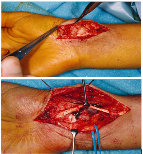
Surgery revealed an infiltrative solid tumor surrounding the flexor carpi radialis tendon and extending towards the radial artery, median nerve and flexor pollicis longus tendon (Figures 1 & 2). Macroscopic irradical excision with preservation of the median nerve was performed. Intraoperative frozen section revealed a inflammatory pseudotumor. The postoperative course was uneventful.

Figure 1 & 2 Solid tumor surrounding the flexor carpi radialis tendon and extending towards the radial artery, median nerve and flexor pollicis longus tendon.
Histological examination demonstrated fascicular arranged spindle cells with nuclear atypia and pleomorphism with high cellularity. Mitotic figures were seen frequently, some being atypical. Tumor cells were embedded in a collagenous matrix with lymphocytic and plasmacellular infiltration. Focal myxoid changes were also seen. Lymphocytic follicles were found at the border of the proliferative lesion. Immunohistochemical staining was postive for: vimentin, HHF35 and focal positive staining for SMA. Cytokeratin, sarcomeractin, CD34, S100, desmin, and MAC 387 stains were negative.
Local pathologists in collaboration with “the Dutch Sarcoma Panel” debated the differential diagnosis: nodular fasciitis or fibrosarcoma. However, later a definitive diagnosis: intermediate grade spindle cell sarcoma, most likely of myofibroblastic origin was made after revision by a pathologist abroad (Harvard Medical School).
The patient was screened for metastasis by means of a plain chest radiograph, CT-scan of the thorax and physical examination. These showed no evidence for metastasis resulting in a Stage IIa (G2 T1 N0 M0) spindle cell sarcoma of the wrist according to the AJCC classification.2 (moet dit nog nakijken). Six weeks later a debulking procedure was performed. Now the radial artery and flexor pollicis tendon (?) were also involved by tumor and therefore resected. Furthermore, tumor was also seen at to the flexor retinaculum and bone (welke?). She went on to receive curative radiotherapy (dosering?/schema?). A follow up protocol (?) was made after obtaining a baseline MRI. Approximately one year after initial diagnosis a new somewhat painful swelling was found at the right wrist. Physical examination revealed a firm swelling at the (site of prior resection?) volar-radial side of the right wrist approximately 3 cm in diameter. No palpable (lymph-) nodes were found.
Magnetic Resonance Imaging revealed a new lesion at volar-radial side of the wrist, 1 cm in diameter located in the subcutaneous fat and closely related to the underlying muscle group with increased signal strength on T2 images (foto?). CT-scan of the chest showed no evidence for metastasis. Following this an uncomplicated forearm amputation, 13 cm proximal to the wrist, was performed. The specimen showed tumor recurrence/residu at the styloid process of the radius with a maximal diameter of 2 cm histologically identical to the prior resection specimen. The resection was radical.
The patient has remained well without recurrence (kan dit na amputatie; wel bij stomp toch?) or distant metastasis for 7 years.
Ganglion cysts of the wrist are usually diagnosed based on clinical finding only. Their typical appearance at characteristic locations at the dorsum of the hand (60-70%) and volar wrist (18-20%) makes them well recognized.2 If the diagnosis based on clinical grounds seems clear, no additional diagnostic investigation is necessary. However, ultrasonography could be helpful to differentiate cystic from non-cystic lesions and establish the relationship of the tumor with the surrounding structures.3 Also, needle aspiration could be diagnostic if typical mucin-like fluid is aspirated. In this case a rather typical presentation did not yield any concern towards a malignancy and therefore no imaging or needle aspiration was obtained.
It is well recognised that sarcomas can mimic benign lesions in their clinical presentation. Additionally, classification of certain sarcomas appears to be difficult as seen in this case where primarily the differential diagnosis of inflammatory pseudotumor or (inflammatory) fibrosarcoma was made.
Spindle cell sarcoma of myofibroblastic origin is a rare diagnosis. They are fascicular spindle cell neoplasms resembling fibrosarcoma or leiomyosarcoma which infiltrate deep into soft tissue and can recur locally but do not tend to metastasize.4 Several studies demonstrated the improved survival for malignancies of the hand and wrist in comparison to musculoskeletal malignancies elsewhere (5, alle?). In a retrospective single institution study of 123 sarcomas of the hand and wrist which were compared to similar tumors in other musculoskeletal sites (n=6591) showed improved survival for sarcomas of the hand and wrist. The authors hypothesize that symptoms occur earlier due to the restricted anatomic confines intrinsic to the hand resulting in a relatively early diagnosis.5
This case illustrates that a sarcoma mimicking a benign lesion is a diagnostic pitfall. Therefore we conclude that not all wrist masses are ganglion cysts and should yield a high index of suspicion.
Prof, Daniel Altaha. Critical review.
None.

©2016 Acherman, et al. This is an open access article distributed under the terms of the, which permits unrestricted use, distribution, and build upon your work non-commercially.