MOJ
eISSN: 2374-6939


This article describes a case report of a Bosworth fracture-dislocation with review of the current literature (Table 1) and makes recommendations as to the timing of surgical fixation to help avoid future complications such as post-traumatic arthritis. A Bosworth fracture-dislocation is a rare type of ankle fracture where the proximal portion of the fibula fracture is dislocated posterior to the incisura fibularis.1–5 In most instances the distal syndesmotic ligaments are disrupted allowing for this fracture variant.5 This type of ankle fracture is challenging to reduce if not irreducible because of the posterolateral osseous portion of the tibia preventing close reduction of the fibula.6,7 Our case shows how this type of fracture can be close reduced. With disruption of the syndesmotic ligaments the proximal portion of the distal fibula was unstable which then allowed it to re-dislocate posterior behind the tibia after being close reduced. If this type of fracture-dislocation is not identified it can be detrimental to the function and prognosis of the patient.2,8 Up to this point literature has not described whether this rare variant should be treated differently than typical ankle fractures as to the timing of surgical fixation.
|
Author |
Article |
Summary |
Current recommendation |
|
Bosworth DM 2 |
Fracture-dislocation of the ankle with fixed displacement of the fibula behind the tibia. JBJS (Am) |
Original description of fracture-dislocation of an ankle with the proximal portion of the distal fibula locked posterior to the tibia |
Awareness of a rare variant ankle fracture that doesn’t reduce may be this type of fracture-dislocation. |
|
Perry et al.5 |
Posterior fracture-dislocation of the distal part of the fibula. Mechanism and staging of injury. JBJS (Am) |
Mechanism of Bosworth fracture-dislocation is a supination injury with external rotation |
An ankle fracture with supination and external rotation injury pattern that is difficult to reduce should raise suspicions of an Bosworth variant |
|
Cecil et al.3 |
Fracture-dislocation of the Ankle with Fixed Displacement of the fibula behind the tibia. Orthopaedic Review |
Reviewed literature up to that date and encouraged prompt treatment with full length tibia-fibula films. Most are irreducible ankle fractures. |
Prompt treatment with full length tibia-fibula films. |
|
Szalay & Roberts10 |
Compartment Syndrome After Bosworth Fracture-Dislocation of the Ankle. JOT |
Anterior compartment syndrome after ORIF with a Bosworth’s variant |
Awareness of compartment syndrome with Bosworth fracture-dislocations |
|
White & Pallister7 |
Fracture-dislocation of the ankle with fixed displacement of the fibula behind the tibia- a rare variant. Injury |
Anterior inferior tibiofibular ligament was entrapped causing an irreducible ankle fracture. |
Urgent surgical fixation (within 24 hours). Close attention to the syndesmotic ligaments w/ repair. |
|
Beekman & Watson12 |
Bosworth Fracture-Dislocation and Resultant Compartment Syndrome. JBJS |
Compartment syndrome after Failed reduction of a Bosworth fracture-dislocation. Pt was sent home. |
Continue awareness of Compartment syndrome with suggestion of possible hospitalization for urgent fixation. |
|
Bartonicek et al.1 |
Bosworth-type fibular entrapment Injuries of the ankle- The Bosworth Lesion. JOT |
Review of literature up to that date. |
After close reduction failure article recommends open reduction with internal fixation. Syndesmotic fixation with medial malleolus and/or deltoid ligament repair. |
|
Lui et al.8 |
Ankle stiffness after Bosworth fracture dislocation of the ankle. Arch Orthop Traum Surg |
Failed closed reductions with ORIF 2-4 days after injury led to increased ankle stiffness (verified arthroscopically) and decreased ROM |
Emergent to urgent open reduction with internal fixation (within 24-48 hours) |
|
Khan & Borton9 |
A constant Radiological Sign in Bosworth’s Fractures: “The Axilla Sign”. J Foot & Ankle Int. |
Found a consistent radiographic sign on the mortise view at the axilla of the medial tibial plafond due to internal rotation of the tibia from the posteriorly perched proximal fibula fragment |
Radiographic clue to Bosworth fracture dislocation is a “axilla sign” |
|
Wright et al.11 |
A Contemporary Approach to the Management of a Bosworth Injury. Injury |
Posterolateral incision with patient in prone position and urgent open reduction with internal fixation |
Possibly approach fracture with prone position and posterolateral incision. Consider CT scan. Urgent fixation of fracture. |
|
Schepers et al.6 |
An Irreducible Ankle Fractue Dislocation: The Bosworth Injury. J Foot & Ankle Surgery |
Irreducible ankle fracture that was taken to the OR the day of injury. |
Minimal amount of reduction attempts (< 3 attempts). Urgent surgical fixation (within 24 hours). |
Table 1 Review of current literature with recommendations
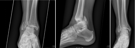
A Sixty-one year old man fell down multiple stairs at his home and sustained an external rotation injury to the right ankle. He was unable to ambulate and had notable deformity of the right ankle. The patient was then taken to the emergency department where he was examined for his injuries. AP, Mortise and Lateral x-rays of the right ankle were obtained. Initial radiographs showed a fracture-dislocation of the right distal fibula with tibiotalar dislocation and a posterior malleolus fracture, the distal fibula being posterior to the tibia. The Bosworth fracture-dislocation was not initially identified radiographically. A radiographical axilla of the medial tibial plafond was visible on x-ray (white arrow in (Figure 1-4)) due to internal rotation of the tibia when the fibula is dislocated posterior to the tibia.9

Figure 1 Mortise, Lateral and AP of initial injury films.
Note: “Axilla” sign on mortise view (white arrow).
.

Figure 3 AP, Mortise & Lateral of Right ankle post-reduction with plating of the distal fibula, syndesmotic screw fixation and Anterior to Posterior screw fixation of the posterior malleolus.

Figure 4 AP, Mortise & Lateral of Right ankle at 6 months post-operatively, 3 months s/p syndesmotic screw removal.
The patient described little to no pain at the time of initial exam. Sensation to light touch was grossly intact in the medial and lateral plantar, deep and superficial peroneal, saphenous and sural nerve distributions. There was an obvious deformity of the ankle with the tibia being more prominent anteriorly at the level of the ankle joint. There were no abrasions or lacerations. An ankle block was performed. The patient’s knee was then flexed and an anterior force was applied to the heel with a counterforce applied to the mid-tibia with traction. A palpable reduction was achieved and the patient was placed in a short leg AO plaster splint. Post-reduction films were obtained and indicated an adequate reduction primarily. The patient was discharged home and instructed to be non-weight bearing and follow up in the office the next day. The patient was taken to surgery five days from the date of injury. The patient was positioned and prepped like a typical ankle injury in the supine position with a bump under the ipsilateral hip. The distal fibula was approached through a direct lateral incision. Identification of the local anatomy was difficult secondary to the re-dislocation of the tibiotalar joint prior to intervention. When the osseous anatomy was identified the proximal fibula segment was found to be dislocated posterior to the tibia and hooked on the posterior tubercle of the incisura.
We performed a reduction maneuver of the tibiotalar joint with the intent of reducing the distal fragment to the proximal. This maneuver successfully brought the two fragments together but also reduced the proximal segment to the tibia. We did not have to perform an independent maneuver for the proximal segment as has been described in other case reports. Once reduced the fibula was clamped and treated like a typical fibula fracture with lag screw and neutralization plate application. The posterior malleolus was clamped and fixed with an anterior to posterior 4.0mm cannulated partially threaded cancellous screw. The syndesmosis was then stressed in the coronal and sagittal planes and found to be unstable. This was then secured with a 3.5mm cortical screw. The wound was closed in standard layered fashion and a well padded splint was applied. The patient was kept non-weight bearing until the syndesmotic screw was removed at 10 weeks post operatively.
The patient was seen two weeks post-operatively. His incisions were well healed and his staples were removed at that time. The patient was then placed in a walker boot and remained non-weight bearing on the right lower extremity. He was seen back at six weeks for a range of motion check and subsequently scheduled for syndesmotic screw removal at 12 weeks from the initial surgery. The syndesmotic screw was removed three months from the original surgery the patient was allowed to place full weight on the right lower extremity with crutches and advance as tolerated. The patient was involved in physical therapy for strengthening after the removal of the syndesmotic screw which was successful. The patient was most recently seen at five months post-operatively and found to have full range of motion in the right ankle and no pain.
Bosworth fracture-dislocations are challenging to recognize. A radiographic marker called the “axilla sign” can help identify the Bosworth variant of fracture-dislocations of the distal fibula on the mortise view of plain x-rays. A radiographical axilla of the medial tibial plafond is visible on x-ray due to internal rotation of the tibia when the fibula is dislocated posterior to the tibia.9 Bosworth’s original paper, as well as subsequent papers describes difficulty with reduction of this type of fracture and the necessity for operative treatment instead of non-operative management.2–8,10,11 Open reduction with internal fixation and close examination of the syndesmosis with increased likelihood of syndesmotic fixation is the treatment of choice.1,6 Posttraumatic arthritis is a known complication from initial non-operative treatment or failure to recognize this type of fracture-dislocation or inadequate fixation. Posttraumatic arthritis from malunion of the unreduced proximal fibula posterior to the tibia has led to arthrodesis as an ultimate pain relieving procedure within two years of injury.2,8 The mechanism for Bosworth’s fracture-dislocations has been demonstrated with a cadaveric study showing that it is by supination and external rotation that the fracture pattern occurs. Initially the investigators divided the anterior and posterior inferior tibiofibular ligaments before applying an external rotation and supination force to the ankle to demonstrate the fracture pattern and described them in stages.5
Others have noted compartment syndrome with Bosworth’s fracture dislocations.12 One report of an anterior compartment syndrome variant has been described as well indicating the importance of recognition this rare fracture-dislocation.10 Comparing the results of early vs. delayed treatment of ankle fractures, it has been shown that delayed treatment was equivocal in wound complications and the patients had a shorter hospital stay. That study excluded any fracture requiring syndesmotic fixation and therefore would have excluded most if not all Bosworth fracture-dislocations.13 Posttraumatic adhesive capsulitis was a complication of the ankle with patients who had failed closed reduction with surgery occurring 2-4 days after injury. The patient who was treated the same day had no ankle stiffness in the short term post operative follow up. Good outcomes have been reported with operative fixation of the Bosworth fracture-dislocation within 24 hours of injury.8 Additional imaging such as a CT scan prior to surgery and posterolateral approach may allow for improved visualization for reduction and fixation of the fracture-dislocation.11 It is our view that Bosworth fracture-dislocations should be treated with urgency when the distal portion of the proximal fibula is not able to be close reduced after a few attempts then open reduction with operative stabilization should be performed. Perry also reported only 27 percent of Bosworth’s fractures have been able to be closed reduced.5 Compartment syndrome may also be an iatrogenic cause from repeated reduction attempts resulting in additional trauma to the soft tissues.8,10,12
Our patient was discharged home from the emergency department after closed reduction but with reports of compartment syndrome associated with a Bosworth fracture-dislocation it may suggest closer monitoring as an inpatient. If two or three initial attempts of close reduction are not successful a CT scan may prove helpful in identifying the Bosworth variant and aid in surgical planning.11 It is also our recommendation after review of the literature that fixation should be performed urgently, within 24-48 hours after injury. Urgent surgery may help decrease the risk of posttraumatic arthritis and by paying close attention to the reduction of the distal fibula fracture-dislocation.1,6-8,10,11 The actual incidence of Bosworth fracture-dislocations appear to be rare but the consequences of non-recognition can be detrimental to the patient. Following these guidelines might help decrease the complications associated with Bosworth fracture-dislocations.
None.
The authors declare there is no conflict of interest.

© . This is an open access article distributed under the terms of the, which permits unrestricted use, distribution, and build upon your work non-commercially.