MOJ
eISSN: 2381-179X


Case Report Volume 2 Issue 2
Department of Pathology Anatomy, Sam Ratulangi University, Indonesia
Correspondence: Poppy M Lintong, Department of Pathology Anatomy, Medical Faculty of Sam Ratulangi University, Manado, Indonesia
Received: January 29, 2015 | Published: April 18, 2015
Citation: Lintong PM, Durry M. Xanthogranulomatous pyelonephritis. MOJ Clin Med Case Rep. 2015;2(2):36-38. DOI: 10.15406/mojcr.2015.02.00016
Xanthogranulomatous pyelonephritis (XGP) is grouped as chronic pyelonephritis that showed kidney enlargement resembling a neoplasm, therefore, it is called pseudotumor. XGP causes destruction of the kidney, disturbance of renal function, and an invasion of infection of renal parenchyme into perinephric adipose tissues and peritoneum. Preoperatively, it is often suspected as a renal cell carcinoma.XGP is occurred rarely, it accounts only 1% of all kidney infection. It can be seen in all ages, but more frequently in middle age female and elderly. The main cause of XGP is chronic obstruction and infection of Proteus or E. Coli. Microscopically, the characteristic feature is the existence of lipid-laden foamy macrophages. A surgery is the first choice and the prognosis is better in unilateral infection cases. We reported a 30-year-old female with a clinical diagnosis of ovarial cyst. Post-operative tissue was found in cystic form tissue. Microscopical examination showed a pyelonephritis and foci of lipid-laden foamy macrohages. It was concluded as an XGP case. The XGP case in female 30years old is very rare. In this case, the causes of XGP were chronic obstruction and infection. The destructed kidney showed a cystic form that looked like an obstructive hydronephrosis.
Keywords: XG, pyelonephritis, lipid-laden foamy macrophages
XGP, xanthogranulomatous pyelonephritis ; RCC, renal cell carcinoma
XGP usually appears as a kidney enlargement resembling a neoplasm therefore referred as a pseudo tumor. XGP may lead to kidney destruction, kidney malfunction and infection from renal parenchyma to fat tissues and peritoneum.1 Macroscopic appearance of XGP shows yellowish kidney mass with necrosis and hemorrhagic which are similar as renal cell carcinoma.1–3 XGP is a rare variant of pyelonephritis chronic. The incidence can be seen in all ages but more often in middle age female with history of recurrent urinary tract infections. Typical symptoms of XGP included pain, fever, malaise, anorexia, and weight loss.3,4 The main etiology of XGP is a long duration infection of Proteus, E.coli and also Pseudomonas.1 Physical examination reveals a mass in more than 50% cases and in ultrasound examination found kidney enlargement, disappearance of its normal architecture and central stone shadow.
Approximately 80% of XGP patients are associated with nephrolithiasis. Nephrolithiasis may lead to obstruction in renal collecting system and become a predisposition of infection. Inadequate response of host to the acute inflammation may lead to kidney infection and necrosis at the obstruction site.1,3,5,6 The typical macroscopic appearance of XGP shows kidney enlargement with hydronephrosis, renal stone and some other symptoms caused by obstruction. Single or multiple nodules, orange to yellowish color and with central necrosis and abscess. The infection may infiltrate through perinephric fat tissue and through the perinephric connective tissue in two third of cases.3
The characteristic of microscopic features in XGP including a mixture of neutrophils, lymphocytes plasma, histiocytes or macrophages and giant cells. The patognomonic signs are the foamy histiocytes or macrophages (xanthomatous) consist of fat droplets (lipid-laden foamy macrophage). Sometimes, cholesterol crystals and fibrous tissue may reveal. The macroscopic and microscopic appearances of XGP are very similar with renal cell carcinoma (RCC) where the foamy macrophages in XGP contain microvesicular sitoplasm similar with the RCC cells. The difference within both entities is the inflammation background in XGP and there are no pleomorpic cells and mitotic figures. In some difficult cases, Immunohistochemistry staining should be performed. The foamy macrophage in XGP shows negative with cytokeratin and vimentin stains whereas positive for lysozime stain.3 The treatment of XGP is surgery consist of nephrectomy and it have a good prognosis if the infection only affect one kidney.
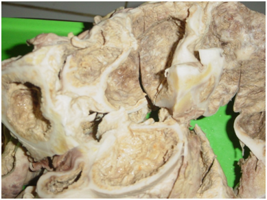
A 30 years old female with previous clinical diagnose of ovarian cyst. During the surgery they revealed that the cyst is not at the ovary but at the kidney, thus the partial nephrectomy was performed. Macroscopic examination reveals a 15x14x10cm tissues consist of multiple small cysts with brown thick septae (Figure 1). Microscopic examination shows kidney tissues with granulomatous inflammation. The glomeruli hialinated, atrophy of tubules and some with thyroidisatio (Figure 2). It also shows abundant foamy macrophages and granulomatous (Figure 3).

Figure 1 Gross features. The kidneys contain yellowish mass with cystic space. Kidney’s normal architecture has disappeared.
XGP is a rare variant of pyelonephritis which is more common in women, usually affects unilateral kidney and associated with nephrolithiasis.3 There is one reported case of XGP in 40years old male with staghorn calculus but nephrolithiasis is not found in our case. Macroscopically, the affected kidneys enlarge, contain cysts formation, yellowish masses and dilated calices which can be found in our case. The destruction of affected all renal parenchyma in almost all cases which have been reported. The normal structures of kidney replaced by cystic structures as in hydronephrosis obstruction. The tipycal microscopic appearance of XGP consists of large macrophages with vacuolated sitoplasm, mononuclear inflammatory cells including plasma cells and lymphocyte. In this case we found a large number of foamy macrophage with vacuolated sitoplasm, arrange in granulomatous structures and several giant cells (Figure 4). We also found the features of classic chronic pyelonephritis which are interstitial inflammatory infiltration and tubules thyroidisation (Figure 2).2,3,7,8
XGP shares many characteristic with RCC and may coexist with RCC as reported in two cases and two other cases with squamous cell carcinoma at the same kidney. In some cases which is difficult to distinguish between XGP and RCC the immunohistochemical stain is needed. There have been reported two cases XGP which positive PAS stain for the foamy macrophages.8–10 XGP is a rare case and most of the cases are unilateral, but bilateral disease has also reported. The treatment of XGP is nephrectomy and hemodialysis.11–14
We have reported a rare case of XGP, variants of chronic pyelonephritis, in a 30year old woman which has been previously misdiagnosed as ovarian cyst but later in macroscopic and microscopic obviously revealed an XGP.
None.
The author declares no conflict of interest.

©2015 Lintong, et al. This is an open access article distributed under the terms of the, which permits unrestricted use, distribution, and build upon your work non-commercially.