MOJ
eISSN: 2381-179X


Case Report Volume 2 Issue 3
Basildon Hospital NHS Trust, UK
Correspondence: Nirupam Purkayastha, Basildon Hospital NHS Trust, Nethermayne Basildon SS16 5NL, UK,
Received: April 17, 2015 | Published: April 30, 2015
Citation: Purkayastha N, Nandagudi A, Bharadwaj A, et al. We present a case of cardiac and ear involvement as atypical manifestations of microscopic polyangiitis. MOJ Clin Med Case Rep. 2015;2(3):56-57. DOI: 10.15406/mojcr.2015.02.00020
A 43 year old Caucasian lady was admitted with palpitations and a non-productive cough. Two months before she had symptoms of left sided otalgia, otorrhoea and progressive hearing loss. This was treated with antibiotics with grommet inserts. Apart from sensorineural deafness, there was no other neurological abnormality. Cardiovascular and respiratory examination was unremarkable. Urine analysis showed + blood and ++ protein, with no casts. Blood tests showed a CRP of 254mg/L, white cell count of 14.6x10⁹/L, haemoglobin 99g/dl, creatinine 166µmol/L (eGFR 29mL/min) and troponin 538mmol/L, with subtle T wave flattening on ECG. An unclear picture led to a CT pulmonary angiogram, where incidental large bilateral renal haematomas were found, with no evidence of pulmonary emboli. Autoimmune tests showed ANCA positive with a raised MPO titre of 134U/ml. A renal angiogram showed renal artery aneurysms. A cardiac MRI showed features of myocarditis. A diagnosis of Microscopic Polyangiitis was made. She was treated with high dose steroids and oral cyclophosphamide with good resolution. In the absence of typical cardiovascular symptoms and normal ECG, cardiac MRI is a useful imaging technique to aid diagnosis. It is important to emphasise the need for classification of vasculitis for prognostic value and treatment duration.
Keywords: ECG, cardiac, MRI, MPA
ECG, electrocardiogram; MRI, magnetic resonance imaging; RAA, renal artery aneurysm; MPA, microscopic polyangiitis; GPA, granulomatosis with polyangiitis
We present a case of cardiac and ear involvement as atypical manifestations of Microscopic Polyangiitis (MPA). Furthermore, ruptured renal artery aneurysm (RAA) is also an uncommon finding in MPA with only two adult cases described in the literature.1,2 A previously fit and well 43year old Caucasian lady was admitted with a sudden onset episode of palpitations and a non-productive cough. She denied any chest pain or breathlessness. There was no history of asthma, heart disease, foreign travel or TB exposure. It was revealed that two months prior to this she had symptoms of left sided otalgia, otorrhoea and progressive hearing loss. This was treated with a course of oral antibiotics by her GP and subsequently required grommet insertion. She denied any headache or visual disturbance, but suffered weight loss of half a stone in two months with recurrent night sweats, lethargy and myalgia. On examination, there was no evidence of a rash. Apart from sensorineural deafness, there was no other neurological abnormality. Cardiovascular and respiratory examinations were unremarkable.
Urine analysis showed + blood and ++ protein, with no casts. Blood tests showed a CRP of 254mg/L, white cell count of 14.6x10⁹/L, haemoglobin 99g/dl, creatinine 166µmol/L (eGFR 29mL/min) and troponin 538mmol/L, with subtle T wave flattening on ECG.
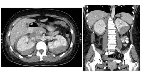
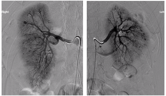
An unclear picture led to a CT pulmonary angiogram, which did not reveala pulmonary embolism but it was incidentally found that there were bilateral large renal haematomas (Figure 1). Subsequent blood tests revealed worsening renal function, inflammatory markers, and anaemia requiring blood transfusions. The patient was also found to be ANCA positive with a raised MPO titre of 134U/ml. A renal angiogram was performed and showed several renal artery aneurysms (Figure 2).

Figure 1 A) CT contrast abdomen, axial view showing bilateral capsular and subcapsular renal haematomas. B) CT abdomen sagittal view.

Figure 2 Renal angiogram right and left showing extensive renal artery aneurysms, with compression of left renal artery as a result of the haematoma.
A cardiac MRI showed myocardial scar tissue in keeping with features of myocarditis with regional wall abnormalities and reduced ejection fraction. A renal biopsy was difficult to achieve given the surrounding renal haematomas. She was treated with high dose steroids and oral cyclophosphamide. This resulted in a good recovery with complete cardiac resolution, improved hearing and progressive renal improvement. All these features suggested a diagnosis of MPA, according to the Revised Chapel Hill Consensus definition 2012. Ear involvement is a common manifestation of patients with Granulomatosis with Polyangiitis (GPA) but the absence of lung findings, and the lack of histology makes it less likely. On the other hand, a presentation of weight loss, myalgia, sensorineural deafness, raised creatinine and renal aneurysms would fulfil the classification criteria for classical PAN according to the ACR 1990 criteria. However, the positive MPO–ANCA makes it unlikely according to the Revised Chapel Hill Consensus definition.3
This case demonstrates that patients with MPA can present atypically, with cardiac and ear involvement and ruptured renal aneurysms. It is also important to emphasise the need for classification of vasculitis for prognostic value and treatment duration. Finally, in the absence of typical cardiovascular symptoms and a normal ECG; Cardiac MRI is a useful imaging technique to aid diagnosis of myocardial involvement.
None.
The author declares no conflict of interest.

©2015 Purkayastha, et al. This is an open access article distributed under the terms of the, which permits unrestricted use, distribution, and build upon your work non-commercially.