MOJ
eISSN: 2381-179X


Case Report Volume 12 Issue 2
1Dental Surgeon, Christus University Center, Fortaleza-Ceará/ Brazil
2Doctorate in Oral Rehabilitation Bauru Dentistry School/ University of São Paulo, Adjunct Professor Prosthodontics and Occlusion Federal University of Ceará-Sobral, Brazil
3Doctorate in Oral Rehabilitation Bauru Dentistry School/ University of São Paulo, Professor Christus University - Unichristus/Brazil
4Post-Graduate student in Dental Implant, Dental Prosthesis and Periodontics at Ceará Institute of Dental Specialties, Undergraduate degree at Harvard Medical School Fundamentals Program Online Learning - Course Pharmacology
Correspondence: Jaqueline Alves do Nascimento, Department of Dental Implant, Prosthodontics and Periodontics at Ceará Institute of Dental Specialties, Street Pe. Valdevino 276, Fortaleza-CE, 60135-040/Brazil
Received: August 31, 2022 | Published: September 7, 2022
Citation: Delfino MCH, Lima JFM, de Castro DSM, et al. Use of lithium disilicate veneers with dual cementation protocol for anterior rehabilitation: case report. MOJ Clin Med Case Rep. 2022;12(2):27-31. DOI: 10.15406/mojcr.2022.12.00414
The need to perform restorations with a natural appearance is one of the most challenging aspects of Dentistry, as well as the reproduction of the color of natural teeth in restorations is a clinical challenge, due to the complex optical characteristics of dentition Natural. As use of ceramics the need to use more opaque restorative materials or in different thicknesses, there is a great difficulty in obtaining adequate results in relation to the final color of the restoration. The objective of this work is to report a clinical case of smile rehabilitation in the anterior region with facets made of lithium dissilicate using the double cementation protocol, aiming at the final uniformization of color, and showing the advantages of use of this protocol in obtaining the aesthetics of the smile. A 47-year- old female patient with Diabetes Melitus, with main complaint the aesthetics of anterosuperior teeth, diagnosed in a pure ceramic crown in tooth 21 and a resin facet in tooth 11. With this, a planning was carried out to exchange these restorations and correction of the gingival margin of element 11, because the elements in question were different zeniths. The double cementation protocol allows a better aesthetic integration of restorations, besides allowing a reduction in the number of parts corrections by the laboratory, the preparation of facets simultaneously, ensuring a color and appropriate shape. Therefore, given the diversity of cementing agents in the dental market, it is important that the dentist has extensive scientific knowledge to know which cement to use in each prosthetic restoration, in addition to practical knowledge to know which technique to use and obtain a satisfactory result approved by the patient.
Keywords: pure ceramics, cementing agents, ceramic systems, dental materials
In dentistry, the presence of any abnormality in the anterior teeth, such as alterations in color, shape, size or position, can adversely affect the patient's smile. The beauty standards imposed as normal have been sought after by people in the different branches of esthetics. In this sense, the preparation of work that takes into account the aesthetic as well as the functional reestablishment brings back self-esteem to the individual.1
Pegoraro Bonfante, Valle et al.2, reported that states that to carry out a rehabilitative treatment it is necessary to seek oral health, the restoration of function, esthetics, and comfort of the patient, and not only to be based on the technical possibilities available, clearly without forgetting the patient's financial availability.
Among the various esthetic restorative materials that the dental market currently makes available, ceramics present themselves as an excellent alternative for reproducing natural teeth. Their wide use has promoted a great change in dentistry, allowing a more promising phase in the aesthetic treatments.3
Conrad4 related that pure porcelain crowns are extremely attractive, because besides aesthetics, they have biocompatibility, improved chemical inactivity, adequate physical and mechanical properties, with their optical properties combined with their natural characteristics, making them the synthetic material that most faithfully reproduces the dental structure.
Among the possibilities of rehabilitative treatments, ceramic laminates or veneers are one of the most conservative alternatives for restoring the shape and color of weakened teeth. One of the primary precautions that must be taken when using this restorative option is the thickness, which, being smaller than that of a full crown, interferes with the final color of the restoration, both because of the color of the tooth substrate, as well as the color of the cement used.
In view of this, using fully light-cured cements is recommended as they have a variety of shades and different degrees of opacity. In addition, the light-cured luting agent has greater color stability than the dual- cured luting agent, due to the absence of the tertiary amine that can undergo long-term color change. The light- cured luting agent provides a thin bond line along with high fluidity and excellent flowability, facilitating the removal of excess cement.5
Moreover, a determining factor for the clinical success of ceramic restorations is the achievement of reliable adhesive strength between the luting agent and the internal surface of the restoration. Glass-ceramic systems, such as feldspathic porcelains and lithium disilicate-reinforced ceramics, have similar strengths after cementation. The explanation for this is the principle of the adhesive system that ensures bond strength through transfer from one substrate to the other.6
Therefore, it is necessary a good individualized planning of cases in order to meet all the criteria requested, bearing in mind that the patient's satisfaction is very important and that all steps must be followed carefully to obtain the desired success. The chief complaint is also extremely important during the anamnesis, a summary of its history must be evaluated in order to bring a viable solution to the patient.2
The aim of this paper is to report a clinical case of smile rehabilitation in the anterior region, with the use of ceramic laminates made of lithium disilicate, using a double cementation protocol.
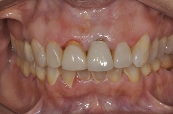
Patient I. A., female, 47 years old, attended the dental office with the chief complaint of dissatisfaction with her anterior teeth and smile. On clinical examination a ceramic veneer was found on tooth 11 with altered color, irregular shape and contour, and on tooth 21 an all-ceramic crown with altered color was found. Both restorations had different colors and volumes, as well as the gingival apex on both teeth was discrepant (Figures 1-3).

Figure 1 Initial photograph showing the discrepancy in color, shape and size between the restorations.
During the initial anamnesis, the patient reported having diabetes, but taking medication and controlling the disease. She also reported having a pacemaker and a history of problems with dental anesthesia, thus making more complex procedures impossible.
The initial planning of the case consisted of a minor intervention in the gum tissue of tooth 11, and the replacement of two ceramic restorations in the central incisors (11 and 21).
To perform the gingivoplasty, an intervention was planned only in soft tissue, without removal of bone tissue and with the use of anesthetics with less dental risk. The anesthetic solution used was 2% Mepivaicaine with adrenaline 1:100.000. Gingivoplasty is an essential part of smile transformations, especially in dental contact lens treatments that rely on this type of surgery to level and align gums, a mandatory procedure to achieve more harmonious results.
After the initial healing period of periodontal surgery, the veneer of tooth 11 and the crown of tooth 21 were removed with the aid of diamond burs in high rotation, making a longitudinal cut of the portion on the buccal surface, then with the aid of a spatula, the fragments of each restoration were removed. This technique was used to optimize working time, but it is worth noting the risk of tooth fracture with this method (Figure 4).
With the removal of the ceramic restorations, it was possible to analyze the color of the dental substrate in each of the teeth to be prepared. Tooth 21 had a cast metal core and a brown dentin substrate, tooth 11 had no enamel, and the old restoration was cemented to the dentin. The dental preparations were refined, with the preparation for a full crown left wider, with 1.5 mm of wear of the axial portion and 2 mm of the incisal portion, and with the cervical endplate positioned 0.7 mm below the gingival level. The veneer preparation was adjusted by keeping the distal contact point and breaking the mesial contact point in order to allow the ceramic to reestablish the same contact point more effectively (Figure 5).
The impression of the preparations was taken using the double-wire technique, in 2 stages, first positioning the thinner #OO retractor wire (Ultrapak, Ultradent Products Inc.®) and taking the impression with heavy addition silicone (Express XT, 3M Espe®) with relief accomplished through a PVC film positioned on the impression material. The second stage of the molding process began with the placement of a #0 wire over the first wire, in order to promote a greater distance from the marginal gingiva. After a 4-minute waiting period, the second wire was removed and the light impression material was injected onto the preparation surface. After the silicone had fully set (5'30"), the tray was removed and the mold was disinfected with 1% sodium hypochlorite (immersion for 10 minutes). In the same impression taking session the provisional crowns were made with the aid of stock tooth veneers (Figure 6).
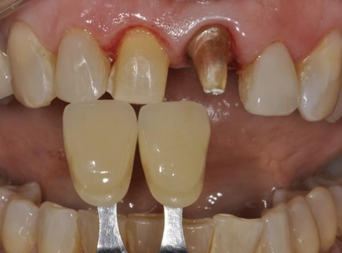
In this particular case, due to the different types of restorations to be fabricated, the correct shade selection is extremely important, as the final shade selection of the restoration must be made, as well as taking the shade of the dentin substrate. With the patient's help, shade A1 from the Vitapan Classical scale (Vita Zanhfabrik GmbH®) was chosen (Figure 7).
The shade selected for the dental substrate varied between A3.5 and A4 of the same scale. This information was sent to the laboratory in order to select the most appropriate ceramic material for the case (Figure 8).

Figure 8 Substrate color selected between A3.5 and A4 of the Vitapan Classical scale (Vita Zanhfabrik GmbH®).
To solve this case it was planned to fabricate two types of restorations, a ceramic veneer on tooth 11 and a full ceramic crown on tooth 21. Since these two types of restorations have different thicknesses (0.7 millimeters and 1.5 millimeters respectively), their aesthetic behavior would be different with respect to the final shade of the case, The infrastructure of the crown of tooth 21 would mimic the shape and color of the preparation for laminate present on tooth 11, thus allowing the two ceramic laminates, now with the same thickness, to be cemented with the same cementing agent, enabling a better uniformity of the final color (Figure 9).
With the restorations ready, a so-called dry try-in was conducted, in which the restorations are placed on the preparations without any cementing agent. The purpose of this step is to check the adaptation of the ceramic restorations on the dental preparations. After the try-in and their respective adjustments, the cement to be used was selected, choosing color A1 (Allcem Veneer, FGM Prod. Odontológicos S/A®), following the color of the ceramic material (Figure 10).
The cementation procedure began by attaching the ceramic laminate on the coping of tooth 21 crown, for this procedure, its buccal surface was conditioned with hydrofluoric acid 5% for 20 seconds, after this period, the surface was washed in tap water and a layer of phosphoric acid 37% was applied, remaining for 60 seconds, this second conditioning aims to remove the precipitate left by the hydrofluoric acid. After the application of acids, the coping received two layers of bonding agent Silane (Angelus Ind. Com. Prod. Odontológicos S/A®). The internal portion of the ceramic veneer received the same surface conditioning protocol, and was then ready to be cemented. The cement was applied to the inner portion of the veneer and it was positioned and seated on the ceramic coping, the excess was removed with a soft bristled brush and the restoration was light-cured for 40 seconds on each side (Figure 11).
After complete polymerization of the cement, the interface was polished with finishing rubbers. The cementation of the complete crown on the dental preparation was performed using a self-adhesive dual resin cement (SetPP, SDI®). This type of cement does not require any previous conditioning of the tooth structure, only cleaning with pumice and drying. The cement was applied inside the restoration and it was positioned on the tooth, the excess was removed and the cement was light-cured for 40 seconds on each side.
Cementation of the ceramic veneer on tooth 11 was performed following the same procedures described for the cementation of the ceramic restoration on tooth 21, differing only in the cement used, since only light- curing cement (Allcem Veneer, FGM Prod. Odontológicos S/A®) was required for this restoration. After the entire protocol of surface treatment of the ceramic restoration, the tooth surface was etched with 37% phosphoric acid for 15 seconds, then washed with a layer of dentin adhesive (Ambar APS, FGM Prod. Odontológicos S/A®). The cement was then positioned inside the veneer and it was seated on the preparation, the excess was removed with a thin brush and the restoration was light-cured for 40 seconds on each side. Cementation of the ceramic veneer on tooth 11 was performed following the same procedures described for the cementation of the ceramic restoration on tooth 21, differing only in the cement used, since only light-curing cement (Allcem Veneer, FGM Prod. Odontológicos S/A®) was required for this restoration. After the entire protocol of surface treatment of the ceramic restoration, the tooth surface was etched with 37% phosphoric acid for 15 seconds, then washed with a layer of dentin adhesive (Ambar APS, FGM Prod. Odontológicos S/A®). The cement was then positioned inside the veneer and it was seated on the preparation, the excess was removed with a thin brush and the restoration was light-cured for 40 seconds on each side.
After complete polymerization of the cement, occlusion was checked with the aid of occlusal carbons and the cervical margins were cleaned with an exploratory probe. The cementation interface was finished and polished with abrasive rubbers of different granulations (Figure 12, 13 and 14).
In the present article, a clinical sequence of esthetic rehabilitation of the anterior sector was reported, using all-ceramic restorations, individualized and planned in order to achieve the highest possible esthetic result. Due to the differences in thickness of both restorations, a double cementation strategy was chosen, whereby the crown of tooth 21 would simulate a preparation for a ceramic veneer identical to that of tooth 11, which would subsequently be cemented using the same light-curing cement, thus enabling a uniformity of the final color of the restorations.
The success rate of indirect restorations, such as veneers, fixed crowns, and partial restorations depends on correct planning, broad technical and theoretical knowledge, and adequate prosthetic preparation, thus achieving the ideal characteristics for each type of work.
Therefore, aesthetics is a highly relevant factor in anterior rehabilitation of the maxilla and should be taken into consideration when choosing the cement, since ideally these should not interfere with the optical properties of fixed crowns. Therefore, light-cured resin cements are widely used by professionals for their color stability characteristics in the final restoration product. However, they are expensive, require a critical handling technique, require absolute isolation during cementation, and are difficult to remove excess material, especially in proximal areas.
The composition of most resin cements is similar to that of composite resins for restoration (resin matrix with inorganic fillers treated with silane). Therefore, the final result of the color of the restorations depends on some factors such as the color of the substrate and the cement to be used. The selection of these cements should be determined by the clinical conditions of each case, the physical properties of the indirect restorative material, and the physical and biological characteristics of the cementing material, such as: adhesiveness, solubility, strength and biocompatibility. An additional desirable characteristic of a dental cement is that it should have a film thickness that provides satisfactory adaptation between the tooth and restoration surfaces. They must also have adequate marginal sealing, high tensile and compressive strength, adequate setting and working times, be radiopaque and have good optical properties.7
Furthermore, it is worth mentioning that some factors may directly interfere with the final appearance of indirect restorations. Therefore, aesthetics should be taken into consideration when choosing the cementing agent, as it should not interfere with the optical properties demonstrated by ceramic and glass polymer restorative materials. The shade stability of resin cements is another important factor, and for this reason, many professionals prefer to use light-cured cementation systems for veneers and pure crowns on anterior teeth, as these present greater shade stability.
In this context, the dual cementation protocol allows for a better esthetic integration of the restorations, as well as a reduction in the number of corrections by the laboratory, and the simultaneous fabrication of veneers, ensuring an adequate shade and shape.5 So we can state that the dual cementation protocol is a viable alternative when we have ceramic restorations that have different thicknesses, and that may have a final color change due to differences in light reflection of the materials.
The final cementation of each prosthetic rehabilitation is interfered by the choice of each material, and the wrong choice jeopardizes the clinical success of the rehabilitation. Given the diversity of cementing agents in the dental market, it is important that the dentist has a broad scientific knowledge to know which cement to use in each prosthetic restoration, as well as practical knowledge to know which technique to use and obtain a satisfactory result approved by the patient.
The dual cementation protocol in ceramic crowns is a viable means of achieving aesthetics in cases where the professional has the challenge of working with different substrate colors and thicknesses of ceramic restorations, thus being able to achieve greater uniformity through equalization of the useful cement.
None.
Authors declare that there is no conflict of interest.

©2022 Delfino, et al. This is an open access article distributed under the terms of the, which permits unrestricted use, distribution, and build upon your work non-commercially.