MOJ
eISSN: 2381-179X


Case Report Volume 5 Issue 4
Department of Pathology, Jawaharlal Nehru Medical College, India
Correspondence: Kafil Akhtar, Professor, Department of Pathology, Jawaharlal Nehru Medical College, Aligarh Muslim University, India
Received: October 30, 2015 | Published: December 28, 2016
Citation: Akhtar K, Rafey M, Arif SH, et al. Tuberculosis of the lip: an interesting case presentation. MOJ Clin Med Case Rep. 2016;5(4):253-254. DOI: 10.15406/mojcr.2016.05.00141
Tuberculosis of oral cavity is relatively less common as compared to the other parts of the body. The most common site is the tongue in the oral cavity, but tuberculosis of lip is a rare entity. Mostly labial tuberculosis presents as a non-healing ulcerative lesion, but sometimes it may present as a cystic swelling. The disease may be primary or secondary to pulmonary tuberculosis. We present herewith a case of 50year female with primary tuberculosis of the lip.
Keywords: lip, tuberculosis, histopathology
Tuberculosis is troubling mankind from ancient times and still it forms a major chunk of patients in developing and underdeveloped countries. It is estimated that one-third of the world’s population are infected with Mycobacterium tuberculosis.1
Although tuberculosis may involve any part of the body, but tuberculosis of lip occurs rarely. Tuberculosis of lip can be primary or secondary. Primary tuberculosis of lip is a diagnostic challenge for clinicians as well as for pathologists.2
Oral tuberculosis usually results from secondary inoculation of the breached oral mucosa by infected sputum, or by haematogenous dissemination from other infected sites3,4 0.1%–0.5% of subjects with pulmonary tuberculosis usually develops secondary oral tuberculosis affecting most commonly the tongue, followed by the palate, the lips, the buccal mucosa, and the gingival.5,6 It usually manifests as a single non-healing ulcers but may also occur as nodules and granulomata or fissures.7,8
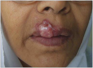
A 50year old female presented to the plastic surgery clinics with complaints of irregular, 2x1.5cm, solid to cystic swelling with excoriation at the upper lip, situated between the midline and angle of the mouth since 2months (Figure 1). There was no history of fever, cough, loss of appetite, weight loss, anti-tubercular treatment intake or contact and any lymphadenopathy. Fine needle aspiration biopsy showed typical epithelioid cell clusters with foci of caseous necrosis (Figure 2). Excision biopsy of the lesion was performed and sent for the histopathological examination. Grossly multiple small irregular brownish white soft to firm tissue pieces, 1.8cm in aggregate was received. Microscopically dense lymphocytic infiltrate in the underlying dermis with fibrosis and well defined epithelioid granulomas with langhan’s giant cells and widespread foci of caseous necrosis was seen (Figure 3). AFB stains revealed magenta colored acid fast tubercular bacilli. A histopathological diagnosis consistent with tuberculosis of lip was given. A nine-month anti-TB drug regimen of isoniazid, rifampicin, pyrazinamide, and ethambutol, was prescribed and within four weeks after initiation of the anti-TB treatment, the labial lesion strikingly improved.

Figure 1 Grossly an irregular 2x1.5cm, solid to cystic swelling with excoriation at the upper lip, situated between the midline and angle of the mouth.
Tuberculosis is a very ancient disease, even identified in mummies of pyramids.3 It’s still troubling the humanity and forms a major chunk of morbidities and mortalities. However with improved sanitation, cases of tuberculosis has declined in incidence and prevalence in developed countries but third world countries are still crippled with tuberculosis.4,5 But with increasing incidence of HIV infection, tuberculosis is on the rise again in developed countries.9,10 In India around 10million cases of tuberculosis are present.11 Tuberculosis infects primarily the respiratory system, however it may involve any part of the body. Oral tuberculosis is relatively rare and constitute approximately 1% of the tubercular lesions, affecting commonly the tongue and palate, but the tuberculosis of the lip is a rare variety.4,5,11
Tuberculosis of lip may be primary or it may be secondary, but mostly secondary tuberculosis is found. Secondary tuberculosis is due to spread of the disease directly from the primary site or through the blood or infected sputum. In case of oral tuberculosis, it spread either through sputum or it may spread through hematogenous route.6 Our patient presented with a solid to cystic swelling at the upper lip, without any symptoms of tuberculosis. Histopathological examination revealed the diagnosis of primary tuberculosis of lip, which is a rare manifestation of tuberculosis.
Tuberculosis is a devastating disease and still it’s a challenge for medical science. Tuberculosis should be kept in the differential diagnosis in any oral lesion and keen workup for tuberculosis should be done, including all ancillary investigations with chest radiograph and other microbiological investigations, and treated promptly with antitubercular regimen.
None.
The author declares no conflict of interest.

©2016 Akhtar, et al. This is an open access article distributed under the terms of the, which permits unrestricted use, distribution, and build upon your work non-commercially.