MOJ
eISSN: 2381-179X


Case Report Volume 10 Issue 1
Advisor in Pediatrics and Pediatric Psychiatry, Children Teaching Hospital of Baghdad Medical City, Iraq
Correspondence: Aamir Jalal Al Mosawi, Advisor in Pediatrics and Pediatric Psychiatry, Children Teaching Hospital of Baghdad Medical City, Iraq
Received: December 27, 2019 | Published: February 11, 2020
Citation: Mosawi AJAI. The use of cerebrolysin in pediatric Wohlfart Kugelberg Welander syndrome. MOJ Clin Med Case Rep . 2020;10(1):20-23. DOI: 10.15406/mojcr.2020.10.00335
Background: Juvenile spinal muscular atrophy which is also called Wohlfart Kugelberg Welander syndrome, is an autosomal recessive condition which appears within the first few years of life. Most of the patients are able to walk during early life, but difficulties in walking appears gradually leading to a variable degree of disability. There is no known satisfactory therapy for the treatment of Wohlfart Kugelberg Welander syndrome. The aim of this paper is to describe the novel use of cerebrolysin in the treatment of pediatric Wohlfart Kugelberg Welander syndrome.
Patients and methods: Two unrelated Iraqi boys aged four years with pediatric Wohlfart Kugelberg Welander syndrome were observed, and the one who was more severely affected was treated with intramuscular cerebrolysin. The less affected boy received no treatment. The father of the boy with less severe condition mainly complained that his son can not run as fast as his peers and he didn’t have obvious gait abnormality, but he was unable to stand on foot which is a milestone commonly achieved at the age of three years. The more severely affected boy had noticeable gait abnormalities and obvious difficulty with squatting, and was also unable to stand on foot.
Results : Cerebrolysin treatment was not associated with any side effects. Cerebrolysin treated boy experienced improvements in gait and squatting, and was able to stand on one foot momentarily for 2-3 seconds, while the untreated boy with the less severe condition couldn’t .
Conclusion: A study enrolling more patients is recommended.
Keywords: Wohlfart-Kugelberg-Welander syndrome, treatment, cerebrolysin
Juvenile spinal muscular atrophy which is also called Wohlfart Kugelberg Welander syndrome, is generally an autosomal recessive condition which appears within the first few years of life. Most of the patients are able to walk during early life, but difficulties in walking appears gradually leading to a variable degree of disability.1–3
Wohlfart Kugelberg Welander syndrome is a genetic disorder results from degeneration of the anterior horn cells and motor fibers to the proximal muscles of the limbs. It presents with lower motor neuron signs including weakness, wasting, and loss of tendon reflexes especially of the quadriceps.
The disorder is associated with a steady, slowly progressive course, and weakness affects first the large muscles of the buttock and thighs. Weakness may affect later the muscles of the upper arm. Late in the course of this condition, the more distal limb muscles may also be affected. Muscles of the face and neck are not affected.1–3
There is no known satisfactory therapy for the treatment of juvenile spinal muscular atrophy (Wohlfart Kugelberg Welander syndrome). The aim of this paper is to describe the novel use of cerebrolysin in the treatment pediatric Wohlfart Kugelberg Welander syndrome.
Two unrelated Iraqi boys pediatric with Wohlfart Kugelberg Welander syndrome were observed, and the one who was more severely affected was treated with intramuscular cerebrolysin. The less affected boy received no treatment.
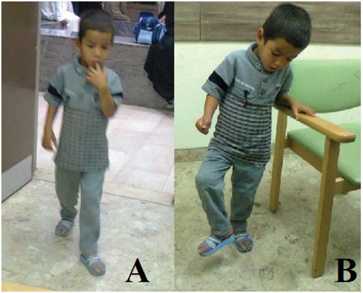
The less severely affected boy was first seen at the age of about four years during April, 2018 at the Children Teaching Hospital of Baghdad Medical City. The parents were relatives. They noted that he can not run as fast as his peer. He walked at the clinic and no obvious abnormality with his gait could be noticed (Figure-1A). Sensory examination was normal. Gower sign was negative and he didn’t have obvious difficulty in squatting and standing from the squat position.
However, the boy was unable to stand on one foot without holding the furniture (Figure-1B), A normal child can stand on one foot momentarily at three years. Serum creatine phosphokinase was not significantly elevated.

Figure 1 The less severely affected patient. (A) The boy walked at the clinic and no obvious gait abnormality. (B) The boy was unable to stand on one foot without holding the furniture.
Right and left median nerve.
Right ulnar nerve.
Right and left sural nerve.
Right and left common peroneal nerves.
Tibial nerves.
Reduced amplitude of the compound action potentials.
Normal sensory parameters.
Prolonged distal motor latencies.
Lower border and reduced motor conduction velocities.
Prolonged F-wave latencies.
No responses were obtained from EDB and abductor hallucis muscles.
No evidence of focal conduction block.
Sensory |
Motor |
||||||
Nerve |
Latency msec/cm |
Amplitudeuv |
SNCV m/sec |
Muscle |
DML msec/cm |
WNCVmsec/cm |
F-wave Latency |
Right median |
2.5 |
22.6 |
55 |
APB |
4.2 |
50.3 |
25.6 |
Right ulnar |
2.3 |
25.6 |
56.3 |
FDI |
4.3 |
51.6 |
26.5 |
Right common peroneal |
Tibialis Ant |
4.6 |
|||||
EDB |
No response |
||||||
Left common peroneal |
Tibialis Ant |
4.6 |
|||||
EDB |
No response |
||||||
Right tibial |
Gastroenemious |
4.4 |
|||||
Abductor Hallucis |
No response |
||||||
Tibialis Post |
5.9 |
||||||
Table 1 The findings of nerve conduction study and needle electromyography of the less severely affected patient
Right FDI.
Right deltoid.
Right brachioradialis
Right and left vastus medialis.
Right and left tibialis anterior.
EDB muscles.
Spontaneous activity grade 1-2 in the form of fibrillation, positive sharp,
myotonic discharges, and no peripheral fasciculations.
The average duration of 20 motor units:
Right deltoid=22.8msec (n=10.5msec)
Right biceps=22.9msec (n=10.3msec)
Right vastus medialis=22.2msec (n=10.5msec)
Left vastus medialis=22.5 msec (n=10.5msec)
Right tibialis anterior=14.2 msec (n=10.5 msec)
Polyphasia of long duration high amplitude was observed in 50-60%.
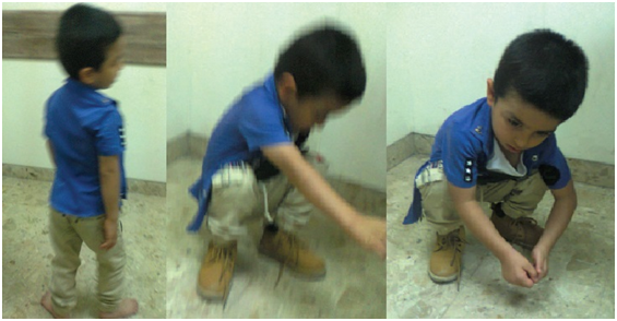
The second patient was more severely affected than the first patient. The boy was also first seen at the age of four years at the Children Teaching Hospital of Baghdad Medical City. The parents thought that his walking was rather awkward, and he was running rather slowly. He also had difficulty when climbing stairs.
When the patient walked at the clinic, his gait was rather awkward, and he was about to fall when he squatted (Figure 2). However, Gower’s sign was negative, and the sensory examination was normal. Like the first patient, the second patient was also unable to stand on one foot momentarily.

Figure 2 The more severely affected boy had noticeable gait abnormality and was about to fall when he squatted.
Serum creatine phosphokinase was measured during February, 2018 and it was 185u/L (Normal: 40-200u/L). However, Serum creatine phosphokinase was measured twice during July, 2018 and it was mildly elevated (210, and 255u/L).
Right and left median nerve.
Right ulnar nerve.
Right and left sural nerve.
Right and left common peroneal nerves.
Tibial nerves.
Reduced amplitude of the compound action potentials on proximal and distal stimulation site.
Normal sensory parameters.
Normal distal motor latencies, motor conduction velocities, and also normal
F-wave latencies.
|
|
|
Sensory |
|
|
|
Motor |
Nerve |
Latency msec/cm |
Amplitudeuv |
SNCV m/sec |
Muscle |
DML Msec/cm |
WNCVmsec/cm |
F-wave Latency |
Right median |
2.5 |
22.6 |
55 |
APB |
3.4 |
53.3 |
20.7 |
Right ulnar |
2.3 |
25.6 |
56.3 |
FDI |
3.7 |
53.6 |
21.7 |
Right common peroneal |
Tibialis Ant |
3.7 |
|||||
EDB |
4.8 |
44.3 |
42.6 |
||||
Left common peroneal |
Tibialis Ant |
3.9 |
|||||
EDB |
4.9 |
||||||
Tibialis post |
5.5 |
||||||
Tibialis post |
5.6 |
||||||
Table 2 The findings of nerve conduction study and needle electromyography of the more severely affected patient
Right deltoid.
Right biceps.
Right brachio-radials.
Right and left vastus medialis.
Right and left tibialis anterior.
Spontaneous activity grade 1-2 in the form of fibrillation, positive sharp, myotonic discharges, no peripheral fasciculations.
The average duration of 20 motor units:
Right deltoid=14.8msec (n=8.5msec).
Right biceps=15.9msec (n=8.3msec).
Right vastus medialis=15.2msec (n=8.5msec).
Left vastus medialis=14.3msec (n=8.5msec).
Right tibialis anterior=14.2msec (n=10.5msec).
Left tibialis anterior=15.3msec (n=10.5msec).
Polyphasia of long duration high amplitude was observed in 30-40%.
Reduced recruitment pattern of high amplitude.
The findings of the nerve conduction and electromyography (EMG) study were consistent with chronic diffuse anterior horn disease of moderate severity. The lumbo-sacral proximal segments were maximally involved especially on the right side. Both Tibialis posterior muscles were well functioning.
The findings of the nerve conduction and electromyography (EMG) study showed no evidence of peripheral polyneuropathy or muscle disease.
At about the age of four years and half year, during July, 2018, the boy was treated with ten intramuscular cerebrolysin given at a dose of 3ml on alternate days.
The protocol for this therapeutic trial was approved by the scientific committee of Iraq headquarter of Copernicus Scientists International Panel and conforms to the provisions laid out in the Declaration of Helsinki (as revised in Edinburgh 2000).
The treatment was not associated with any side effects. Treatment didn’t affect muscle mass and atrophy of the upper thighs remained obvious. After treatment improvement in his stance and gait was reported by the mother and was also observed at the clinic.
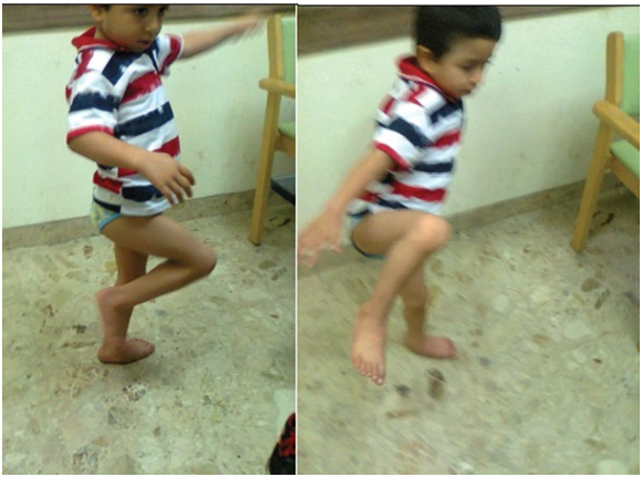
Before treatment the boy had undeniable difficulty in squatting and was about to fall when he squatted, while after treatment the boy was easily squatting without any difficulty (Figure 3).

Figure 3 The cerebrolysin treated boy was definitely able to stand momentarily on one foot for 2-3 seconds after treatment.
Both the first boy who was less affected and the cerebrolysin treated patient had difficulties in standing on one foot without support. However, although the second boy experienced some difficulty in standing on one foot without support after treatment, he was definitely able to stand momentarily on one foot for 2-3 seconds (Figure 3).
Recent research evidence suggests that cerebrolysin is endowed with interesting pharmacological properties that can make it useful in the treatment of various childhood neurologic and psychiatric disorders including mental retardation, pervasive developmental disorders including autism , brain atrophy, cerebral palsy, Rett syndrome and myelomeningocele.4–8
Cerebrolysin is a peptidergic therapeutic agent containing mainly biologically active neuro-peptides including brain-derived neurotrophic factor, glial cell line-derived neurotrophic factor, nerve growth factor, and ciliary neurotrophic factor. It has a nerve growth factor like activity on neurons, and growth promoting efficacy in different neuronal populations from peripheral and central nervous system.4-8 Cerebrolysin has been shown to have neurotrophic and neuroprotective properties effects in vitro and in vivo.
The effects of cerebrolysin seems to be similar to the pharmacological activities of naturally occurring nerve growth factors.4–8
Inhibition of apoptosis.
Improving synaptic plasticity and induction of neurogenesis.
Augmenting the proliferation, differentiation, and migration of adult subventricular zone neural progenitor stem cells, contributing to neurogenesis.
Induction of stem-cell proliferation in the brain.
This study suggested that intramuscular cerebrolysin is a promising agent for a new effective treatment of Wohlfart Kugelberg Welander syndrome.
A study enrolling more patients is recommended.
The author would like to express his gratitude for the parents of the patients for accepting publishing the photos of their children.
The authors report no conflicts of interest.
None.

©2020 Mosawi. This is an open access article distributed under the terms of the, which permits unrestricted use, distribution, and build upon your work non-commercially.