MOJ
eISSN: 2381-179X


Case Report Volume 13 Issue 4
1Investigación Terapia Celular y Metabolismo, Facultad de Medicina, Universidad de la Sabana, Colombia
2Unidad de Genética Médica, Policlínica Metropolitana de Caracas, Venezuela
Correspondence: Valentina Villarreal, Universidad de la Sabana, Campus del puente Comun Km. 7, Autopista Norte de Bogotá, Chía, Cundinamarca, Colombia, Tel 6018615555
Received: November 21, 2023 | Published: December 6, 2023
Citation: Valentina VH, Mariana S, Luis GC, et al. The new karyotypic alteration in a patient with cleft lip and palate-ectrodactyly and ectodermal dysplasia syndrome. MOJ Clin Med Case Rep. 2023;13(4):84-86. DOI: 10.15406/mojcr.2023.13.00445
Ectodermal dysplasias, a diverse group of disorders affecting ectodermal structures, represent challenges in diagnosis and management due to their varied clinical presentations. This article presents an exceptional case of Cleft Lip and Palate-Ectrodactyly and Ectodermal Dysplasia syndrome, highlighting a previously unreported chromosomal alteration (deletion on chromosome 21) in the absence of hereditary genetic backgrounds up to the third generation. The main syndrome, EEC, manifests with characteristic features such as ectrodactyly, cleft lip, and varied ectodermal anomalies. Genetic counseling addressed autosomal dominant inheritance, incomplete penetrance, and variable expression, emphasizing the importance of multidisciplinary follow-up for tailored medical care. A systematic literature review and comparison with the presented case underscored the significance of timely diagnosis and the necessity of genetic studies to inform parents about potential risks. The confirmed deletion on chromosome 21 adds a unique dimension to the case, reinforcing the need for a comprehensive approach involving a multidisciplinary healthcare team for optimal patient outcomes.
Keywords: ectrodactyly-ectodermal dysplasia-clefting syndrome, ectodermal dysplasias, embryonic ectoderm, ectodermal syndrome, developmental defects, epithelial-mesenchymal interaction
EEC, ectrodactyly-ectodermal dysplasia-clefting syndrome
Ectodermal dysplasias are a complex and heterogeneous group of disorders characterised by abnormalities in ectodermal structures, such as teeth, hair, nails, sweat glands, tear conduct among others. These malformations result from a defect in the development of tissues whose progenitor cells originate from the embryonic ectoderm. However, to be considered an ectodermal syndrome, at least 3 or 2 systems must be affected simultaneously.1-5
Regarding the incidence, it is estimated to be 0.7 to 1 per 100,000 live births, with more than 170 different subtypes that can be classified into two groups. Group 1 includes diseases in which a defect in the regulation of development and epithelial-mesenchymal interaction can be recognized, and group 2 includes cases where a defect in a protein involved in the maintenance of the cytoskeleton and other cellular structures.6
One of the main ectodermal syndromes is ectrodactyly-ectodermal dysplasia-clefting syndrome (EEC), which is the most common. Apart from affecting hair, skin, and teeth, patients may present with cleft lip and palate and ectrodactyly. Additionally, this syndrome is accompanied by characteristic facial features such as maxillary hypoplasia, a broad nasal tip, and thin nails, among others. Despite the wide variety of clinical presentations, ectodermal dysplasias seem to have a genetic origin.7-9
The following article aims to present a clinical case of a patient with cleft lip and palate syndrome, ectrodactyly, and ectodermal dysplasia, who has no history of hereditary genetic disease for up to 3 generations. Additionally, the patient has a chromosomal alteration that has not been reported for this syndrome thus far.
Ethics statement, consent, and permissions
This study was conducted in accordance with the Helsinki Declaration, and approved by the Institutional Review Board of Sabana University, Chia, Colombia. Written informed consent was obtained from the legal guardian of the infant.
Subject
The patient under study is a male infant, identified as E.C.A (Figure 1), born on March 20, 2022, and evaluated on August 17, 2023, at the age of 17 months. The patient was referred for a genetic evaluation due to multiple congenital anomalies observed during the neonatal period. Genealogical information was collected, and blood samples were taken for karyotyping, laboratory tests, including genetic analyses, and diagnostic imaging.

Figure 1 Clinical photographs of the infant (referred to as E.C.A) with Cleft Lip and Palate - Ectrodactyly and Ectodermal Dysplasia, whose karyotype revealed a deletion on the short arm of chromosome 21, in the region p11;pter.
Clinical history and physical examination
Relevant clinical history of the patient, including genealogical information, pregnancy, birth, and the first weeks of life, was collected. Notable findings include bilateral complete cleft lip and palate, ectrodactyly, adactyly in hands and feet, as well as other physical features and age-appropriate neurological development. A comprehensive physical examination of the patient was performed, with an emphasis on observed phenotypic characteristics. Craniofacial features, limb anomalies, ectodermal conditions such as a flat interciliary hemangioma, and any other observed anomalies were documented.
Genetic analysis and counseling
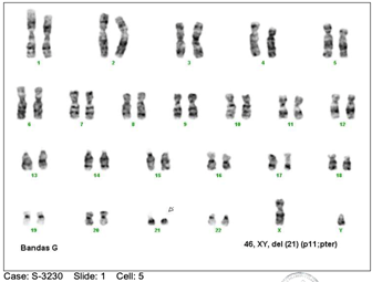
Genetic analysis was performed through a lymphocyte karyotype in peripheral blood on August 17, 2023, with the aim of identifying potential chromosomal abnormalities. The karyotype revealed a deletion on the short arm of chromosome 21, specifically in the region p11;pter (Figure 2).10 Genetic counselling was provided to the patient's parents to inform them about the genetic nature of the syndrome and its implications for future pregnancies. The discussion covered autosomal dominant inheritance, incomplete penetrance, and variable expression of the syndrome. The 50% risk of transmitting the mutation to offspring was mentioned, along with additional risks due to germline mosaicism.

Figure 2 Lymphocyte Karyotype in Peripheral Blood 46, XY, of (21) (p11;pter), an abnormal male karyotype with deletion of the short arm of chromosome 21.
Multidisciplinary follow-up and evaluation
The need for multidisciplinary follow-up for the patient was emphasised, involving assessment and treatment by orthopaedic surgeons, plastic surgeons, and dentists. This is essential for addressing the malformations and specific medical care needs associated with the syndrome.
Literature search and discussion
A systematic review was conducted on scientific articles using databases such as PubMed, MedLine, ScienceDirect, and The Cochrane Library Plus, with no date restrictions, in both Spanish and English languages. Study type was not limited, incorporating case reports and literature reviews. Abstracts were screened, and full articles were examined when necessary, ultimately including all articles featuring Labio-palatine Cleft Syndrome-Ectrodactyly and Ectodermal Dysplasia. These articles were then compared and discussed in relation to the case presented.
The physical examination revealed a male patient with normal brain development, broad forehead, ocular hypertelorism, flat interciliary hemangioma, palpebral obliquity, complete bilateral cleft lip and palate, ectrodactyly- adactyly with syndactyly in the right hand, third finger of the left hand with an amniotic band, amniotic band in the right leg with an increase in the volume of the right leg below the same, syndactyly in the left foot, nail dysplasia of the hands and feet, nail dysplasia of the hands and feet, third finger of left hand with amniotic band, amniotic band in right leg with increase of volume of this leg below it, adactyly in left foot, nail dysplasia of hands and feet, neurological development according to age. Lymphocyte karyotype in peripheral blood was 46, XY, del (21) (p11;pter), abnormal male karyotype with deletion of the short arm of chromosome 21, as the most relevant findings in the case.11-18
Presents an exceptional case of the described syndrome, emphasising the uniqueness of a chromosomal alteration not previously reported in this condition, coupled with the absence of hereditary genetic backgrounds up to the third generation. The importance of maintaining an open mind towards genetic and phenotypic diversity is underscored, even in the absence of apparent family history. Genetic counselling is provided to the parents, delving into the genetic nature of the syndrome, autosomal dominant inheritance, incomplete penetrance, and variable expression. The significance of multidisciplinary follow-up is highlighted to address the malformations and specific medical needs associated with the syndrome. Throughout the case, a literature search and discussion were conducted, comparing the presented case with existing literature on cleft lip palate syndrome, ectrodactyly, and ectodermal dysplasia. The physical characteristics of the patient are described in this case, and the deletion in the short arm of chromosome 21 is confirmed through lymphocytic karyotyping.15-18
EEC syndrome is an autosomal dominant disorder with incomplete penetrance and variable expression. It presents three cardinal signs characterised by ectrodactyly and syndactyly of the hands and feet, cleft lip with or without cleft palate, and various anomalies in ectodermal structures such as the skin, hair, teeth, and nails, genitourinary abnormalities, sensorineural hearing loss, among other possibilities.19-23
Given the above case, it highlights the importance of making a timely diagnosis, which includes taking an appropriate medical history and conducting relevant tests in patients showing alterations compatible with the cleft lip and palate, ectrodactyly, and ectodermal dysplasia syndrome. It underscores the significance of conducting a genetic study to advise parents of the risk that an affected person has of transmitting the mutation (50%) and the possibility of having another child affected by the same syndrome (4%). In this particular case, an unreported chromosomal alteration was observed (deletion of the short arm of chromosome 21) in a patient with no family history of the condition for up to three generations. However, based on the morphological characteristics, a diagnosis was made, aiming to improve the patient's quality of life through treatment guided by a multidisciplinary team, including a geneticist, paediatrician, orthopaedic surgeon, plastic surgeon, paediatric dentist, and nephrologist, among others.23-25
None.
The authors declare no conflict of interest.

©2023 Valentina, et al. This is an open access article distributed under the terms of the, which permits unrestricted use, distribution, and build upon your work non-commercially.