MOJ
eISSN: 2381-179X


Case Report Volume 13 Issue 1
1Professor of the Pneumology Service, Department of Clinical Medicine, Antonio Pedro University Hospital, Fluminense Federal University, Brazil
2Medical Doctor of Pneumology Service, Antonio Pedro University Hospital, Fluminense Federal University, Brazil
3Resident of Pneumology Service, Department of Clinical Medicine, Antonio Pedro University Hospital, Fluminense Federal University, Brazil
4Professor of Discipline of the Medical Clinic, Department of Clinical Medicine, Antonio Pedro University Hospital, Fluminense Federal University, Brazil
Correspondence: Marcos César Santos de Castro, Professor of the Pneumology Service. Department of Clinical Medicine, Antonio Pedro University Hospital, Fluminense Federal University, Rua Marques do Paraná, 303, 6o andar (MMC), Niterói, Rio de Janeiro, Brazil–CEP 24033-900, Tel +55-21- 26299210
Received: January 08, 2023 | Published: January 18, 2023
Citation: Castro MCS, Moreira VB, Nani ASF, et al. The importance of tomographic findings for the diagnosis of silicotuberculosis: case report. MOJ Clin Med Case Rep. 2023;13(1):1-3. DOI: 10.15406/mojcr.2023.13.00424
Silicosis is a pneumoconiosis characterized by fibrosis of the lung parenchyma caused by the inhalation of silica dust. Tuberculosis is an important and frequent infectious complication of silicosis, called silicotuberculosis. The risk of pulmonary tuberculosis in patients with silicosis is up to 40 times higher when compared to the general population. As the radiological findings are similar in both diseases, the diagnosis of silicotuberculosis is not always simple. We describe the case of a patient with silicosis associated with clinical and radiological findings compatible with tuberculosis.
Silicosis is the most prevalent pneumoconiosis in Brazil and in the world and is characterized by fibrosis of the lung parenchyma caused by the inhalation of silica dust.1,2 Several professional activities are related to silicosis such as, the mineral extraction industry (mining), the processing of minerals (grinding, cutting of semi-precious stones and quartz, crushing and grinding), the transformation industry (glass, foundry, ceramics) the cosmetic industry and mixed activities, involving prosthetics, well diggers and sandblasters.3-5 Recently, new activities have been described such as, textile fabric blasting (jeans)6 and working with artificial stones, widely used in the manufacture of kitchen and bathroom countertops.3,7-10
Several diseases are associated with silicosis such as, tuberculosis,11 mycobacterial infection, lung cancer,7,12 chronic obstructive pulmonary disease,5,7 autoimmune diseases (scleroderma, rheumatoid arthritis, systemic lupus erythematosus, ANCA vasculitis, in addition to renal impairment.3,7,13 The association of tuberculosis in a patient with silicosis is called silicotuberculosis. The risk of pulmonary tuberculosis is greater in individuals with a current or previous history of exposure to silica, with or without silicosis, with a risk up to 40 times greater than when compared to the general population.14 The diagnosis of silicotuberculosis is not always simple. This occurs because tuberculosis and silicosis both present clinically with cough and dyspnea and has a radiological characteristic of upper lobe predominant centrilobular nodules.5,10,15,16 In this article, we describe the case of a patient with silicotuberculosis, where the radiological aspect was fundamental for the diagnostic suspicion.
We describe the case of a 60-year-old male patient with hypertension and a previous history of smoking. The patient worked with sandblasting at a naval shipyard in the State of Rio de Janeiro (Brazil) for 20 years. He is regularly monitored at the occupational pneumopathies outpatient clinic of the Hospital Universitário Antônio Pedro (HUAP-UFF) due to his diagnosis of silicosis.
The patient reported the onset of fever, predominantly in the late afternoon, for approximately 2 months, associated with night sweats. The patient even related worsening of dry cough with progression to productive cough after 1 month of onset of fever, in addition to worsening of dyspnea. He reported unintentional weight loss of 6 kg in 4 months. Physical examination revealed diffuse wheeze in both lungs.
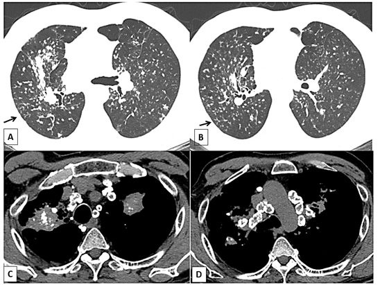
Leukogram and biochemistry showed normal results except elevated C-reactive protein. Computed tomography showed diffuse interstitial pattern, with small nodules and conglomerated masses in the upper lobes of both lungs. Hilar and mediastinal lymph nodes were enlarged with extensive calcification, with the characteristic eggshell pattern. In addition, the tree-in-bud sign was more observed in right upper lobe of the lung (Figure 1).
In view of the clinical history and the radiological finding of the tree-in-bud sign on chest computed tomography, acid-fast bacilli and microbial cultures were performed from spontaneous sputum. Sputum examination demonstrated acid-fast bacilli and subsequently culture for mycobacteria showed the presence of Mycobacterium tuberculosis. We started bronchodilators, rifampicin, isoniazid, pyrazinamide, and ethambutol and patient improved thereafter.
The diagnosis of tuberculosis in a patient with silicosis is called silicotuberculosis. The risk of pulmonary tuberculosis is greater in individuals with a current or previous history of exposure to silica, with or without silicosis, with a risk up to 40 times greater than when compared to the general population.14,17
This association has already been observed and described many years ago. In 1911, Domenico Cesar-Bianchi and Luigi Devoto observed the causal association between exposure to silica and tuberculosis in experiments with animals exposed to silica.18-20 In 1961, Paul et al. conducted a study of copper miners with silicosis in Zimbabwe. The authors observed a 30 times greater risk of developing tuberculosis in patients with silicosis when compared to non-silicotic patients.21
In 1990, Sherson and Lander conducted a longitudinal study on silicotic and non-silicotic patients of 5,579 foundry workers. The authors concluded that tuberculosis in this sample was 3 times more prevalent among workers with silicosis.22 In 1998, Hnizdo and Murray studied 2,255 gold miners in South Africa. They observed that the diagnosis of tuberculosis occurred after the diagnosis of silicosis in 90% of the cases.22
Several mechanisms seem to be involved in the increased risk of tuberculosis in patients with silicosis. Silica dust modify the cellular immune response of the lungs, impairing the metabolism and function of pulmonary macrophages. This exposure also promotes the death of macrophages in addition to compromising the lymphatic drainage of the pulmonary parenchyma consequent to fibrosis.23
In 1968, Allison and Hart suggested that the change in the permeability of the phagosome membrane by silica would allow the bacilli to penetrate and multiply more quickly in the cytoplasm and that intracellular silica would trigger a metabolic alteration in the macrophage, facilitating bacillary growth. The same authors also described a possible chemical effect of silica on bacillary growth, causing macrophage toxicity and leading to a decrease in bacillary lysis. Another mechanism described by the authors refers to the fibrosis caused in the lung parenchyma in silicosis, altering the local lymphatic drainage and allowing the bacilli to stay longer in the lung tissue.24
In a study conducted by Pasula in 1999 and in another work by Gold, published in 2004, both groups demonstrated that the higher prevalence of tuberculosis in patients with silicosis could be related to surfactant protein A, which was shown at high levels in the Bronchoalveolar lavage of silicotics. Excess of this protein seems to be associated with greater susceptibility to tuberculosis, possibly because it allows mycobacteria to enter alveolar macrophages without triggering cytotoxicity and because it inhibits the formation of reactive nitrogen intermediates by activated macrophages.25,26
In patients with silicosis and concomitant signs and symptoms compatible with tuberculosis, the diagnosis of tuberculosis should always be considered and, preferably, ruled out.27,28 Some findings should serve as a warning for the presence of tuberculosis, especially in cases where there are few symptoms, or a slight worsening of silicosis symptoms. Therefore, the presence of some radiological findings is important for the suspicion of tuberculosis in cases of silicosis.
Simple silicosis is radiologically characterized by the presence of bilaterally symmetrical micronodular interstitial infiltrates, predominantly in upper lobes. These nodules have a centrilobular pattern of distribution, but may also have a perilymphatic distribution. The presence of small airway filling, with images of a tree-in -bud, is not characteristic of simple silicosis and should always suggest an associated mycobacterial infection, as demonstrated in the case presented (Figure 1).7,18,20

Figure 1 Typical case of complicated silicosis (massive progressive fibrosis) in a 60-year-old man. (A) and (B) High resolution tomography scan of the chest (window of parenchyma): presence of small nodules and opacities in the posterior of the upper lobes. (arrows): Tree-in-bud pattern. (C) Mediastinal window illustrating a conglomerated mass with punctiform calcifications in the posterior region of the upper lobes, and adjacent pleural thickening of the right lung and the calcifications of the lymph nodes. (D) Mediastinal window illustrating egg-shell-type calcifications on the periphery of the lymph nodes.
Another important fact is that pleural involvement in silicosis is extremely uncommon in silicosis alone. The presence of pleural effusion in a patient exposed to silica crystals for dust, with or without silicosis, should not be justified by silicosis alone. The possibility of pleural tuberculosis or associated neoplastic disease must always be ruled out.7,18,20
The radiological progression is bilateral and slow in silicosis. Rapid progression, mainly associated with new signs and/or symptoms, or asymmetrical radiological progression of opacities, should lead to the possibility of associated tuberculosis.7,18,20
In addition, in silicosis, cavitation of conglomerate masses is rarely observed due to simple progression of the disease. Although there are authors who describe a 10% prevalence of conglomerated masses cavitation in patients with complicated silicosis without tuberculosis,20 in countries with a high incidence and prevalence of tuberculosis, cavitation of silicotic masses should always lead to the possibility of associated tuberculosis.7,18,20
It is also important to point out that exposure to silica, even in the absence of silicosis, increases the risk of tuberculosis by up to 4 times. In the workers with increase risk of exposure and inhalation of silica dust and the presence of an infectious syndrome of respiratory origin associated with pulmonary parenchymal infiltrate, the suspicion of pulmonary tuberculosis should also be considered.23,28
Tuberculosis is an important infectious complication in patients with silicosis. In addition to the clinical picture, radiological findings are essential for suspecting associated tuberculosis. Small nodules and large opacities can be observed in patients with silicosis, but tree-in-bud pattern is not characteristic of this disease. Tuberculosis should always be investigated in a patient with silicosis and infectious respiratory symptoms with a tree-in-bud pattern.
None.
The authors declare that they have no conflict of interest.

©2023 Castro, et al. This is an open access article distributed under the terms of the, which permits unrestricted use, distribution, and build upon your work non-commercially.