MOJ
eISSN: 2381-179X


Case Report Volume 8 Issue 1
1Orthopedic Surgery Resident, Postgraduate School in Naval Health, Centro Medico Naval, Universidad Naval, Mexico
2Orthopedic Surgery Chief, Professor of the residence of Traumatology and orthopedics, High Specialty Naval Health Hospital, Centro Medico Naval, Universidad Naval, Mexico
3Orthopedic Surgeon with high specialty in acetabulum, hip and pelvis, Postgraduate School in Naval Health, Centro Medico Naval, Universidad Naval, Mexico
4Anesthesiologist, Hospital General Regional #2 of Traumatology and Orthopedics, Mexico
5Postgraduate School in Naval Health, Centro Medico Naval, Universidad Naval, Mexico
6Orthopedic Surgeon, Centro Medico Dalinde, Mexico
7General Surgery Department, Hospital General Regional 1A, Rodolofo de Mucha Macias, IMSS, Mexico
Correspondence: Luis Angel Medina Andrade, General Surgery Department, Hospital General Regional, México
Received: February 07, 2018 | Published: February 15, 2018
Citation: Arroyo-Aparicio JY, Jesús BD, Cornejo MIG, et al. Stress fracture of the femoral neck in a marine trainee female: case report. MOJ Clin Med Case Rep. 2018;8(1):26-28. DOI: 10.15406/mojcr.2018.08.00233
Stress fractures of the femoral neck are uncommon, accounting for 5% of all stress fractures, being one of the most severe and disabling complications in training military personnel. This could bring potential serious consequences if an early diagnosis and treatment is not accomplished.
We present the case of a neck stress fracture in a young 25-year-old military female, treated by reduction and internal fixation. She was followed up for two years and is reported without a vascular necrosis of the femoral head, without osteoarthrosis data and adequate bone consolidation.
This article aims to provide the first contact physician with the necessary tools for the early recognition of this type of injury so that it could predict the progression and displacement of the fracture, as well as significantly reduce the risk of devastating complications such as a vascular necrosis.
Keywords: femoral fracture, marine trainee, stress fracture
A stress fracture is the one secondary to overuse, that results for repetitive damage with load cycles lower than the stress required fracturing the bone, resulting in micro fractures. They are related to intense exercise, especially that involving repetitive weight-bearing loads, like military training or running. They occur when bone is unable to repair the repetitive damage.1
This kind of fractures is especially frequent in military training compared with athletic programs and represents the most important cause of hospital admission during military training with the subsequent loss of life quality.1
The risk factors are multiple, but some of them had been clearly identified as intrinsic or extrinsic. The first include aerobic fitness, hormonal and nutrition factors and gender. The gender and physical conditioning prior to the time of beginning training have been identified as an important risk factor, and extrinsic factors include factors related to physical training, especially volume and intensity of training.1
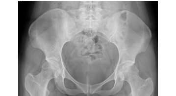
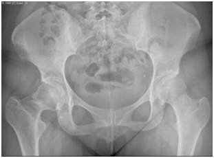
Female patient of 25years-old, active military, size 165cm, weight 52kg, BMI 19.1, with antecedent of military training for about 5 to 6hours a day, 6days a week, for 2months prior to the onset of symptomatology. She refers with discrete right inguinal pain, which increased with physical effort, radiating towards the proximal third of the thigh and right buttock. It is evaluated by a general physician and diagnosed as a muscle contraction, without abnormalities in the x-ray exam (Figure 1) indicating treatment with analgesics and non-steroidal anti-inflammatory drugs, without suspension of physical activities. She comes back to the emergency department of the Naval General Hospital of High Specialty one week later, referring an acute increase in the symptoms and limitation of movement due to pain, an antero posterior pelvic radiograph is performed, without data of apparent bone lesion (Figure 2).

Figure 1 24/08/2015 anteroposterior pelvic radiograph, taken on the day of admission, with no data of apparent bone lesion.

Figure 2 01/09/2015 anteroposterior pelvic radiograph, taking in the follow up, without no data of apparent bone lesion.
General surgery service is consulted by the inguinodynia diagnosis, but the need for surgical treatment is discarded, and patients are discharged with a diagnosis of muscle contracture of the hip abductors, going under treatment with physical therapy, rehabilitation, and analgesics.
After a month without improvement, the orthopedics service is consulted again. At physical exam, the patient presented shortening of about 2cm of the right pelvic limb and limitation due to pain in the right hip mobility arches. A new antero posterior pelvic radiograph is requested, finding a right femoral trans-cervical continuity solution, without displacement, with varus angulation and cervical-diaphyseal angle of 117º (Figure 3).

Figure 3 14/10/2015 pelvic anteroposterior radiography one month after the initial date of admission. A right femoral transcervical continuity solution was observed, with no varus angulation and a cervical-diaphyseal angle of 17º.
During the hospital stay, hormonal profile and bone densitometry were performed and bone metabolism alterations discarded. Magnetic resonance image of the lumbar spine was performed to rule out a neuropathic pathology but was negative too.
After study protocol patient was treated with surgical reduction and internal fixation with two cannulated screws, recovering diaphyseal cervical angle at 130º (Figure 4), and starts with partial discharge after two weeks of the surgery. Bone callus formation is identified two months after surgery (Figure 5), initiating total support of the limb 3months after surgery, and at 6months he fully reincorporates her activities with excellent clinical evolution. After two years of follow-up on his control radiographs, no avascular necrosis of the femoral head was observed and adequate bone remodeling appreciated (Figure 6).
A stress fracture is the one that occurs secondary to the inability of the bone to support a repetitive applied force of a lower magnitude than the fracture threshold but generating gradual alteration of the bone structure. The osseous tissue has the capacity to remodel and adapt to changes in the magnitude and distribution of the forces acting on it. However, this process is insufficient compared to the repeated application of high loads, which are capable of generating trabecular microfractures.2
Fatigue fracture of the femoral neck mainly affects young patients, it appears in the military during periods of intensive instruction and in athletes who perform an increase in their frequent physical activity, although stress fractures are relatively common in this type of patient, it is estimated that only 1% of these fractures affect the femoral neck, there is usually no previous traumatic event and when there is no displacement of the fracture the symptoms can be very unclear and non-specific, and frequently the initial radiographs are normal, which leads to a delay in the diagnosis and treatment.2,3 The most important for the diagnosis of this injury is to suspect its existence, being the most common symptoms the inguinal pain that is exacerbated by physical activity and decreases with rest, with an special increase incidence in females.1,3
The delay in the diagnosis of this condition is common, several studies report an average of 14weeks of delay for the diagnosis, two weeks more than in the present case.4,5 The displacement of stress fractures is the factor of greatest determination and prognosis for patient evolution, is responsible for 60% of the reduction of physical and sports activities in the patient, and carries a 30% incidence for a vascular necrosis, one of the most devastating results for this kind of population and their lifestyle, with the subsequent importance of medical staff knowledge about this pathology and initial presentation, for an early suspect and promptly treatment.6
Early recognition of stress fractures in high risk population could predict the progression and displacement of the fracture and significantly reduce the risk of devastating complications such as a vascular necrosis, especially in young patients with the previously mentioned lifestyle. This document highlights the significance of stress fractures of the femoral neck in the military in intense training, alerting first-contact physicians to suspect this type of injury in a high-risk population, which could prevent serious sequelae in these patients.
None.
The author declares no conflict of interest.

©2018 Arroyo-Aparicio, et al. This is an open access article distributed under the terms of the, which permits unrestricted use, distribution, and build upon your work non-commercially.